SLC27A2 / Long-chain fatty acid transport protein 2 · IHC design guide

Design Immunohistochemistry for SLC27A2

Plan SLC27A2 chromogenic IHC in paraffin sections using cytoplasmic proximal tubule and hepatocyte staining as reference patterns (HPA tissue IHC). This guide covers consistent fixation and the catalog antibody’s 2–5 μg/ml IHC range (datasheet A07140-1).

Evidence assembled Oct 2026 · For research use; verify linked source records and product datasheet before use
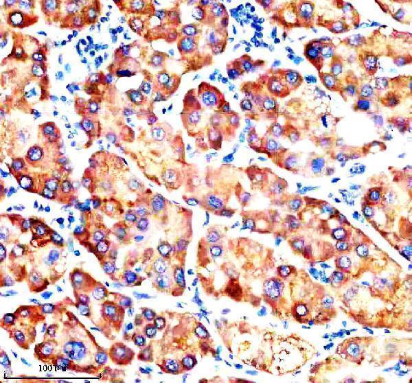
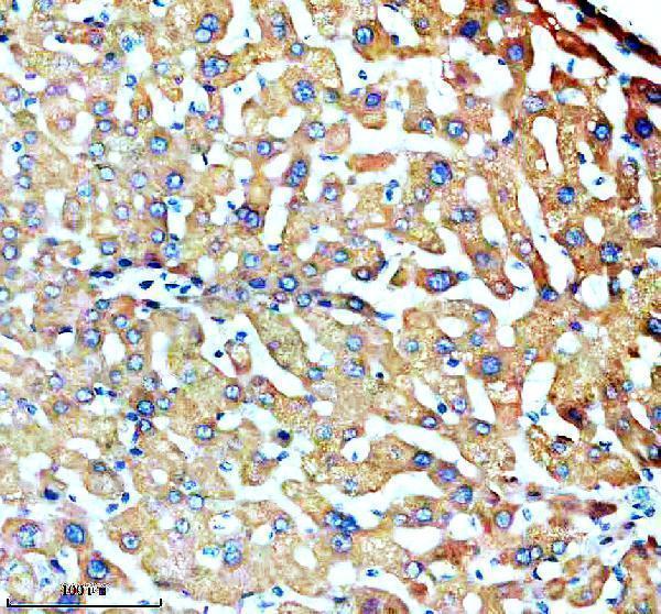
Immunohistochemistry protocol sheet for SLC27A2 (IHC for SLC27A2): expected localisation Cytoplasmic in kidney and liver (HPA tissue IHC); ER and peroxisome membranes (UniProt), antibody A07140-1, validated IHC image, and IHC protocol steps
Printable SLC27A2 IHC protocol sheet — expected localisation Cytoplasmic in kidney and liver (HPA tissue IHC); ER and peroxisome membranes (UniProt), antibody A07140-1, controls and protocol steps. Open the full SLC27A2 IHC guide →

SLC27A2 Immunohistochemistry Experimental Design Guide

Expected localisation, validated protocols, controls and antibodies — the at-a-glance facts below, then the full design guide.

Must know before staining
Expected localisation Cytoplasmic in kidney and liver (HPA tissue IHC); ER and peroxisome membranes (UniProt)
Staining pattern Cytoplasmic proximal tubules (high); hepatocytes (medium) (HPA tissue IHC)
Antigen retrieval EDTA pH 8.0 HIER, heat-mediated (datasheet A07140-1)
Positive control ⓘ Kidney+4 more · see all
Negative control ⓘ Adipose tissue+4 more · see all
Important caveats
Reasons your staining may differ from the expected pattern.
Fixation Keep fixation consistent across sections (standard IHC practice; not target-specific)
Caveat Hepatocytes stain less strongly than proximal tubules (HPA tissue IHC)
Regulation Kidney and liver are tissue enhanced (HPA tissue IHC)
Isoform / epitope 2 isoforms; check epitope position against lumenal and cytoplasmic regions (UniProt)
Section 1

Recommended SLC27A2 IHC & IF Protocols

Compare the catalog antibody’s IHC-P protocol (datasheet: EDTA pH 8.0 retrieval) with published protocols for ccRCC (PMC12249168) and breast tissue (PMC11098904).

Recommended immunohistochemistry (IHC-P) protocol parameters
SampleParaffin-embedded human liver cancer tissue; fixative not specified (datasheet A07140-1)
FixationImage fixative and duration unreported (datasheet A07140-1); verify before use.
Sectioning4–5 µm sections on charged slides (standard)
DeparaffinisationXylene, graded ethanol series to water (standard)
Antigen retrievalHeat retrieval: EDTA pH 8.0 (datasheet A07140-1); 20 min, 95–100 °C (standard)
Peroxidase block3% H2O2, 10 min, room temperature (standard)
Blocking10% goat serum (datasheet A07140-1)
Primary antibodyRabbit anti-SLC27A2, 2-5 μg/ml (datasheet A07140-1)
Primary incubationOvernight at 4 °C (datasheet A07140-1)
DetectionHRP-conjugated secondary, DAB chromogen (datasheet A07140-1)
CounterstainHematoxylin, blue, dehydrate and mount (standard)
Expected resultSLC27A2-positive staining in proximal tubules (cell body) of kidney (HPA tissue IHC: High). HPA tissue profile: Cytoplasmic expression mainly in renal tubules of kidney and hepatocytes in liver. No signal in the no-primary control.
💡Decision noteStart with EDTA pH 8.0 heat retrieval (datasheet). Published alternatives are high-pH retrieval for ccRCC (PMC12249168) and microwave citric acid retrieval for breast tissue (PMC11098904).
Section 2

What Is the Expected SLC27A2 Staining Pattern?

SLC27A2 is annotated at the endoplasmic reticulum, peroxisome and cell membranes, with three transmembrane segments (UniProt O14975 topology). In paraffin section IHC, expect chiefly cytoplasmic staining in kidney renal tubules and liver hepatocytes (HPA: tissue IHC profile). HPA rates the tissue staining pattern Enhanced, citing consistency between antibody staining and RNA expression; that rating does not establish the pattern in every specimen (HPA: reliability description).

What am I looking at on my slide?
Strong cytoplasmic staining in kidney proximal tubule cell bodies; moderate staining in hepatocytes.This matches the strongest listed tissue and a second expected positive tissue (HPA: proximal tubules High; hepatocytes Medium). Compare the cell bodies with neighboring structures, and score each cell population separately rather than assigning one intensity to the whole section (general IHC practice).
Signal is confined to nuclei, or a uniform rim outlines nearly every cell.Treat this as a compartment mismatch until checked: the reported tissue IHC profile is chiefly cytoplasmic, although SLC27A2 has membrane locations (HPA: tissue IHC profile; UniProt O14975 subcellular location). Check a positive control and the staining distribution before interpreting either appearance as specific.
Strong staining appears in adipocytes, adrenal glandular cells or bone marrow hematopoietic cells.Those listed cell populations were not detected in HPA tissue IHC (HPA: adipose tissue, adrenal gland and bone marrow). Unexpected signal may reflect cross-reactivity or chromogenic detection activity (general IHC practice). Evaluate its location and run a no-primary control before assigning it to SLC27A2.
A diffuse wash of color covers positive and expected-negative structures alike.A widespread wash is less persuasive than staining confined to the expected cells (HPA: kidney and liver tissue IHC profile). Excess background can arise from blocking, washes or detection chemistry (general IHC practice). Compare the no-primary control and assess signal against local background.
Kidney proximal tubules show no convincing staining.This fails an expected strong-positive reference (HPA: kidney proximal tubules High). Check that the relevant tubules are present, then review the catalog antibody's IHC-P instructions, retrieval and detection controls (general IHC practice). Absence alone does not establish that the specimen lacks SLC27A2.
💡Expected SLC27A2 appearanceCall the result positive when kidney proximal tubule cell bodies show strong cytoplasmic signal, with hepatocytes providing a moderate comparator (HPA: kidney High; liver Medium); widespread nuclear or expected-negative-cell staining warrants investigation (HPA: tissue IHC profile; general IHC practice).
How each factor affects the staining
Membrane topology and IHC appearanceSLC27A2 has three transmembrane segments and annotated endoplasmic reticulum, peroxisome and cell membrane locations (UniProt O14975). Its tissue IHC profile is chiefly cytoplasmic (HPA: tissue IHC profile). Interpret that profile at the light-microscopy level; it does not identify an individual organelle.
Tissue and cell selectionKidney proximal tubules are High and hepatocytes Medium; several other listed cell populations range from Medium to Low or are not detected (HPA: tissue IHC levels). Use the named cell population when comparing sections: a whole-organ label does not imply uniform staining throughout that organ.
Antibody validationHPA026089 is rated Enhanced for IHC, and HPA describes high consistency between antibody staining and RNA expression (HPA: antibody IHC status; reliability description). This supports the reported tissue pattern, but unexpected staining in a new specimen still needs slide controls (general IHC practice).
IF/ICC Q&A: what pattern can this guide predict?HPA summarizes the subcellular location as Membrane but supplies no main location or cell-line ICC-IF images (HPA: subcellular record). UniProt lists several membrane locations (UniProt O14975). These records do not establish a specific IF/ICC puncta pattern or an IF/ICC protocol.
Why is my staining missing, weak or wrong?
SituationLikely causeNext action
No signal in the expected kidney positive control.The tissue or target cell population may be absent from the section, or an IHC workflow step may have failed (general IHC practice); proximal tubules are an expected High population (HPA: kidney).Confirm proximal tubules on the counterstained section. Review the catalog antibody's IHC-P instructions and check retrieval and detection controls before interpreting the specimen as negative (general IHC practice).
Signal appears mainly in nuclei.The compartment differs from HPA's chiefly cytoplasmic tissue pattern (HPA: tissue IHC profile); nonspecific staining or detection background is possible (general IHC practice).Compare nuclear color with cytoplasmic color in proximal tubules and hepatocytes, then inspect a no-primary control. Do not score isolated nuclear signal as the expected pattern (HPA: tissue IHC profile; general IHC practice).
Unexpected cells stain as strongly as proximal tubules.A cell population reported as not detected may show cross-reactivity or detection activity (HPA: listed negative populations; general IHC practice).Identify the stained cell type, compare its distribution with HPA's cell-specific tissue entries, and check the no-primary control. Keep unexpected staining separate from the positive-cell score (general IHC practice).
Color is diffuse across the section.Background from blocking, washing or chromogenic detection may obscure the cell-specific pattern (general IHC practice; HPA: tissue IHC profile).Inspect the no-primary control, review blocking and wash steps, and assess whether proximal tubule cell bodies remain distinct from adjacent background (general IHC practice; HPA: kidney proximal tubules High).
Liver stains, but less intensely than kidney.That ordering is compatible with HPA's listed levels: hepatocytes Medium and kidney proximal tubules High (HPA: liver and kidney tissue IHC).Score hepatocytes and proximal tubules as separate cell populations. Interpret each against its own background and section controls rather than requiring equal intensity (HPA: tissue IHC levels; general IHC practice).
A faint signal appears in colon glandular cells.Colon glandular cells are listed as Low, so a faint result may fit the reported pattern (HPA: colon tissue IHC); background can also produce faint color (general IHC practice).Compare glands with nearby background and a no-primary control. Use kidney proximal tubules as the stronger reference before calling faint colon color target-associated (HPA: colon Low; kidney High; general IHC practice).

Sample controls for SLC27A2 IHC & IF

🧪Run kidney first: proximal tubule cell bodies should stain strongly (HPA: High in kidney proximal tubules). Use adipose tissue as the negative tissue, with adipocytes expected to be unstained (HPA: Not detected in adipocytes); on the kidney slide, assess adjacent cells without visible staining as relative internal background rather than assuming every non-proximal cell is negative (HPA: High in proximal tubules).
Positive control tissue: Kidney (Proximal tubules (cell body), HPA High)
Negative control tissue: Adipose tissue (HPA Not detected)
ICC-IF cell lines (HPA subcellular resource): HPA carries no ICC-IF cell line for SLC27A2; derive a cell-line control from the positive tissue's cell type (Proximal tubules (cell body)) and confirm it by RNA or western blot first.
Technical controls: Include a no-primary, secondary-only control, a host- and clonality-matched isotype control, and a SLC27A2 knockout sample or peptide-block control if the immunizing peptide is available (standard IHC practice; selected-SKU caption: rabbit primary antibody). Block endogenous peroxidase in kidney sections before HRP/DAB detection, and assess tubular background separately from specific staining (standard IHC practice; selected-SKU caption: HRP/DAB detection).
⚠️Feasibility: A target-specific fixation window or fixation effect is unreported, and the selected-SKU paraffin-section caption does not state the fixative (selected-SKU caption: fixative not stated). The reported IHC procedure uses heat-mediated EDTA retrieval at pH 8.0, so begin with that condition while checking whether retrieval is needed in the actual specimens (selected-SKU caption: EDTA retrieval, pH 8.0). The supplied evidence does not establish whether frozen sections or IF are easier; kidney tubular background may complicate interpretation, and IF requires checking autofluorescence (HPA: High in kidney proximal tubules; standard IHC/IF practice).

HPA tissue IHC evidence for SLC27A2

Comprehensive Human Protein Atlas IHC scoring per tissue (reliability: Enhanced — High consistency between antibody staining and RNA expression data.). Rows are taken directly from the HPA tissue chart — click any row's HPA link to view the source.

Positive expression · recommended positive controls

TissueCell typeLevelEvidenceSource
Kidney Proximal tubules (cell body) High Protein (IHC) HPA →
Bronchus Ciliated cells (cell body) Medium Protein (IHC) HPA →
Epididymis Glandular cells Medium Protein (IHC) HPA →
Fallopian tube Ciliated cells (ciliary rootlets) Medium Protein (IHC) HPA →
Liver Hepatocytes Medium Protein (IHC) HPA →

Undetected expression · recommended negative controls

TissueCell typeLevelEvidenceSource
Adipose tissue Adipocytes Not detected Protein (IHC) HPA →
Adrenal gland Glandular cells Not detected Protein (IHC) HPA →
Bone marrow Hematopoietic cells Not detected Protein (IHC) HPA →
Breast Adipocytes Not detected Protein (IHC) HPA →
Caudate Glial cells Not detected Protein (IHC) HPA →
Section 3

Advanced SLC27A2 IHC Tips

Troubleshoot SLC27A2 staining in paraffin sections by checking retrieval, cellular pattern, controls, and scoring before interpreting chromogenic signal.

What retrieval should I start with for SLC27A2 in paraffin sections?
Start with heat-mediated retrieval in EDTA at pH 8.0 for the catalog antibody (datasheet A07140-1). The product image documents this condition in a paraffin-embedded human liver cancer section, followed by 2 μg/ml primary antibody overnight at 4°C (datasheet A07140-1). If staining is weak, compare a modestly adjusted heating time on matched sections while keeping buffer, antibody concentration, and detection constant (standard IHC practice). Check hepatocytes or renal proximal tubules as expected positive cell populations, and record whether improved signal retains a cellular pattern (HPA: hepatocytes medium; proximal tubules high). Excessive retrieval can damage morphology and complicate compartment-level interpretation (standard IHC practice).
Could fixation explain weak or uneven SLC27A2 staining?
Target-specific fixation sensitivity is unknown: the selected paraffin-section image does not state a fixative (datasheet A07140-1: fixative not stated). Record the fixative, fixation duration, section age, and processing history before comparing staining across specimens (standard IHC practice). If signal varies, stain adjacent sections from comparably processed blocks in the same run using EDTA pH 8.0 retrieval (datasheet A07140-1). Include a positive tissue with identifiable hepatocytes or renal proximal tubules, then compare signal and preserved morphology together (HPA: hepatocytes medium; proximal tubules high). Neither the reported tissue pattern nor the protein’s membrane topology establishes a fixation requirement (HPA tissue IHC; UniProt O14975 topology).
Where should convincing SLC27A2 staining appear within a section?
Assess signal in the cell body of renal proximal tubules and in hepatocytes; HPA reports high and medium staining, respectively (HPA tissue IHC). A cytoplasmic-appearing chromogenic pattern is compatible with the reported tissue stain and the protein’s endoplasmic-reticulum and peroxisome membrane locations (HPA tissue IHC; UniProt O14975 subcellular location). UniProt also lists cell membrane and microsome locations, so a single sharply defined compartment is not required by the record (UniProt O14975 subcellular location). Compare candidate signal with cell boundaries and nuclear counterstain at the same magnification, and reject pigment or deposits lacking a cellular pattern (standard IHC practice). Document compartment and cell type separately when scoring mixed tissue (standard IHC practice).
Can this antibody distinguish SLC27A2 isoforms or reveal epitope masking?
The record lists 2 SLC27A2 isoforms, but the supplied product caption does not identify the antibody epitope or establish isoform selectivity (UniProt O14975 isoforms; datasheet A07140-1). The protein has 3 transmembrane segments and alternating lumenal and cytoplasmic regions, making epitope position relevant to access in sections (UniProt O14975 topology). Its annotated N6-acetyllysine at residue 291 and phosphothreonine at 577 do not establish an antibody-specific staining effect (UniProt O14975 modified residues). If an isoform-specific claim matters, obtain the immunogen sequence and map it against both isoforms before assigning identity (standard antibody validation practice). Compare retrieval conditions only after preserving the documented EDTA pH 8.0 condition as the reference (datasheet A07140-1).
How should I pilot SLC27A2 immunofluorescence alongside a cell-type marker?
Treat IF as a separate validation experiment; the supplied product evidence demonstrates chromogenic staining in a paraffin section, not IF performance (datasheet A07140-1). Multiplex with a validated hepatocyte or renal proximal-tubule marker so the SLC27A2 channel can be assessed in expected cells (HPA tissue IHC; standard IF practice). Choose a fluorophore channel with low tissue autofluorescence, and collect unstained and single-stain controls before interpreting overlap (standard IF practice). Optimise permeabilisation against the mapped epitope: residues 28–106 and 283–620 are cytoplasmic, whereas 1–4 and 128–261 are lumenal (UniProt O14975 topology). Because the antibody epitope is unspecified, test access empirically and avoid inferring colocalisation from merged images alone (datasheet A07140-1; standard IF practice).
How can I reduce diffuse brown signal without losing true SLC27A2 staining?
The documented tissue protocol used 10% goat serum blocking, 2 μg/ml primary antibody overnight at 4°C, and a peroxidase-linked secondary for 30 minutes at 37°C (datasheet A07140-1). If background rises, compare a primary-omission control, shorten chromogen development, and titrate the primary on adjacent sections (standard IHC practice). Block endogenous peroxidase before HRP/DAB detection and inspect unstained tissue for pigment that could resemble reaction product (standard chromogenic IHC practice). Preserve an expected positive compartment while adjusting one variable at a time; renal proximal tubules provide a high-expression reference (HPA: proximal tubules high). Diffuse staining in nuclei or extracellular spaces should prompt a control review before biological interpretation (standard IHC practice).
How should I score SLC27A2 across regions with different cell composition? ⚠ ANSWER MARKED FOR VERIFICATION
Define the cell population and region before scoring; HPA reports high staining in renal proximal tubules and medium staining in hepatocytes (HPA tissue IHC). For hepatocyte-rich or tubular regions, record the percentage of positive target cells and a prespecified intensity score, then calculate an H-score if intensity grading is reproducible (standard IHC scoring practice). Where cell counts are impractical, measure positive area or optical density per mm² of viable target-cell area using one threshold across slides (standard image-analysis practice). Normalise to the relevant target-cell population, rather than total section area, when tissue composition differs (standard IHC scoring practice). Exclude folds, edges, necrosis, and blank spaces using identical rules across specimens (standard IHC scoring practice).
What separates genuine SLC27A2 signal from staining artefact?
Give greatest weight to reproducible cell-associated staining in renal proximal tubules or hepatocytes, which match the reported tissue pattern (HPA: proximal tubules high; hepatocytes medium). A cytoplasmic-appearing pattern can fit membrane-associated SLC27A2, whose annotated sites include endoplasmic reticulum and peroxisome membranes (HPA tissue IHC; UniProt O14975 subcellular location). Signal confined to nuclei, section edges, necrotic areas, or unexpected cell populations needs independent validation before assignment to SLC27A2 (standard IHC interpretation practice). Check primary-omission and endogenous-peroxidase controls when brown deposits persist outside intact cells, because the documented detection uses HRP and DAB (datasheet A07140-1; standard IHC practice). Report cell type, compartment, intensity, and control behaviour together, especially in heterogeneous sections (standard IHC reporting practice).
Boster reagents

Best SLC27A2 / Long-chain fatty acid transport protein 2 IHC Antibodies

A07140-1 has IHC images from paraffin-embedded human liver and liver cancer sections (image captions). Listed reactivity covers human, mouse, and rat (catalog reactivity); no IF image is supplied (catalog IF image list).

Real IHC data IHC analysis of FATP2/SLC27A2 using anti-FATP2/SLC27A2 antibody (A07140-1). FATP2/SLC27A2 was detected in a paraffin-embedded section of human liver cancer tissue. Heat mediated antigen retrieval was performed in EDTA buffer (pH 8.0, epitope retrieval solution). The tissue section was blocked with 10% goat serum. The tissue section was then incubated with 2 μg/ml rabbit anti-FATP2/SLC27A2 Antibody (A07140-1) overnight at 4°C. Peroxidase Conjugated Goat Anti-rabbit IgG was used as secondary antibody and incubated for 30 minutes at 37°C. The tissue section was developed using HRP Conjugated Rabbit IgG Super Vision Assay Kit (Catalog # SV0002) with DAB as the chromogen.
Anti-FATP2/SLC27A2 Antibody ®
Cat # A07140-1

A07140-1 was shown on paraffin-embedded human liver and liver cancer sections (A07140-1 image captions). IHC is a listed application, and reactivity includes human, mouse, and rat; IF/ICC is not listed (catalog applications and reactivity).

Which to pick: For paraffin-section IHC, choose A07140-1: its images document human liver and liver cancer sections, EDTA retrieval at pH 8.0, and 2 μg/ml primary antibody; the fixative is unreported (A07140-1 image captions). No SKU in this payload has IF/ICC validation or an IF image, so there is no supported IF/ICC pick (catalog applications; IF image list). For mouse or rat IHC, A07140-1 lists both species as reactive, but its supplied IHC images show human sections only (catalog reactivity; A07140-1 image captions).

Each figure is that product's own IHC / IF validation image from its datasheet.

References

  1. UniProt Consortium. UniProt entry O14975 (S27A2_HUMAN, Long-chain fatty acid transport protein 2).
  2. Human Protein Atlas. SLC27A2 tissue IHC expression (reliability: Enhanced).
  3. Human Protein Atlas. SLC27A2 subcellular location (ICC-IF): Membrane.
  4. Human Protein Atlas. SLC27A2 antibody validation summary (1 antibodies).
  5. Integrating machine learning and experimental validation identifies a post-translational modification gene signature for prognosis and treatment response in breast cancer. Scientific reports 2025 — PMC12618648.
  6. Immunohistological Examination of HEATR1 and SLC27A2 Expression in ccRCC Samples to Evaluate Their Potential as Prognostic Markers-A Preliminary Study. Cancers 2025 — PMC12249168.
  7. Integrative Proteomics and Machine Learning Identify SLC27A2 as a Candidate Biomarker and Potential Mediator of Pyrotinib Response in HER2-Positive Breast Cancer. Cancers 2026 — PMC13510513.
  8. Risk assessment model based on nucleotide metabolism-related genes highlights SLC27A2 as a potential therapeutic target in breast cancer. Journal of cancer research and clinical oncology 2024 — PMC11098904.
  9. PubMed PMID:10198260 — UniProt-cited evidence.
  10. PubMed PMID:14702039 — UniProt-cited evidence.
  11. PubMed PMID:16572171 — UniProt-cited evidence.