SLC27A4 / Long-chain fatty acid transport protein 4 · IHC design guide

Design Immunohistochemistry for SLC27A4

Plan SLC27A4 paraffin-section IHC around cytoplasmic staining that is high in duodenal glandular cells (HPA tissue IHC). Start the IHC-validated antibody at 2–5 μg/ml (datasheet A05299-2), and compare with adipocytes reported as undetected (HPA tissue IHC).

Evidence assembled Oct 2026 · For research use; verify linked source records and product datasheet before use
Immunohistochemistry protocol sheet for SLC27A4 (IHC for SLC27A4): expected localisation Cytoplasmic tissue staining; ER membrane molecularly (HPA tissue IHC) (UniProt), antibody A05299-2, validated IHC image, and IHC protocol steps
Printable SLC27A4 IHC protocol sheet — expected localisation Cytoplasmic tissue staining; ER membrane molecularly (HPA tissue IHC) (UniProt), antibody A05299-2, controls and protocol steps. Open the full SLC27A4 IHC guide →

SLC27A4 Immunohistochemistry Experimental Design Guide

Expected localisation, validated protocols, controls and antibodies — the at-a-glance facts below, then the full design guide.

Must know before staining
Expected localisation Cytoplasmic tissue staining; ER membrane molecularly (HPA tissue IHC) (UniProt)
Staining pattern General cytoplasmic staining; high in duodenal glandular cells (HPA tissue IHC)
Antigen retrieval EDTA pH 8.0 HIER, heat-mediated (datasheet A05299-2)
Positive control ⓘ Bone marrow+4 more · see all
Negative control ⓘ Adipose tissue+2 more · see all
Important caveats
Reasons your staining may differ from the expected pattern.
Fixation Keep fixation consistent across sections. (standard IHC practice; not target-specific)
Caveat Adipocytes lack detectable staining despite adipose expression (HPA tissue IHC) (UniProt)
Regulation Intensity varies by tissue and cell type (HPA tissue IHC)
Isoform / epitope 2 isoforms; epitope side and splice impact unknown (UniProt)
Section 1

Recommended SLC27A4 IHC & IF Protocols

Start with the catalog antibody’s IHC-P protocol (datasheet: A05299-2), then compare the two published porcine IHC protocols below (PMC13328565; PMC11507490).

Recommended immunohistochemistry (IHC-P) protocol parameters
SampleParaffin-embedded human colorectal adenocarcinoma tissue; fixative not specified (datasheet A05299-2)
FixationImage fixative and duration unreported (datasheet A05299-2); verify before use.
Sectioning4–5 µm sections on charged slides (standard)
DeparaffinisationXylene, graded ethanol series to water (standard)
Antigen retrievalHeat retrieval: EDTA pH 8.0 (datasheet A05299-2); 20 min, 95–100 °C (standard)
Peroxidase block3% H2O2, 10 min, room temperature (standard)
Blocking10% goat serum (datasheet A05299-2)
Primary antibodyRabbit anti-SLC27A4, 2-5 μg/ml (datasheet A05299-2)
Primary incubationOvernight at 4 °C (datasheet A05299-2)
DetectionHRP-conjugated secondary, DAB chromogen (datasheet A05299-2)
CounterstainHematoxylin, blue, dehydrate and mount (standard)
Expected resultSLC27A4-positive staining in hematopoietic cells of bone marrow (HPA tissue IHC: High). HPA tissue profile: General cytoplasmic expression with high levels in the gastrointestinal tract. No signal in the no-primary control.
💡Decision noteTry EDTA pH 8.0 heat retrieval first (datasheet: A05299-2); the published protocols used citrate pH 6.0 (PMC13328565; PMC11507490).
Section 2

What Is the Expected SLC27A4 Staining Pattern?

SLC27A4 is an endoplasmic reticulum membrane protein with two transmembrane segments (UniProt Q6P1M0 topology). In paraffin IHC, expect chiefly cytoplasmic staining, including high staining in duodenal glandular cells and pancreatic exocrine glandular cells (HPA tissue IHC). HPA rates its tissue IHC profile Approved, with medium consistency between staining and RNA data (HPA tissue IHC).

What am I looking at on my slide?
Cytoplasmic staining is prominent in duodenal glands or pancreatic exocrine cells (HPA tissue IHC).This fits the reported high staining in those cells (HPA tissue IHC) and the intracellular membrane location (UniProt Q6P1M0). Compare cells within the same section; an entire tissue need not stain uniformly because the HPA observations are cell specific (HPA tissue IHC).
Staining appears exclusively nuclear, with no convincing cytoplasmic signal.A nuclear only pattern conflicts with the general cytoplasmic IHC profile (HPA tissue IHC) and endoplasmic reticulum membrane location (UniProt Q6P1M0). Treat it as suspect and check the antibody control and detection steps before assigning it to SLC27A4 (general IHC practice).
Strong staining appears in adipocytes, ovarian stromal cells, or vaginal squamous cells.HPA reports SLC27A4 as not detected in these specific cell types (HPA tissue IHC). Unexpected staining could reflect cross reactivity or endogenous detection activity; compare a negative reagent control and the known positive cells on the same run (general IHC practice).
Color spreads across stroma, empty spaces, and cells without a clear cellular pattern.Diffuse background does not match the reported cytoplasmic, cell specific profile (HPA tissue IHC). Examine reagent controls and review blocking, washes, detection time, and counterstain so a technical deposit is not scored as cellular staining (general IHC practice).
There is no signal in duodenal glandular cells or pancreatic exocrine cells.Both are reported as high staining sites (HPA tissue IHC). Check whether the relevant cells are present, then inspect positive controls and the IHC workflow (general IHC practice). A failed run is possible; one absent sample signal alone does not establish loss of SLC27A4.
💡Expected SLC27A4 appearanceCall positive when staining is chiefly cytoplasmic and strong in a reported high staining cell type, such as duodenal glandular cells (HPA tissue IHC); isolated nuclear color or diffuse deposits warrant control review (general IHC practice).
How each factor affects the staining
Compartment and topologyUniProt places SLC27A4 in the endoplasmic reticulum membrane and annotates transmembrane segments at residues 20–42 and 139–156 (UniProt Q6P1M0 topology). HPA describes tissue IHC more broadly as cytoplasmic (HPA tissue IHC); these labels have different spatial resolution.
Cell type and tissue selectionHPA reports high staining in duodenal glandular, pancreatic exocrine, respiratory epithelial, and bone marrow hematopoietic cells (HPA tissue IHC). Its adipocyte result is not detected (HPA tissue IHC), although UniProt reports expression in adipose tissue (UniProt Q6P1M0 tissue specificity); avoid treating a tissue level statement as an adipocyte IHC prediction.
Evidence strengthThe tissue IHC profile is Approved but has medium consistency with RNA data (HPA tissue IHC). The listed HPA antibodies, HPA007293 and CAB009771, are each IHC Approved; neither is listed as IHC Enhanced (HPA antibodies). Interpret departures from the reference pattern with controls rather than as definitive biology.
Isoforms and antibody recognitionUniProt lists two isoforms, 1 and 2 (UniProt Q6P1M0). The supplied record gives no antibody epitope or isoform recognition data, so an isoform specific staining prediction cannot be made; consult the IHC validated antibody's stated recognition information before drawing that conclusion.
IF/ICC Q&A: Should its puncta match paraffin IHC?HPA calls the ICC-IF main location vesicles, with images from A-431, U-251MG, and U2OS (HPA subcellular ICC-IF). Paraffin tissue IHC is described as generally cytoplasmic (HPA tissue IHC). Compare each assay with its own reference pattern; the ICC-IF result does not specify a paraffin IHC punctate pattern.
Why is my staining missing, weak or wrong?
SituationLikely causeNext action
Known high staining cells are blank (HPA tissue IHC).The relevant cells may be absent, or the run may have failed (general IHC practice).Confirm cell identity and inspect an appropriate positive control, primary antibody step, and detection reagents (general IHC practice).
Only nuclei stain.This conflicts with cytoplasmic IHC and endoplasmic reticulum membrane location (HPA tissue IHC; UniProt Q6P1M0).Review reagent controls and score nuclear only staining as unconfirmed (general IHC practice).
Low or unreported cells stain as strongly as expected positive cells.Cell type mix up or nonspecific signal is possible (general IHC practice).Identify cells morphologically and compare with a negative reagent control and HPA cell type observations (HPA tissue IHC; general IHC practice).
Color obscures cell boundaries across the section.Excess background or overdeveloped chromogen may obscure localization (general IHC practice).Review blocking, washes, detection time, and counterstain using matched controls (general IHC practice).
Adipocytes stain strongly.HPA reports adipocytes as not detected, despite UniProt reporting expression in adipose tissue (HPA tissue IHC; UniProt Q6P1M0).Verify cell identity and controls before interpreting the adipocyte signal as SLC27A4 (general IHC practice).
A vesicular IF image seems inconsistent with diffuse cytoplasmic IHC.HPA assigns vesicles in ICC-IF and a general cytoplasmic profile in tissue IHC (HPA subcellular ICC-IF; HPA tissue IHC).Judge the paraffin result against tissue IHC controls and cell types; use the ICC-IF location only for that assay (general IHC practice; HPA tissue IHC).

Sample controls for SLC27A4 IHC & IF

🧪Run duodenum first and expect glandular cells to stain (HPA: High in duodenal glandular cells). Run vagina as the negative tissue (HPA: Not detected in vaginal squamous epithelial cells); on the duodenum slide, any unstained neighboring cells should remain at background levels, but HPA does not identify a specific internal negative cell type.
Positive control tissue: Bone marrow (Hematopoietic cells, HPA High)
Negative control tissue: Adipose tissue (HPA Not detected)
ICC-IF cell lines (HPA subcellular resource): HPA ICC-IF images show SLC27A4 in A-431, U-251MG, U2OS, with annotated localisation: Vesicles (approved) (HPA subcellular).
Technical controls: Include no-primary (secondary-only) and concentration-matched rabbit IgG isotype controls for the rabbit catalog antibody, plus SLC27A4 knockout tissue as a biological negative if available (selected-SKU IHC caption: rabbit primary; standard IHC practice). Quench endogenous peroxidase and compare staining with the no-primary control in duodenum when using HRP–DAB detection (selected-SKU IHC caption: HRP–DAB; standard IHC practice).
⚠️Feasibility: A target-specific fixation window or fixation effect is unreported, and the selected-SKU paraffin-section caption does not state the fixative (selected-SKU IHC caption: fixative unreported). The reported tissue-IHC example uses heat retrieval in EDTA at pH 8.0, but it does not establish whether retrieval is required under other conditions (selected-SKU IHC caption). The supplied evidence does not establish whether frozen sections or IF are easier; IF interpretation should account for the HPA vesicular ICC-IF pattern and tissue autofluorescence (HPA: vesicles; standard IF practice).

HPA tissue IHC evidence for SLC27A4

Comprehensive Human Protein Atlas IHC scoring per tissue (reliability: Approved — Medium consistency between antibody staining and RNA expression data.). Rows are taken directly from the HPA tissue chart — click any row's HPA link to view the source.

Positive expression · recommended positive controls

TissueCell typeLevelEvidenceSource
Bone marrow Hematopoietic cells High Protein (IHC) HPA →
Bronchus Respiratory epithelial cells High Protein (IHC) HPA →
Cervix Glandular cells High Protein (IHC) HPA →
Duodenum Glandular cells High Protein (IHC) HPA →
Epididymis Glandular cells High Protein (IHC) HPA →

Undetected expression · recommended negative controls

TissueCell typeLevelEvidenceSource
Adipose tissue Adipocytes Not detected Protein (IHC) HPA →
Ovary Ovarian stroma cells Not detected Protein (IHC) HPA →
Vagina Squamous epithelial cells Not detected Protein (IHC) HPA →
Section 3

Advanced SLC27A4 IHC Tips

Troubleshoot SLC27A4 staining in paraffin sections by checking retrieval, compartment, cell type and controls (datasheet A05299-2; UniProt Q6P1M0; HPA tissue IHC).

Which retrieval conditions should I try first if SLC27A4 staining is weak?
Use heat-mediated EDTA at pH 8.0 for paraffin sections (datasheet A05299-2). This is the retrieval used for the catalog antibody’s colorectal adenocarcinoma image, followed by 2 μg/ml primary antibody overnight at 4°C (datasheet A05299-2). If staining remains weak, compare a shorter and a longer heating interval on matched sections while holding antibody concentration and detection constant (standard IHC practice). Include a gastrointestinal section as a tissue control, since HPA reports high gastrointestinal expression (HPA tissue IHC). Excessive heating can damage morphology, so judge signal alongside intact tissue architecture and background (standard IHC practice).
How should I troubleshoot possible fixation effects on SLC27A4 IHC?
The selected paraffin-section caption does not report its fixative, so target-specific fixation sensitivity is unknown (datasheet A05299-2). Record each specimen’s fixative and fixation duration, then compare similarly processed sections before changing retrieval or antibody concentration (standard IHC practice). If a section stains weakly, check a control section processed in the same run and review morphology for poor preservation or uneven processing (standard IHC practice). Keep heat-mediated EDTA at pH 8.0 as the initial retrieval condition for this antibody while testing one processing variable at a time (datasheet A05299-2; standard IHC practice). Do not assign a fixation effect from staining intensity alone (standard IHC practice).
Should SLC27A4 staining look membranous, cytoplasmic or vesicular?
Assess the dominant signal in the cytoplasm and its intracellular distribution: UniProt places SLC27A4 at the endoplasmic reticulum membrane, while HPA reports general cytoplasmic tissue staining and approved vesicular localisation in ICC/IF (UniProt Q6P1M0 subcellular location; HPA tissue IHC; HPA subcellular). Its 2 annotated transmembrane segments support membrane association but do not establish that a DAB rim at the cell surface is specific (UniProt Q6P1M0 topology; standard IHC interpretation). Compare staining in expected cells with adjacent tissue and a matched control section (HPA tissue IHC; standard IHC practice). Treat isolated nuclear, extracellular or uniformly surface-only signal cautiously until an independent control supports it (UniProt Q6P1M0 subcellular location; standard IHC practice).
Could isoforms or epitope access explain discordant staining?
UniProt lists 2 SLC27A4 isoforms, but the supplied antibody caption does not identify its epitope or isoform coverage (UniProt Q6P1M0 isoforms; datasheet A05299-2). Consequently, a negative section cannot establish absence of both isoforms without independent validation (standard IHC interpretation). SLC27A4 has transmembrane segments at residues 20–42 and 139–156, so epitope position would matter when assessing accessibility, but its position is unreported here (UniProt Q6P1M0 topology; datasheet A05299-2). Compare matched sections using the documented EDTA pH 8.0 retrieval and an independently validated antibody to a disclosed region, if available (datasheet A05299-2; standard IHC practice).
How can IF help verify a doubtful chromogenic SLC27A4 pattern?
Use IF as an orthogonal localisation check, recognizing that the supplied catalog image documents paraffin-section chromogenic IHC rather than IF performance for that antibody (datasheet A05299-2). Multiplex SLC27A4 with a validated marker for the cell population being examined; HPA reports high staining in duodenal glandular cells, for example (HPA tissue IHC; standard IF practice). Select fluorophores after inspecting unstained tissue autofluorescence and include single-stain controls to assess spectral spillover (standard IF practice). If the antibody epitope is intracellular, permeabilise sufficiently to expose its membrane-facing side, but establish epitope orientation before attributing a negative IF result to biology (UniProt Q6P1M0 topology; standard IF practice). HPA’s approved vesicular ICC/IF location offers a pattern for comparison (HPA subcellular).
What should I check when SLC27A4 DAB staining is diffuse?
First inspect a section without primary antibody to distinguish detection-system background from primary-dependent signal (standard IHC practice). The catalog image used 10% goat serum before 2 μg/ml primary antibody overnight at 4°C, followed by peroxidase detection and DAB (datasheet A05299-2). Use an endogenous peroxidase block and check the secondary reagent against the no-primary control; these are general chromogenic IHC steps, not evidence of SLC27A4 specificity (standard IHC practice). If background persists, titrate primary concentration and incubation while keeping EDTA pH 8.0 retrieval fixed (datasheet A05299-2; standard IHC practice). Reject apparent signal dominated by tissue edges, folds or damaged areas (standard IHC practice).
How should I score SLC27A4 across heterogeneous sections? ⚠ ANSWER MARKED FOR VERIFICATION
Define the cell population and compartment before scoring, because HPA reports general cytoplasmic staining with high levels in the gastrointestinal tract (HPA tissue IHC). For a cell-based readout, record the percentage of positive cells and an intensity-weighted H-score; use positive-cell density per mm² when cell counts per tissue area are the relevant endpoint (standard IHC practice). Normalize comparisons to the same tissue compartment, viable area, staining run and image-analysis threshold (standard IHC practice). Exclude folds, necrosis and edge staining using prespecified rules, and assess a matched control section in each run (standard IHC practice). Report the scoring threshold and number of sections so comparisons can be reproduced (standard IHC practice).
When is a positive SLC27A4 IHC result convincing?
A convincing result combines the expected cell population with predominantly intracellular staining and a clean no-primary control (HPA tissue IHC; UniProt Q6P1M0 subcellular location; standard IHC practice). HPA reports high staining in duodenal glandular cells and no detection in adipocytes, providing useful comparisons while its tissue-IHC reliability is Approved with medium RNA–protein consistency (HPA tissue IHC). Be cautious with isolated nuclear staining, strong tissue-edge deposits, necrotic regions or signal reproduced without primary antibody (UniProt Q6P1M0 subcellular location; standard IHC practice). Endogenous peroxidase can produce DAB signal independently of the target, so verify the peroxidase block and control section before interpreting a positive field (standard IHC practice).
Boster reagents

Best SLC27A4 / Long-chain fatty acid transport protein 4 IHC Antibodies

A05299-2 has a human paraffin-section IHC image (catalog image caption). The catalog lists human, mouse, and rat reactivity, but provides no IF image for the listed IF/ICC antibodies (catalog reactivity; catalog IF images).

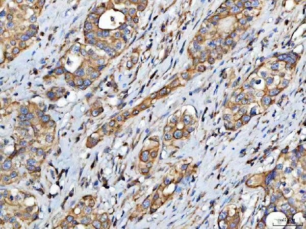
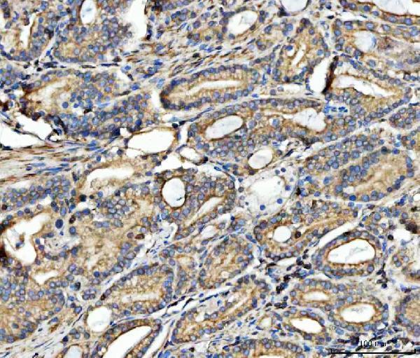
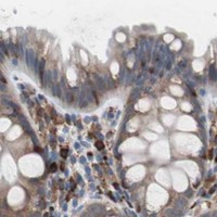
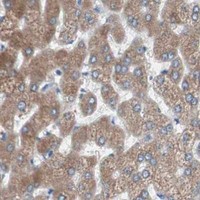
Real IHC data IHC analysis of FATP4/SLC27A4 using anti-FATP4/SLC27A4 antibody (A05299-2). FATP4/SLC27A4 was detected in a paraffin-embedded section of human colorectal adenocarcinoma tissue. Heat mediated antigen retrieval was performed in EDTA buffer (pH 8.0, epitope retrieval solution). The tissue section was blocked with 10% goat serum. The tissue section was then incubated with 2 μg/ml rabbit anti-FATP4/SLC27A4 Antibody (A05299-2) overnight at 4°C. Peroxidase Conjugated Goat Anti-rabbit IgG was used as secondary antibody and incubated for 30 minutes at 37°C. The tissue section was developed using HRP Conjugated Rabbit IgG Super Vision Assay Kit (Catalog # SV0002) with DAB as the chromogen.
Anti-FATP4/SLC27A4 Antibody ®
Cat # A05299-2

The sole rendering card is A05299-2, shown on a paraffin-embedded human colorectal adenocarcinoma section (catalog image caption). Its caption documents EDTA pH 8.0 retrieval, 2 μg/ml primary antibody, and DAB detection; it does not report the fixative (catalog image caption).

Which to pick: Choose A05299-2 for tissue IHC when the documented human paraffin-section procedure is useful; its fixative is unreported (catalog image caption). For IF/ICC, M05299-1 is a rabbit monoclonal that lists both IHC and IF/ICC for human samples, while M05299 is a rabbit monoclonal that lists IF/ICC without IHC; neither has an IF image in the payload (catalog host, clone, applications, reactivity, and IF images). For cross-species planning, A05299-2 and M05299 list human, mouse, and rat reactivity, but the supplied IHC image for A05299-2 shows human tissue only (catalog reactivity; catalog image caption).

Each figure is that product's own IHC / IF validation image from its datasheet.

References

  1. UniProt Consortium. UniProt entry Q6P1M0 (S27A4_HUMAN, Long-chain fatty acid transport protein 4).
  2. Human Protein Atlas. SLC27A4 tissue IHC expression (reliability: Approved).
  3. Human Protein Atlas. SLC27A4 subcellular location (ICC-IF): Localized to vesicles..
  4. Human Protein Atlas. SLC27A4 antibody validation summary (2 antibodies).
  5. Fatty acid transporters in the porcine conceptus and early placenta and the effects of polyunsaturated fatty acids on trophoblast cells. Scientific reports 2026 — PMC13328565.
  6. Expression Profiles of Fatty Acid Transporters and the Role of n-3 and n-6 Polyunsaturated Fatty Acids in the Porcine Endometrium. International journal of molecular sciences 2024 — PMC11507490.
  7. Mice with targeted disruption of the fatty acid transport protein 4 (Fatp 4, Slc27a4) gene show features of lethal restrictive dermopathy. The Journal of cell biology 2003 — PMC2173002.
  8. Fatty acid transport protein 1 can compensate for fatty acid transport protein 4 in the developing mouse epidermis. The Journal of investigative dermatology 2015 — PMC4289464.
  9. PubMed PMID:9878842 — UniProt-cited evidence.
  10. PubMed PMID:14702039 — UniProt-cited evidence.
  11. PubMed PMID:15164053 — UniProt-cited evidence.