This website uses cookies to ensure you get the best experience on our website.


- Table of Contents
Plan chromogenic IHC on paraffin sections using A30575 at 1:100–1:300 (datasheet: IHC dilution). Assess selective cytoplasmic staining in hepatocytes and score it against appropriate controls (HPA tissue IHC).

Expected localisation, validated protocols, controls and antibodies — the at-a-glance facts below, then the full design guide.
| Expected localisation | Hepatocyte cytoplasm (HPA tissue IHC) | |
| Staining pattern | Selective cytoplasmic staining in hepatocytes (HPA tissue IHC) | |
| Antigen retrieval | Citrate pH 6.0 HIER, 95–98 °C, 20 min (rule: cytoplasmic / membrane antigen) | |
| Positive control | Liver | |
| Negative control | Adipose tissue+4 more · see all |
| Fixation | Keep fixation consistent across sections (standard IHC practice; not target-specific) | |
| Caveat | Endogenous liver peroxidase may raise DAB background (standard IHC practice) | |
| Regulation | Liver-enriched expression; other regulation unreported (HPA tissue IHC) | |
| Isoform / epitope | 2 isoforms; cytoplasmic epitope coverage unverified (UniProt) |
Compare the catalog antibody's IHC-P protocol with the published SLC27A5 liver-section protocol (PMC10787101).
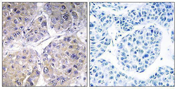
| Sample | Paraffin-embedded human liver carcinoma tissue; fixative not specified (datasheet A30575) |
| Fixation | Image fixative and duration unreported (datasheet A30575); verify before use. |
| Sectioning | 4–5 µm sections on charged slides (standard) |
| Deparaffinisation | Xylene, graded ethanol series to water (standard) |
| Antigen retrieval | Heat-induced epitope retrieval in citrate buffer, pH 6.0, 20 min at 95–98 °C (standard rule: cytoplasmic / membrane antigen) |
| Peroxidase block | 3% H2O2, 10 min, room temperature (standard) |
| Blocking | 10% normal serum of the secondary host, 30 min, room temperature (standard) |
| Primary antibody | Rabbit anti-SLC27A5, 1:100 - 1:300 (datasheet A30575) |
| Primary incubation | Overnight at 4 °C (standard) |
| Detection | HRP-polymer secondary, DAB chromogen 5–10 min (standard) |
| Counterstain | Hematoxylin, blue, dehydrate and mount (standard) |
| Expected result | SLC27A5-positive staining in hepatocytes of liver (HPA tissue IHC: High). HPA tissue profile: Selective cytoplasmic expression in hepatocytes. No signal in the no-primary control. |
SLC27A5 is predominantly expressed in liver (UniProt Q9Y2P5), and the expected IHC signal is selective, high cytoplasmic staining in hepatocytes (HPA: liver hepatocytes High; tissue IHC Enhanced). Its annotated locations include endoplasmic reticulum, microsomal and cell membranes; two transmembrane segments anchor the protein, while most of its sequence is cytoplasmic (UniProt Q9Y2P5 topology).
| Strong cytoplasmic staining in hepatocytes, with surrounding cells much less conspicuous. | This matches the observed liver pattern (HPA: selective cytoplasmic expression in hepatocytes; High). Score the hepatocyte signal and its distribution separately from background (general IHC practice). HPA's Enhanced reliability reflects consistency between staining and RNA data; it does not establish the specificity of every new staining run (HPA: tissue IHC Enhanced). |
| Predominantly nuclear staining, with little hepatocyte cytoplasmic signal. | A nuclear dominant pattern conflicts with the reported cytoplasmic liver staining (HPA: selective cytoplasmic expression in hepatocytes) and the annotated membrane locations (UniProt Q9Y2P5). Treat it as suspect until a no primary control and an independent IHC validated antibody help assess nonspecific detection (general IHC practice; HPA: multiple antibodies IHC Enhanced). |
| Strong staining in liver cells other than hepatocytes, or in a listed negative tissue. | This departs from the selective hepatocyte pattern (HPA: liver hepatocytes High). Adipocytes in adipose tissue and hematopoietic cells in bone marrow were not detected in the supplied tissue profile (HPA: Not detected). Cross reactivity or endogenous detection activity are possibilities to investigate, rather than diagnoses from appearance alone (general IHC practice). |
| Diffuse color over many compartments, with no clear hepatocyte contrast. | The expected signal is selective cytoplasmic hepatocyte staining (HPA: tissue IHC). Widespread haze is insufficient to call SLC27A5 positive; excess antibody, incomplete blocking or detection background can produce a similar appearance (general IHC practice). Compare a no primary control and inspect whether identifiable hepatocytes remain stronger than adjacent cells (general IHC practice). |
| No visible hepatocyte signal in an otherwise interpretable liver section. | This conflicts with the supplied high liver hepatocyte staining (HPA: High). First check tissue identity, staining controls and whether the IHC validated antibody's prescribed conditions were followed (general IHC practice). A negative slide alone cannot distinguish failed detection from a genuine difference in the specimen; HPA reports a reference tissue pattern, not a guarantee for every sample (HPA: tissue IHC). |
| Which tissue and cells set the reference pattern? | Liver hepatocytes provide the positive comparison (HPA: High; tissue enriched RNA). The supplied adipose tissue adipocytes, adrenal glandular cells and bone marrow hematopoietic cells were not detected (HPA: Not detected). Those findings are cell specific observations, not a claim that every cell in those organs lacks SLC27A5 (HPA: supplied tissue profile). |
| How should membrane localization affect scoring? | The IHC observation is cytoplasmic in hepatocytes (HPA: tissue IHC), while UniProt also annotates endoplasmic reticulum, microsomal and cell membranes (UniProt Q9Y2P5). Membrane accentuation within a hepatocyte can be assessed in that context, but surface staining alone does not establish antibody specificity (general IHC practice). |
| What does antibody validation support? | Three supplied antibodies have IHC Enhanced status: HPA007292, CAB056160 and CAB068239 (HPA: antibody validation). The tissue profile also has Enhanced reliability based on agreement with RNA expression (HPA: tissue IHC). These support the reference pattern; they do not validate an unlisted antibody, a particular dilution or a new specimen (HPA: supplied validation scope). |
| Can isoforms or processing explain a different pattern? | UniProt lists two isoforms and a full length chain with no annotated signal peptide, propeptide or glycosylation site (UniProt Q9Y2P5). The supplied record gives no antibody epitope or isoform specific staining data, so it cannot assign a changed IHC pattern to an isoform or processing event (UniProt Q9Y2P5; HPA: supplied antibody data). |
| What should IF/ICC show? | HPA summarizes the subcellular location as membrane but supplies no main location, ICC/IF image cell line or ICC validation for these antibodies (HPA: subcellular summary; antibody validation). An IHC hepatocyte pattern therefore gives no resolved IF/ICC staining prediction for a particular cell model (HPA: supplied IF/ICC data). |
| Situation | Likely cause | Next action |
|---|---|---|
| Liver hepatocytes are blank. | The result disagrees with the reference positive pattern (HPA: liver hepatocytes High); tissue selection or an IHC detection step may be responsible (general IHC practice). | Confirm hepatocytes are present, review the IHC validated antibody's instructions, and run an appropriate positive control beside the sample (general IHC practice). |
| The no primary control also develops color. | Signal without primary antibody indicates background from the detection workflow, including possible endogenous enzyme activity in chromogenic IHC (general IHC practice). | Address the detection background using controls and the detection system's blocking procedure before interpreting SLC27A5 staining (general IHC practice). |
| Negative reference tissue cells stain as strongly as hepatocytes. | That conflicts with their supplied Not detected observations (HPA: adipose adipocytes; adrenal glandular cells; bone marrow hematopoietic cells). Nonspecific antibody binding remains possible (general IHC practice). | Verify cell identity and compare a no primary control; repeat with an independent IHC Enhanced antibody if available (general IHC practice; HPA: antibody validation). |
| Nuclei dominate the staining. | Nuclear dominant signal conflicts with the cytoplasmic hepatocyte pattern (HPA: tissue IHC) and annotated membrane locations (UniProt Q9Y2P5). | Recheck counterstain versus chromogen, then compare a no primary control and an independent IHC validated antibody before scoring nuclei as positive (general IHC practice). |
| Diffuse haze obscures hepatocyte boundaries. | Poor contrast prevents assessment of the selective cytoplasmic pattern (HPA: tissue IHC); antibody concentration, blocking or detection background may contribute (general IHC practice). | Use the antibody's IHC guidance to review dilution and blocking, then assess controls and hepatocyte to adjacent cell contrast (general IHC practice). |
| Two IHC validated antibodies give different liver patterns. | Their shared IHC Enhanced status does not establish equivalent epitopes or identical performance in every specimen (HPA: antibody validation; supplied data omit epitopes). | Compare each antibody's controls and staining conditions, document the discrepancy, and base interpretation on the reproducible hepatocyte pattern supported by the tissue reference (general IHC practice; HPA: tissue IHC). |


Comprehensive Human Protein Atlas IHC scoring per tissue (reliability: Enhanced — High consistency between antibody staining and RNA expression data.). Rows are taken directly from the HPA tissue chart — click any row's HPA link to view the source.
| Tissue | Cell type | Level | Evidence | Source |
|---|---|---|---|---|
| Liver | Hepatocytes | High | Protein (IHC) | HPA → |
| Tissue | Cell type | Level | Evidence | Source |
|---|---|---|---|---|
| Adipose tissue | Adipocytes | Not detected | Protein (IHC) | HPA → |
| Adrenal gland | Glandular cells | Not detected | Protein (IHC) | HPA → |
| Appendix | Endocrine cells | Not detected | Protein (IHC) | HPA → |
| Bone marrow | Hematopoietic cells | Not detected | Protein (IHC) | HPA → |
| Breast | Adipocytes | Not detected | Protein (IHC) | HPA → |
Troubleshoot chromogenic IHC for SLC27A5 in paraffin sections using hepatocyte staining, membrane topology and appropriate controls as guides (HPA tissue IHC; UniProt Q9Y2P5).
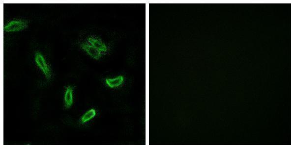
A30575 has IHC data from paraffin-embedded human liver carcinoma tissue and IF data from A549 cells; its listed reactivity is human, mouse and rat (catalog: A30575 images and reactivity).
A30575 will render with an IHC image of paraffin-embedded human liver carcinoma tissue with a peptide-blocked comparison (catalog: A30575 IHC caption). Its listed applications include IHC, IF and ICC, and its IF image shows A549 cells with a peptide-blocked comparison (catalog: A30575 applications and IF caption).
Which to pick: Choose A30575 for paraffin-section IHC: it is a rabbit polyclonal antibody listed for IHC, with an image from paraffin-embedded human liver carcinoma tissue (catalog: A30575 applications and IHC caption). The caption does not report the fixative (catalog: A30575 IHC caption). For IF/ICC or work across human, mouse and rat samples, A30575 is the listed option; its application list includes IF and ICC, its IF image shows A549 cells, and its stated reactivity covers those three species (catalog: A30575 applications, IF caption and reactivity).