SLC2A3 / Solute carrier family 2, facilitated glucose transporter member 3 · IHC design guide

Design Immunohistochemistry for SLC2A3

Plan SLC2A3 paraffin IHC using the reported cytoplasmic and membranous staining in testis, hematopoietic and lymphoid tissue (HPA tissue IHC). Compare a positive tissue with an HPA tissue reported as undetected, using the catalog antibody’s documented paraffin IHC workflow (HPA tissue IHC; datasheet M03259).

Evidence assembled Oct 2026 · For research use; verify linked source records and product datasheet before use
Immunohistochemistry protocol sheet for SLC2A3 (IHC for SLC2A3): expected localisation Membranous and cytoplasmic tissue staining (HPA tissue IHC), antibody M03259, validated IHC image, and IHC protocol steps
Printable SLC2A3 IHC protocol sheet — expected localisation Membranous and cytoplasmic tissue staining (HPA tissue IHC), antibody M03259, controls and protocol steps. Open the full SLC2A3 IHC guide →

SLC2A3 Immunohistochemistry Experimental Design Guide

Expected localisation, validated protocols, controls and antibodies — the at-a-glance facts below, then the full design guide.

Must know before staining
Expected localisation Membranous and cytoplasmic tissue staining (HPA tissue IHC)
Staining pattern Testis, hematopoietic and lymphoid tissue: cytoplasmic/membranous (HPA tissue IHC)
Antigen retrieval EDTA pH 8.0 HIER, heat-mediated (datasheet M03259)
Positive control ⓘ Bone marrow+4 more · see all
Negative control ⓘ Adipose tissue+4 more · see all
Important caveats
Reasons your staining may differ from the expected pattern.
Fixation Keep fixation consistent across sections (standard IHC practice; not target-specific)
Caveat HPA antibody may recognize proteins from more than one gene (HPA tissue IHC)
Regulation No expression regulator specified (UniProt)
Isoform / epitope 0 isoforms annotated; epitope side matters across 12 spans (UniProt)
Section 1

Recommended SLC2A3 IHC & IF Protocols

The catalog antibody’s IHC-P protocol (datasheet M03259) is complemented by three published SLC2A3 IHC methods for colon, testicular and epididymal, and oral tumor tissues (PMC11271793; PMC4438928; PMC11020985).

Recommended immunohistochemistry (IHC-P) protocol parameters
SampleParaffin-embedded human colon cancer tissue; fixative not specified (datasheet M03259)
FixationImage fixative and duration unreported (datasheet M03259); verify before use.
Sectioning4–5 µm sections on charged slides (standard)
DeparaffinisationXylene, graded ethanol series to water (standard)
Antigen retrievalHeat retrieval: EDTA pH 8.0 (datasheet M03259); 20 min, 95–100 °C (standard)
Peroxidase block3% H2O2, 10 min, room temperature (standard)
Blocking10% goat serum (datasheet M03259)
Primary antibodyRabbit monoclonal (clone ABHB-19) anti-SLC2A3, 1:50 recommended; image 1:200 (datasheet M03259)
Primary incubationOvernight at 4 °C (datasheet M03259)
DetectionHRP-conjugated secondary, DAB chromogen (datasheet M03259)
CounterstainHematoxylin, blue, dehydrate and mount (standard)
Expected resultSLC2A3-positive staining in hematopoietic cells of bone marrow (HPA tissue IHC: High). HPA tissue profile: Cytoplasmic and membranous expression mainly in testis, hematopoietic and lymphoid tissue. No signal in the no-primary control.
💡Decision noteStart with heat-mediated EDTA pH 8.0 retrieval for the catalog antibody (datasheet M03259). Check secondary antibody compatibility before adapting the goat-primary method (PMC4438928).
Section 2

What Is the Expected SLC2A3 Staining Pattern?

SLC2A3 is a 12-transmembrane glucose transporter at the cell membrane, perikaryon and neuronal projections (UniProt P11169 topology and subcellular location). In tissue IHC, expect membranous and cytoplasmic staining chiefly in testis and hematopoietic or lymphoid cells (HPA tissue IHC). Treat the pattern as provisional: HPA rates its tissue IHC Approved but reports medium staining–RNA consistency, possible recognition of proteins from multiple genes and pending external verification (HPA tissue IHC reliability).

What am I looking at on my slide?
Membranous staining, with some cytoplasmic staining, in hematopoietic cells of bone marrow or cells of splenic red pulp.This matches the reported compartments and High staining in those cell populations (HPA tissue IHC). Score the identified cells and their compartments; a positive area alone does not establish which cells stained.
Staining is predominantly nuclear, with no discernible membranous pattern in the cells being assessed.A predominantly nuclear result is discordant with the reported membrane, perikaryon and projection locations (UniProt P11169 subcellular location) and with the membranous/cytoplasmic tissue profile (HPA tissue IHC). Review morphology and controls before calling it SLC2A3.
Strong chromogen appears in an unexpected cell population, such as bronchial respiratory epithelium.HPA reports SLC2A3 Not detected in bronchial respiratory epithelial cells (HPA tissue IHC). Consider antibody cross-reactivity or endogenous detection activity; the tissue IHC validation cautions that antibodies may target proteins from multiple genes (HPA tissue IHC reliability).
A broad, diffuse deposit obscures cell borders and appears in both expected positive and expected negative areas.This cannot be scored as a membrane pattern. Check the negative detection control and staining distribution for background from blocking, washing or detection steps (general IHC practice). Compare interpretable cells with HPA's cell-specific observations, rather than assigning the deposit to SLC2A3.
Bone-marrow hematopoietic cells remain unstained while the slide otherwise appears technically intact.Those cells are reported High by tissue IHC (HPA tissue IHC), so an absent result needs investigation. Check tissue identity, an appropriate positive control and the detection run (general IHC practice). A negative slide alone cannot identify a target-specific fixation effect.
💡Expected SLC2A3 appearanceCall a positive result when membranous, sometimes cytoplasmic, staining is resolved in the expected cells, particularly High bone-marrow hematopoietic cells or splenic red-pulp cells (HPA tissue IHC); nuclear-only signal or strong staining of HPA-negative bronchial respiratory epithelium warrants review (UniProt P11169 subcellular location; HPA tissue IHC).
How each factor affects the staining
Cell population and tissueExpected intensity depends on the cells scored: HPA reports High staining in bone-marrow hematopoietic cells and late spermatids, Medium in cerebral-cortex neuropil and lung macrophages, and Not detected in adipocytes (HPA tissue IHC).
Membrane topology and epitope accessSLC2A3 has 12 transmembrane segments plus cytoplasmic and extracellular regions (UniProt P11169 topology). The supplied record does not identify the catalog antibody's epitope, so topology alone cannot specify its retrieval conditions or predict whether permeabilisation helps.
Confidence in tissue interpretationHPA calls tissue IHC Approved, yet reports medium consistency with RNA data, possible recognition of proteins from multiple genes and pending external verification (HPA tissue IHC reliability). Treat discordant cells or compartments as findings to verify with controls or an independent antibody.
IF/ICC Q: What localisation should be expected?Mainly plasma-membrane localisation is supported in ICC-IF (HPA subcellular). That summary also cautions that its antibodies target proteins from multiple genes (HPA subcellular). IF/ICC images are listed for CACO-2, Hep-G2 and U2OS (HPA subcellular); they do not supply an IHC-P protocol.
Why is my staining missing, weak or wrong?
SituationLikely causeNext action
No stain in the positive-control tissue.A run or reagent problem is possible; bone-marrow hematopoietic cells are reported High (HPA tissue IHC).Confirm the expected cells are present, then review antibody application, retrieval and chromogenic detection against the catalog antibody's IHC-P instructions (general IHC practice). No SLC2A3-specific retrieval condition is supplied.
Nuclear signal dominates.That compartment conflicts with reported cell-membrane, perikaryon and projection localisation (UniProt P11169 subcellular location).Check whether the same cells show resolved membrane staining, examine the negative detection control and repeat with controlled detection conditions if needed (general IHC practice).
Strong staining appears in bronchial respiratory epithelium.HPA reports these cells Not detected; its tissue IHC assessment also warns of recognition of proteins from multiple genes (HPA tissue IHC).Check cell identification and background controls, then seek confirmation with an independent antibody before assigning the signal to SLC2A3 (general IHC practice).
Diffuse chromogen obscures cell boundaries.Detection background can prevent compartment-level scoring (general IHC practice).Inspect a negative detection control; review blocking, washes and detection development, then rescore only cells with distinguishable borders (general IHC practice).
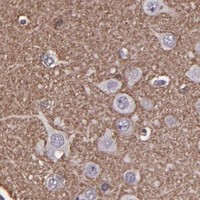
Cerebral cortex looks weaker than bone marrow.HPA reports Medium neuropil staining in cerebral cortex and High hematopoietic-cell staining in bone marrow (HPA tissue IHC).Score the neuropil and hematopoietic cells separately; compare each with its reported level rather than requiring equal intensity across tissues (HPA tissue IHC).
A peripheral IF/ICC signal does not match tissue IHC.ICC-IF mainly supports plasma-membrane localisation (HPA subcellular), while tissue IHC reports membranous and cytoplasmic expression (HPA tissue IHC).Interpret each preparation within its own evidence and controls; use the separate IF/ICC guide for that application. The ICC-IF summary warns of antibodies targeting proteins from multiple genes (HPA subcellular).

Sample controls for SLC2A3 IHC & IF

🧪Run bone marrow first: hematopoietic cells should stain (HPA: High in bone marrow hematopoietic cells). Use adipose tissue adipocytes as the negative comparator (HPA: Not detected in adipocytes); any unstained cells on the bone marrow slide should show counterstain alone, without convincing DAB signal, but should not be assumed target-negative without validation (standard IHC practice).
Positive control tissue: Bone marrow (Hematopoietic cells, HPA High)
Negative control tissue: Adipose tissue (HPA Not detected)
ICC-IF cell lines (HPA subcellular resource): HPA ICC-IF images show SLC2A3 in CACO-2, Hep-G2, U2OS, with annotated localisation: Plasma membrane (supported) (HPA subcellular).
Technical controls: Include a no-primary, secondary-only control, matched rabbit IgG isotype control of the same clonality where applicable, and SLC2A3 knockout material as a biological negative (caption: rabbit primary antibody; standard IHC practice). Block endogenous peroxidase and inspect bone marrow for residual hematopoietic-cell background before interpreting DAB staining (standard chromogenic IHC practice).
⚠️Feasibility: No target-specific fixation window or fixation effect is reported in the supplied evidence, and the selected M03259 paraffin-section caption does not state a fixative (M03259 tissue-IHC caption). That caption uses heat retrieval in EDTA at pH 8.0, but does not establish whether retrieval is required or whether frozen sections or IF are easier (M03259 tissue-IHC caption). Endogenous peroxidase in bone marrow hematopoietic cells can complicate DAB interpretation (standard chromogenic IHC practice).

HPA tissue IHC evidence for SLC2A3

Comprehensive Human Protein Atlas IHC scoring per tissue (reliability: Approved — Medium consistency between antibody staining and RNA expression data. Caution, targets protein from more than one gene. Pending external verification.). Rows are taken directly from the HPA tissue chart — click any row's HPA link to view the source.

Positive expression · recommended positive controls

TissueCell typeLevelEvidenceSource
Bone marrow Hematopoietic cells High Protein (IHC) HPA →
Spleen Cells in red pulp High Protein (IHC) HPA →
Testis Elongated or late spermatids High Protein (IHC) HPA →
Tonsil Non-germinal center cells High Protein (IHC) HPA →
Appendix Lymphoid tissue Medium Protein (IHC) HPA →

Undetected expression · recommended negative controls

TissueCell typeLevelEvidenceSource
Adipose tissue Adipocytes Not detected Protein (IHC) HPA →
Bronchus Respiratory epithelial cells Not detected Protein (IHC) HPA →
Caudate Glial cells Not detected Protein (IHC) HPA →
Cerebellum Cells in granular layer Not detected Protein (IHC) HPA →
Cervix Glandular cells Not detected Protein (IHC) HPA →
Section 3

Advanced SLC2A3 IHC Tips

Troubleshoot SLC2A3 staining in paraffin sections using the catalog antibody’s tissue IHC conditions, compartment controls, and cell type aware scoring.

How should I optimize antigen retrieval when SLC2A3 staining is weak?
Start with heat mediated antigen retrieval in EDTA at pH 8.0 for paraffin sections (datasheet M03259). The selected human colon cancer example used this condition, followed by 10% goat serum and 1:200 primary antibody overnight at 4°C (M03259 caption). If staining is weak, compare retrieval duration on adjacent sections while holding antibody dilution and DAB development constant (standard IHC practice). Examine membrane associated signal alongside a no primary control; the HPA tissue staining profile is cytoplasmic and membranous but carries a cross gene antibody caution (HPA tissue IHC). Record tissue damage before increasing retrieval intensity (standard IHC practice).
Could fixation explain weak or uneven SLC2A3 staining?
The selected paraffin section caption does not state a fixative, so target specific fixation sensitivity is unknown for this antibody (M03259 caption). Compare sections with documented processing histories and keep retrieval in EDTA at pH 8.0 constant while investigating fixation (datasheet M03259; standard IHC practice). Inspect morphology and staining at section edges and centers, then review fixation and processing records for uneven handling (standard IHC practice). Use the same 1:200 primary dilution and overnight 4°C incubation during the comparison (M03259 caption). Neither the HPA staining pattern nor the annotated transmembrane segments establish a fixation effect (HPA tissue IHC; UniProt P11169 topology).
What staining pattern fits SLC2A3 in tissue sections?
Prioritize cell surface associated staining when assessing SLC2A3, which UniProt places at the cell membrane and in neuronal projections (UniProt P11169 subcellular location). In neuronal processes, staining may appear in densely spaced patches rather than as a continuous outline (UniProt P11169 subcellular location). HPA reports cytoplasmic and membranous tissue staining and medium neuropil staining in cerebral cortex (HPA tissue IHC). Its supported plasma membrane IF assignment comes with a warning that the antibodies target proteins from multiple genes (HPA subcellular). Compare compartment patterns in intact cells on matched sections, and investigate diffuse signal that also appears in no primary controls (standard IHC practice).
How do topology and an unknown epitope affect my IHC interpretation?
Do not assign this antibody an intracellular or extracellular epitope without an epitope map; the supplied caption identifies no binding region (M03259 caption). SLC2A3 has 12 annotated transmembrane segments, extracellular residue 43 is glycosylated, and its long cytoplasmic loop spans residues 205–269 (UniProt P11169 topology and glycosylation). UniProt annotates phosphorylation at residues 232, 475, 485, and 492, but these sites do not locate this antibody’s epitope (UniProt P11169 modified residues; M03259 caption). No isoforms are annotated in the supplied record, which does not establish that every tissue molecule presents the same accessible epitope (UniProt P11169 isoforms; standard IHC practice). Compare retrieval conditions on adjacent sections if accessibility is in doubt (standard IHC practice).
How can IF help resolve ambiguous chromogenic SLC2A3 staining?
Use IF as a separate localization check while keeping the paraffin section IHC result anchored to EDTA retrieval at pH 8.0 (datasheet M03259). Multiplex SLC2A3 with a validated marker for the cell population being examined; HPA reports high staining in bone marrow hematopoietic cells and medium staining in lung macrophages (HPA tissue IHC). Choose fluorophores after checking unstained tissue for autofluorescence, and include single color controls (standard IF practice). For an extracellular epitope, compare labeling without permeabilization; an intracellular epitope requires access across the membrane, but this antibody’s epitope side is unreported (UniProt P11169 topology; M03259 caption; standard IF practice). Assess membrane overlap before using IF to clarify a diffuse DAB pattern (standard IHC/IF practice).
How should I investigate widespread DAB background?
Run no primary and secondary only controls to distinguish tissue or detection background from primary antibody dependent staining (standard IHC practice). The selected protocol used 10% goat serum, 1:200 rabbit primary overnight at 4°C, and a peroxidase linked secondary for 30 minutes at 37°C (M03259 caption). Review the general peroxidase blocking step before DAB development and compare development times on matched sections (standard IHC practice). Because HPA reports staining in hematopoietic and lymphoid tissue, identify the stained cells before calling all signal in those regions background (HPA tissue IHC). Treat diffuse deposition on damaged or folded areas cautiously, especially when controls show similar color (standard IHC practice).
How should I quantify SLC2A3 across sections with different cell populations? ⚠ ANSWER MARKED FOR VERIFICATION
Define the cell population and compartment before scoring, using HPA’s reported cytoplasmic and membranous pattern as context (HPA tissue IHC). For discrete cells, record percent positive cells and an H score from staining intensity; for dispersed positive cells, report density per mm² (standard IHC quantification practice). Normalize cell counts to the relevant cell population or measured tissue area, and exclude folds, edges, and necrotic areas by a prespecified rule (standard IHC quantification practice). Keep retrieval at pH 8.0, primary dilution at 1:200, and DAB development consistent across the comparison (datasheet M03259; standard IHC practice). Report membrane and cytoplasmic scores separately when both are present (HPA tissue IHC; standard IHC quantification practice).
When should I doubt an apparent SLC2A3 positive result?
A convincing result should fit the assessed cell type and show interpretable membrane associated staining, consistent with UniProt’s cell membrane assignment (UniProt P11169 subcellular location). HPA reports high staining in bone marrow hematopoietic cells but no detected staining in adipocytes, while cautioning that its tissue antibody may recognize proteins from multiple genes (HPA tissue IHC). Question a strong call based solely on the wrong cell population, uniformly diffuse cytoplasm, a cut edge, or necrotic material (standard IHC interpretation practice). Compare no primary and peroxidase controls before attributing brown deposits to SLC2A3, since DAB depends on enzyme detection (standard IHC practice). Resolve discordant patterns with an independent antibody or orthogonal expression evidence (standard IHC validation practice).
Boster reagents

Best SLC2A3 / Solute carrier family 2, facilitated glucose transporter member 3 IHC Antibodies

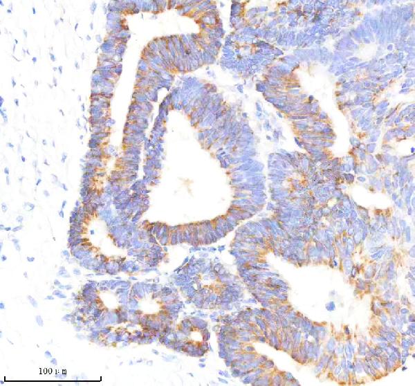
M03259 has IHC images from paraffin sections of human colon and stomach cancer (catalog: IHC captions). Its listed reactivity covers human, mouse, and rat (catalog: reactivity).

Real IHC data IHC analysis of GLUT3/SLC2A3 using anti-GLUT3/SLC2A3 antibody (M03259). GLUT3/SLC2A3 was detected in a paraffin-embedded section of human colon cancer tissue. Heat mediated antigen retrieval was performed in EDTA buffer (pH 8.0, epitope retrieval solution). The tissue section was blocked with 10% goat serum. The tissue section was then incubated with 1:200 rabbit anti-GLUT3/SLC2A3 Antibody (M03259) overnight at 4°C. Peroxidase Conjugated Goat Anti-rabbit IgG was used as secondary antibody and incubated for 30 minutes at 37°C. The tissue section was developed using HRP Conjugated Rabbit IgG Super Vision Assay Kit (Catalog # SV0002) with DAB as the chromogen.
Anti-GLUT3 SLC2A3 Rabbit Monoclonal Antibody
Cat # M03259

M03259 will render with its human colon cancer paraffin-section IHC figure (catalog: figure caption). A second caption documents human stomach cancer paraffin-section IHC; IHC is listed as an application for M03259 (catalog: IHC caption; catalog: applications).

Which to pick: Choose M03259 for tissue IHC: it is a rabbit monoclonal with human paraffin-section images, although the fixative is unreported (catalog: host and clone; catalog: IHC captions). No SKU in the payload lists IF/ICC validation, so there is no supported IF/ICC pick (catalog: applications and IF images). M03259 lists mouse and rat reactivity for cross-species work, but the supplied IHC images show human tissue only (catalog: reactivity; catalog: IHC captions).

Each figure is that product's own IHC / IF validation image from its datasheet.

References

  1. UniProt Consortium. UniProt entry P11169 (GTR3_HUMAN, Solute carrier family 2, facilitated glucose transporter member 3).
  2. Human Protein Atlas. SLC2A3 tissue IHC expression (reliability: Approved).
  3. Human Protein Atlas. SLC2A3 subcellular location (ICC-IF): Mainly localized to the plasma membrane. Caution: Based on antibodies targeting proteins from multiple genes..
  4. Human Protein Atlas. SLC2A3 antibody validation summary (2 antibodies).
  5. Multi-Omics analysis elucidates tumor microenvironment and intratumor microbes of angiogenesis subtypes in colon cancer. World journal of gastrointestinal oncology 2024 — PMC11271793.
  6. In‑depth mapping of human testicular and epididymal proteins and their functional association with spermatozoa. Molecular medicine reports 2015 — PMC4438928.
  7. SLC2A3 promotes tumor progression through lactic acid-promoted TGF-β signaling pathway in oral squamous cell carcinoma. PloS one 2024 — PMC11020985.
  8. TAGLN-RhoA/ROCK2-SLC2A3-mediated Mechano-metabolic Axis Promotes Skin Fibrosis. International journal of biological sciences 2025 — PMC11705643.
  9. PubMed PMID:3170580 — UniProt-cited evidence.
  10. PubMed PMID:14702039 — UniProt-cited evidence.
  11. PubMed PMID:15489334 — UniProt-cited evidence.