SLC2A8 / Solute carrier family 2, facilitated glucose transporter member 8 · IHC design guide

Design Immunohistochemistry for SLC2A8

Plan SLC2A8 IHC-P using the catalog antibody at 1:25 on formaldehyde-fixed paraffin sections (datasheet M06641). Assess Leydig-cell staining in testis and interpret the tissue pattern cautiously because its reliability is uncertain (HPA tissue IHC).

Evidence assembled Sep 2026 · For research use; verify linked source records and product datasheet before use
Immunohistochemistry protocol sheet for SLC2A8 (IHC for SLC2A8): expected localisation Cytoplasmic staining in a few tissues; abundant in testis (HPA tissue IHC), antibody M06641, validated IHC image, and IHC protocol steps
Printable SLC2A8 IHC protocol sheet — expected localisation Cytoplasmic staining in a few tissues; abundant in testis (HPA tissue IHC), antibody M06641, controls and protocol steps. Open the full SLC2A8 IHC guide →

SLC2A8 Immunohistochemistry Experimental Design Guide

Expected localisation, validated protocols, controls and antibodies — the at-a-glance facts below, then the full design guide.

Must know before staining
Expected localisation Cytoplasmic staining in a few tissues; abundant in testis (HPA tissue IHC)
Staining pattern Strong Leydig-cell staining; low cytoplasmic signal elsewhere (HPA tissue IHC)
Antigen retrieval Citrate pH 6 HIER, heat-mediated (datasheet M06641)
Positive control ⓘ Testis
Negative control ⓘ Adipose tissue+4 more · see all
Important caveats
Reasons your staining may differ from the expected pattern.
Fixation Formaldehyde-fixed, paraffin-embedded sections (datasheet M06641)
Caveat Presumed off-target binding; verification pending (HPA tissue IHC)
Regulation Insulin-regulated transport; IHC effect unknown (UniProt)
Isoform / epitope No isoforms annotated; epitope side matters across 12 TMs (UniProt)
Section 1

Recommended SLC2A8 IHC & IF Protocols

The catalog antibody’s citrate pH 6 IHC-P protocol (datasheet M06641) is accompanied by a published mouse testis protocol (PMC2557070 methods).

Recommended immunohistochemistry (IHC-P) protocol parameters
SampleFFPE tissue sections (standard)
Fixation10% neutral-buffered formalin, 24 h, standard processing (standard)
Sectioning4–5 µm sections on charged slides (standard)
DeparaffinisationXylene, graded ethanol series to water (standard)
Antigen retrievalHeat-induced epitope retrieval in Citrate buffer, pH 6 (datasheet M06641); 20 min at 95–100 °C (standard)
Peroxidase block3% H2O2, 10 min, room temperature (standard)
Blocking10% normal serum of the secondary host, 30 min, room temperature (standard)
Primary antibodyRabbit anti-SLC2A8, 1:25 (datasheet M06641)
Primary incubation1 hours at 37°C (datasheet M06641)
DetectionHRP-polymer secondary, DAB chromogen 5–10 min (standard)
CounterstainHematoxylin, blue, dehydrate and mount (standard)
Expected resultSLC2A8-positive staining in leydig cells of testis (HPA tissue IHC: High). HPA tissue profile: Low cytoplasmic expression in a few tissues, highly abundant in testis. No signal in the no-primary control.
💡Decision noteStart with citrate pH 6 heat retrieval (datasheet M06641); the published mouse testis protocol used microwave retrieval in a proprietary solution (PMC2557070 methods).
Section 2

What Is the Expected SLC2A8 Staining Pattern?

SLC2A8 is principally on intracellular vesicle membranes, with possible plasma membrane localization (UniProt Q9NY64). In paraffin IHC, the strongest reported staining is in testis Leydig cells (HPA: High). Interpret this pattern cautiously: HPA rates tissue IHC Uncertain because presumed off-target binding was observed (HPA: Uncertain).

What am I looking at on my slide?
Leydig cell cytoplasmic staining in testis (HPA: High)Consistent with intracellular vesicles (UniProt Q9NY64; HPA: vesicles).
Predominantly nuclear stainingUnexpected for a membrane transporter (UniProt Q9NY64 topology); assess specificity.
Strong staining in adipocytes (HPA: Not detected)Possible off-target binding (HPA: Uncertain) or endogenous detection activity.
Diffuse staining across the sectionPossible background; HPA reports presumed off-target binding (HPA: Uncertain).
No Leydig cell signal in testis (HPA: High)Check tissue preservation and detection before calling the sample negative (HPA: High).
💡Expected SLC2A8 appearanceA positive result is relatively strong Leydig cell cytoplasmic staining with a vesicular distribution (HPA: High; HPA: vesicles); predominantly nuclear or widespread staining warrants scrutiny (UniProt Q9NY64 topology; HPA: Uncertain).
How each factor affects the staining
Fixation and cross-linkingMay mask epitopes in paraffin sections; compare fixation conditions (standard IHC practice).
Antigen retrievalAdjust retrieval if Leydig cell signal is absent (HPA: High; standard IHC practice).
Extracellular epitopeLoop 341–367 contains glycosylation site 349; access may vary (UniProt Q9NY64 topology).
Cytoplasmic epitopeThe 460–477 tail faces cytoplasm; epitope access matters (UniProt Q9NY64 topology).
Endogenous peroxidaseCan mimic chromogenic signal; check a no-primary control (standard IHC practice).
Why is my staining missing, weak or wrong?
SituationLikely causeNext action
No testis Leydig cell staining (HPA: High)Epitope masking or weak detection (standard IHC practice).Review fixation, retrieval and detection with a testis control (HPA: High).
Widespread cytoplasmic stainingPossible off-target binding (HPA: Uncertain).Compare cell types with HPA patterns and a no-primary control (HPA: tissue IHC).
Nuclear staining predominatesLocalization conflicts with membrane topology (UniProt Q9NY64).Recheck specificity and compartment scoring (UniProt Q9NY64 topology).
Signal appears without primary antibodyPossible endogenous detection activity (standard IHC practice).Check peroxidase blocking and detection controls (standard IHC practice).
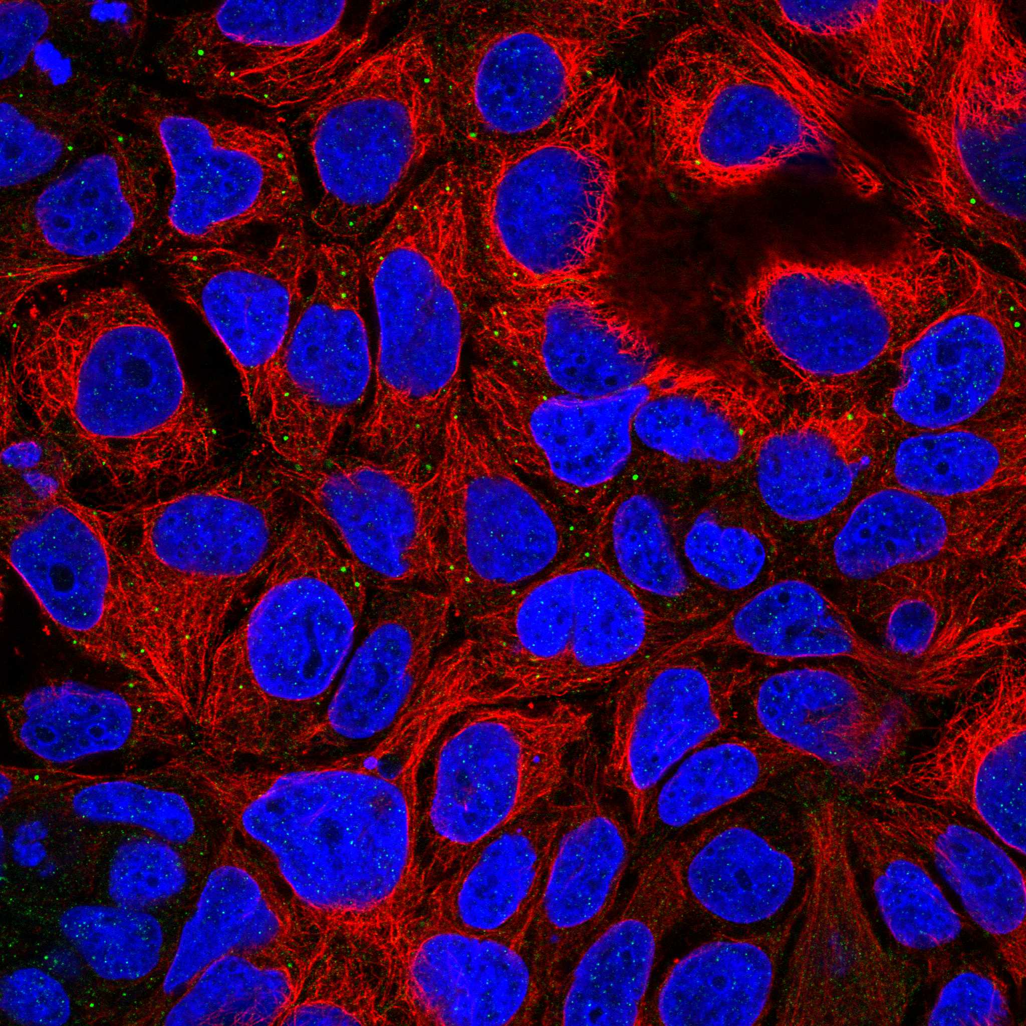
IF/ICC: where should signal appear?HPA reports approved vesicle localization (HPA: ICC-IF).Compare vesicle localization with HPA ICC-IF images; use the separate IF/ICC guide.

Sample controls for SLC2A8 IHC & IF

🧪Run testis first and look for staining in Leydig cells (HPA: High in Leydig cells); neighboring cells can serve as lower-staining internal comparators, but their absence of SLC2A8 is not established by the supplied HPA row. Run adipose tissue as a negative tissue control, focusing on adipocytes (HPA: Not detected in adipocytes).
Positive control tissue: Testis (Leydig cells, HPA High)
Negative control tissue: Adipose tissue (HPA Not detected)
ICC-IF cell lines (HPA subcellular resource): HPA ICC-IF images show SLC2A8 in CACO-2, Hep-G2, U2OS, with annotated localisation: Vesicles (approved) (HPA subcellular).
Technical controls: Include a no-primary (secondary-only) control, an isotype control matched to the primary antibody’s host species, immunoglobulin class and clonality, and an SLC2A8 knockout sample or validated peptide-block control. For chromogenic IHC, quench endogenous peroxidase and check testis background; for IF, assess tissue autofluorescence.
⚠️Feasibility: Keep fixation conditions consistent and optimize antigen retrieval on testis sections, since fixation can mask epitopes and SLC2A8 has 12 transmembrane segments (UniProt Q9NY64 topology). Expect predominantly intracellular, vesicle-associated staining rather than requiring a crisp cell-surface outline (UniProt Q9NY64 subcellular location; HPA: Vesicles). If paraffin-section staining remains weak after retrieval optimization, frozen-section IF can be assessed separately; interpret fluorescence against the tissue autofluorescence control.

HPA tissue IHC evidence for SLC2A8

Comprehensive Human Protein Atlas IHC scoring per tissue (reliability: Uncertain — Presumed off target binding observed and disregarded. Pending external verification.). Rows are taken directly from the HPA tissue chart — click any row's HPA link to view the source.

Positive expression · recommended positive controls

TissueCell typeLevelEvidenceSource
Testis Leydig cells High Protein (IHC) HPA →

Undetected expression · recommended negative controls

TissueCell typeLevelEvidenceSource
Adipose tissue Adipocytes Not detected Protein (IHC) HPA →
Appendix Glandular cells Not detected Protein (IHC) HPA →
Bone marrow Hematopoietic cells Not detected Protein (IHC) HPA →
Breast Adipocytes Not detected Protein (IHC) HPA →
Bronchus Respiratory epithelial cells Not detected Protein (IHC) HPA →
Section 3

Advanced SLC2A8 IHC Tips

Troubleshoot chromogenic SLC2A8 IHC in paraffin sections using its membrane topology, predominantly vesicular localisation, and the limits of available tissue staining evidence.

Which retrieval condition should I try first for weak SLC2A8 staining in paraffin sections?
Start with heat-mediated citrate retrieval at pH 6 for paraffin sections (datasheet M06641). Keep section thickness, heating, cooling, antibody concentration, and detection constant while comparing a short and a longer retrieval run; score intact cellular staining separately from damaged tissue (standard IHC practice). SLC2A8 has 12 transmembrane segments and short extracellular loops, so epitope accessibility may depend strongly on fixation and retrieval (UniProt Q9NY64 topology). If staining remains weak, test an alternative retrieval buffer only as a fallback on adjacent sections, alongside the citrate condition (standard IHC practice). Treat stronger diffuse staining cautiously because the available tissue IHC profile is flagged as uncertain (HPA tissue IHC).
How can I distinguish fixation damage from genuinely low SLC2A8 expression?
Compare sections with documented, consistent formalin fixation and processing, and include a testis section containing Leydig cells in the same staining run (standard IHC practice; HPA tissue IHC: high in Leydig cells). Excessive fixation can mask an epitope, whereas poor fixation can disrupt morphology and make cytoplasmic staining difficult to assign (standard IHC practice). Run adjacent sections with the specified citrate retrieval at pH 6, changing only retrieval duration during the pilot (datasheet M06641; standard IHC practice). Check whether cell borders, nuclei, and vesicle-like cytoplasmic signal remain interpretable before calling a section negative (UniProt Q9NY64 localisation; standard IHC practice). The HPA testis result is a useful comparison, but its tissue IHC reliability is uncertain (HPA tissue IHC).
Should SLC2A8 stain the plasma membrane or cytoplasmic puncta in IHC?
Expect predominantly intracellular, vesicle-associated staining, while allowing some plasma membrane signal because SLC2A8 may move between those compartments (UniProt Q9NY64 localisation). Vesicles are also the approved main location in cell imaging (HPA subcellular). In chromogenic sections, assess punctate or granular cytoplasmic staining within intact cell outlines; do not score diffuse brown precipitate in lumina or damaged areas as vesicular staining (standard IHC practice). SLC2A8 contains a dileucine internalisation motif that is critical for intracellular sequestration, so membrane staining alone is insufficient evidence of specificity (UniProt Q9NY64 localisation). Compare the pattern with a negative reagent control and the expected cell distribution before interpreting unusual compartmental staining (standard IHC practice; HPA tissue IHC).
What should I check if the antibody gives inconsistent SLC2A8 staining across preparations?
Check the catalog antibody's immunogen or mapped epitope before changing staining conditions; the supplied record lists 0 isoforms and one chain spanning residues 1–477 (UniProt Q9NY64 record). Its 12 membrane spans separate cytoplasmic and extracellular regions, so an epitope's position affects access after fixation and retrieval (UniProt Q9NY64 topology; standard IHC practice). A glycosylation site is annotated at residue 349, within the extracellular region spanning 341–367; consider that context if the mapped epitope overlaps it (UniProt Q9NY64 topology and glycosylation). Keep citrate retrieval at pH 6 as the starting condition, then compare serial sections and morphology when troubleshooting epitope masking (datasheet M06641; standard IHC practice).
How should I investigate an SLC2A8 IHC pattern with complementary immunofluorescence?
Use the separate IF/ICC workflow to compare SLC2A8 with a validated marker for Leydig cells when examining testis, while recognising that the reported Leydig-cell IHC signal has uncertain reliability (HPA tissue IHC). Choose a fluorophore channel after inspecting unstained tissue for autofluorescence, and collect single-channel controls before interpreting apparent overlap (standard IF practice). Match permeabilisation to the mapped epitope: intracellular access is needed for cytoplasmic regions, whereas harsh permeabilisation can obscure membrane boundaries (UniProt Q9NY64 topology; standard IF practice). Compare vesicular signal across channels because vesicles are the approved imaging location (HPA subcellular). A matching fluorescent pattern supports further investigation, but cannot by itself validate the chromogenic IHC signal (standard IHC/IF practice).
What causes diffuse brown background when staining SLC2A8 in paraffin sections?
First inspect a section processed without primary antibody to separate detection-system staining from antibody-dependent signal, and check that endogenous peroxidase was blocked before chromogen development (standard chromogenic IHC practice). Titrate the catalog antibody and detection incubation while holding citrate retrieval at pH 6 constant, so a background change has an identifiable cause (datasheet M06641; standard IHC practice). Use appropriate protein blocking and washes, and inspect pigment, folds, edges, and necrotic areas under the counterstain (standard IHC practice). Plausible SLC2A8 staining is cellular and mainly vesicle-associated, not a uniform brown film over tissue (UniProt Q9NY64 localisation; HPA subcellular). Apply a high threshold for specificity because presumed off-target binding was observed in the available tissue IHC assessment (HPA tissue IHC).
How should I score SLC2A8 staining without overcalling nonspecific signal? ⚠ ANSWER MARKED FOR VERIFICATION
Define the eligible cell population and compartment before scoring, then report the percentage of positive cells and an H-score using intensity categories 0–3 (standard IHC scoring practice). Normalise the percentage and H-score to evaluable cells of the same type, excluding folds, necrosis, section edges, and cells without interpretable morphology (standard IHC practice). For spatial comparisons, report positive cells per mm² of viable tissue and record the analysed area (standard IHC quantification practice). Score granular cytoplasmic signal separately from membrane staining because SLC2A8 is principally intracellular but may reach the plasma membrane (UniProt Q9NY64 localisation). State the uncertainty attached to the reference tissue IHC profile when comparing scores across samples (HPA tissue IHC).
What evidence would make a positive SLC2A8 IHC result convincing?
Look for reproducible cellular staining in the expected compartment and cell population, with clean negative reagent controls and intact morphology (UniProt Q9NY64 localisation; HPA tissue IHC; standard IHC practice). In testis, Leydig-cell staining is a reported reference pattern, but the tissue IHC assessment is uncertain because presumed off-target binding was observed (HPA tissue IHC). Predominantly vesicular cytoplasmic signal fits the protein's reported localisation; isolated nuclear staining or widespread extracellular precipitate warrants investigation (UniProt Q9NY64 localisation; HPA subcellular; standard IHC practice). Exclude edge effects, necrosis, pigment, and endogenous peroxidase staining before counting positive cells (standard chromogenic IHC practice). If the conclusion matters biologically, seek agreement from an independently validated antibody or another orthogonal measurement (standard IHC validation practice).
Boster reagents

Best SLC2A8 / Solute carrier family 2, facilitated glucose transporter member 8 IHC Antibodies

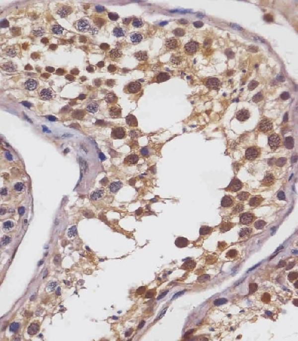
The catalog includes an IHC-P antibody with a human testis staining image (M06641 figure); its listed reactivity is human, mouse and rat (M06641 catalog). No IF image is supplied (M06641 catalog).

Real IHC data M06641 staining SLC2A8 in human testis tissue sections by Immunohistochemistry (IHC-P -paraformaldehyde-fixed, paraffin-embedded sections). Tissue was fixed with formaldehyde and blocked with 3% BSA for 0. 5 hour at room temperature; antigen retrieval was by heat mediation with a citrate buffer (pH6). Samples were incubated with primary antibody (1/25) for 1 hours at 37°C. A undiluted biotinylated goat polyvalent antibody was used as the secondary antibody.
Anti-SLC2A8 Antibody (Center)
Cat # M06641

M06641 has an IHC-P image showing SLC2A8 staining in a human testis section (M06641 figure). It is listed for IHC-P and reacts with human, mouse and rat (M06641 catalog).

Which to pick: For paraffin-section IHC, choose M06641: its figure shows human testis staining, and the listed IHC-P dilution is 1:25 (M06641 figure; M06641 catalog). For mouse or rat samples, M06641 lists reactivity with both species, although the supplied IHC image is from human tissue (M06641 catalog; M06641 figure). No IF/ICC application or IF image is listed, so this payload does not establish an IF/ICC choice (M06641 catalog).

Each figure is that product's own IHC / IF validation image from its datasheet.