SLC34A2 / Sodium-dependent phosphate transport protein 2B · IHC design guide

Design Immunohistochemistry for SLC34A2

Plan paraffin section IHC for SLC34A2 using membranous staining in lung type II pneumocytes as a reference (HPA tissue IHC). Compare tissue patterns with the annotated apical membrane location and account for staining in ciliated cells (UniProt; HPA tissue IHC).

Evidence assembled Oct 2026 · For research use; verify linked source records and product datasheet before use
Immunohistochemistry protocol sheet for SLC34A2 (IHC for SLC34A2): expected localisation Apical membrane (UniProt); type II cell membrane (HPA tissue IHC), antibody A03957-1, validated IHC image, and IHC protocol steps
Printable SLC34A2 IHC protocol sheet — expected localisation Apical membrane (UniProt); type II cell membrane (HPA tissue IHC), antibody A03957-1, controls and protocol steps. Open the full SLC34A2 IHC guide →

SLC34A2 Immunohistochemistry Experimental Design Guide

Expected localisation, validated protocols, controls and antibodies — the at-a-glance facts below, then the full design guide.

Must know before staining
Expected localisation Apical membrane (UniProt); type II cell membrane (HPA tissue IHC)
Staining pattern Membranous type II pneumocytes and female tract epithelium (HPA tissue IHC)
Antigen retrieval EDTA pH 8.0 HIER, heat-mediated (datasheet A03957-1)
Positive control ⓘ Bronchus+4 more · see all
Negative control ⓘ Adipose tissue+4 more · see all
Important caveats
Reasons your staining may differ from the expected pattern.
Fixation Keep fixation consistent across sections. (standard IHC practice; not target-specific)
Caveat Bronchial ciliary rootlets also stain strongly (HPA tissue IHC)
Regulation Expression regulation is unreported (UniProt)
Isoform / epitope 2 isoforms; epitope side is unreported (UniProt)
Section 1

Recommended SLC34A2 IHC & IF Protocols

The catalog antibody’s IHC-P protocol (datasheet: A03957-1) is accompanied by four published SLC34A2 IHC methods (PMC8298178; PMC11168907; PMC11083581; PMC9246846).

Recommended immunohistochemistry (IHC-P) protocol parameters
SampleParaffin-embedded human lung cancer tissue; fixative not specified (datasheet A03957-1)
FixationImage fixative and duration unreported (datasheet A03957-1); verify before use.
Sectioning4–5 µm sections on charged slides (standard)
DeparaffinisationXylene, graded ethanol series to water (standard)
Antigen retrievalHeat retrieval: EDTA pH 8.0 (datasheet A03957-1); 20 min, 95–100 °C (standard)
Peroxidase block3% H2O2, 10 min, room temperature (standard)
Blocking10% goat serum (datasheet A03957-1)
Primary antibodyRabbit anti-SLC34A2, 2-5μg/ml (datasheet A03957-1)
Primary incubationOvernight at 4 °C (datasheet A03957-1)
DetectionHRP-conjugated secondary, DAB chromogen (datasheet A03957-1)
CounterstainHematoxylin, blue, dehydrate and mount (standard)
Expected resultSLC34A2-positive staining in ciliated cells (ciliary rootlets) of bronchus (HPA tissue IHC: High). HPA tissue profile: Membranous expression in lung type II pneumocytes and female genital tract. No signal in the no-primary control.
💡Decision noteStart with EDTA pH 8.0 heat retrieval for the catalog antibody (datasheet: A03957-1); the thyroid and ovarian methods report citrate retrieval (PMC8298178; PMC9246846).
Section 2

What Is the Expected SLC34A2 Staining Pattern?

SLC34A2 is an eight-pass membrane transporter localized to the apical cell membrane, including enterocyte brush borders (UniProt O95436 topology; UniProt O95436 subcellular location). In paraffin-section IHC, expect staining in lung alveolar type II cells and selected ciliated cells of the female genital tract; HPA reports high staining in both settings and rates the tissue profile Enhanced for consistency with RNA expression (HPA tissue IHC).

What am I looking at on my slide?
Distinct membrane-associated staining in alveolar type II cells, with little diffuse background.This matches the reported high lung staining and membrane localization (HPA tissue IHC; UniProt O95436 subcellular location). Score the identified cells and their staining pattern; a uniformly stained lung section does not reproduce the reported cell-specific pattern (HPA tissue IHC).
Apical or microvillar staining in small-intestinal enterocytes.This fits the brush-border localization and HPA's medium enterocyte microvillar staining (UniProt O95436 subcellular location; HPA tissue IHC). Compare intensity within the relevant epithelial compartment; the lung type II cell result is reported as high, while this intestinal result is medium (HPA tissue IHC).
Predominantly nuclear or broadly cytoplasmic staining in the expected positive cells.That is atypical for the main membrane pattern in tissue IHC (HPA tissue IHC; UniProt O95436 subcellular location). Check controls and localization before interpreting it as SLC34A2. HPA also reports supported nucleoplasmic and cytosolic localization in ICC-IF, so those IF observations alone cannot establish an IHC artefact (HPA subcellular ICC-IF).
Strong chromogenic staining in unrelated cells, including adipocytes or bone-marrow hematopoietic cells.Those cells are reported as not detected in the sampled HPA tissues (HPA tissue IHC). Treat an isolated unexpected result as possible nonspecific antibody binding or endogenous detection activity; compare a no-primary control and the expected positive cell compartment before assigning target expression (standard IHC practice).
No membrane-associated staining in a technically interpretable lung section containing alveolar type II cells.This conflicts with the reported high type II cell staining (HPA tissue IHC). First confirm the relevant cells are present and that the run's positive and detection controls worked; an absent signal alone does not distinguish a sample problem from a staining failure (standard IHC practice).
💡Expected SLC34A2 appearanceCall positive when staining is chiefly membrane-associated in alveolar type II cells (high; HPA tissue IHC; UniProt O95436 subcellular location); widespread unrelated-cell or diffuse chromogenic staining warrants a specificity check (standard IHC practice).
How each factor affects the staining
Tissue and cell selectionLung alveolar type II cells are high, while small-intestinal enterocyte microvilli are medium (HPA tissue IHC). Use the reported cell compartment when choosing and reading a positive control; a negative call in unrelated cells cannot replace that comparison (standard IHC practice).
Membrane topology and antibody epitopeUniProt annotates 8 transmembrane segments with cytoplasmic and extracellular regions (UniProt O95436 topology). The supplied evidence does not identify the antibody epitope, so topology cannot predict which retrieval or permeabilisation condition will expose it.
Glycosylation and isoformsUniProt lists 5 glycosylation sites and 2 isoforms (UniProt O95436). No supplied evidence links either feature to a different paraffin-section pattern or to recognition by a particular antibody; do not infer a separate positive cell population from them.
Strength of tissue-pattern evidenceHPA rates its tissue profile Enhanced for agreement between antibody staining and RNA expression; HPA037989 and HPA066474 each have Enhanced IHC validation (HPA tissue IHC; HPA antibodies). These summaries support the reported pattern but do not validate every specimen or chromogenic run (standard IHC practice).
IHC versus ICC-IF localizationPlasma membrane is the main supported ICC-IF location; nucleoplasm and cytosol are additional supported locations (HPA subcellular ICC-IF). Read those as observations from a different preparation, without treating them as an IHC-P staining protocol or as proof that diffuse tissue staining is specific.
Why is my staining missing, weak or wrong?
SituationLikely causeNext action
Lung control shows no signal in alveolar type II cells.The expected high-staining cell population may be absent from the section, or the IHC run may have failed (HPA tissue IHC; standard IHC practice).Confirm type II cells on the section, then review the run's positive control, detection reagents, and recorded retrieval and antibody conditions (standard IHC practice). Do not assign target absence from this slide alone.
Signal appears throughout lung epithelium instead of a cell-specific pattern.Broad signal may reflect background or nonspecific binding; HPA reports high staining in alveolar type II cells, while UniProt says lung expression is absent from bronchiolar epithelium (HPA tissue IHC; UniProt O95436 tissue specificity).Inspect cell identity and membrane localization at higher magnification, and compare the no-primary control (standard IHC practice). Keep HPA's high bronchial ciliary-rootlet observation distinct from bronchiolar epithelial expression (HPA tissue IHC; UniProt O95436 tissue specificity).
Brown signal fills nuclei or extracellular spaces.The dominant expected pattern is membrane-associated (HPA tissue IHC; UniProt O95436 subcellular location); nuclear or extracellular chromogen alone is insufficient to identify SLC34A2 (standard IHC practice).Review the no-primary control, background and counterstain, then judge whether the expected cell membranes also stain (standard IHC practice). Note separately that HPA reports additional nucleoplasmic localization in ICC-IF (HPA subcellular ICC-IF).
Unrelated cells stain as strongly as the expected positive cells.Nonspecific binding or endogenous chromogenic detection activity is possible (standard IHC practice), especially when cells reported as not detected by HPA become prominent (HPA tissue IHC).Compare no-primary and detection controls, check blocking and reagent specificity, and score only staining tied to the expected cell and compartment (standard IHC practice; HPA tissue IHC).
Small intestine is weaker than lung.That ranking matches HPA's medium enterocyte microvillar staining versus high alveolar type II cell staining (HPA tissue IHC).Assess enterocyte microvilli for localized staining before calling the intestinal control negative; compare like cell compartments and the same run's controls (HPA tissue IHC; standard IHC practice).
Can an ICC-IF image define the IHC-P result?The preparations have separate HPA observations: tissue IHC reports membranous cell patterns, while ICC-IF reports a main plasma-membrane location plus additional nucleoplasm and cytosol (HPA tissue IHC; HPA subcellular ICC-IF).Use the tissue IHC pattern to interpret paraffin sections (HPA tissue IHC). Treat ICC-IF localization as context only; IF/ICC methods belong in their own guide, and no IF protocol is specified here.

Sample controls for SLC34A2 IHC & IF

🧪Run lung first: alveolar type II cells should stain (HPA: High in lung alveolar type II cells). Use adipose tissue as the negative tissue, focusing on adipocytes (HPA: Not detected in adipose adipocytes); on the lung slide, bronchiolar epithelium is an internal negative where identifiable (UniProt O95436: absent from bronchiolar epithelium).
Positive control tissue: Bronchus (Ciliated cells (ciliary rootlets), HPA High)
Negative control tissue: Adipose tissue (HPA Not detected)
ICC-IF cell lines (HPA subcellular resource): HPA ICC-IF images show SLC34A2 in RPTEC/TERT1, EFO-21, U2OS, with annotated localisation: Plasma membrane (supported) (HPA subcellular).
Technical controls: Include a no-primary, secondary-only control and a concentration-matched rabbit IgG isotype control (selected-SKU caption: rabbit primary antibody). Use a verified SLC34A2 knockout specimen or, if the immunizing peptide is available, a peptide-block control; quench endogenous peroxidase in lung sections before HRP/DAB detection (selected-SKU caption: lung tissue, HRP/DAB detection; standard IHC practice).
⚠️Feasibility: The selected-SKU lung paraffin-section caption reports heat-mediated EDTA retrieval at pH 8.0, but its fixative is unreported; the supplied evidence gives no target-specific fixation window or fixation effect (selected-SKU tissue-IHC caption). Retrieval was used in that example, but whether SLC34A2 staining depends on it is unreported (selected-SKU tissue-IHC caption). Lung alveolar type II cells provide a high-expression target (HPA: High in lung alveolar type II cells); HPA ICC-IF images support plasma-membrane localization, but the supplied evidence does not establish whether frozen sections or IF would be easier (HPA: plasma membrane supported; HPA: ICC-IF images in RPTEC/TERT1, EFO-21 and U2OS).

HPA tissue IHC evidence for SLC34A2

Comprehensive Human Protein Atlas IHC scoring per tissue (reliability: Enhanced — High consistency between antibody staining and RNA expression data.). Rows are taken directly from the HPA tissue chart — click any row's HPA link to view the source.

Positive expression · recommended positive controls

TissueCell typeLevelEvidenceSource
Bronchus Ciliated cells (ciliary rootlets) High Protein (IHC) HPA →
Endometrium Ciliated epithelial cells High Protein (IHC) HPA →
Fallopian tube Ciliated cells (cilia axoneme) High Protein (IHC) HPA →
Lung Alveolar cells type II High Protein (IHC) HPA →
Small intestine Enterocytes - Microvilli Medium Protein (IHC) HPA →

Undetected expression · recommended negative controls

TissueCell typeLevelEvidenceSource
Adipose tissue Adipocytes Not detected Protein (IHC) HPA →
Adrenal gland Glandular cells Not detected Protein (IHC) HPA →
Appendix Endocrine cells Not detected Protein (IHC) HPA →
Bone marrow Hematopoietic cells Not detected Protein (IHC) HPA →
Breast Adipocytes Not detected Protein (IHC) HPA →
Section 3

Advanced SLC34A2 IHC Tips

Troubleshoot SLC34A2 staining in paraffin sections by checking retrieval, cell identity, membrane pattern and controls before interpreting signal intensity.

What retrieval should I use when SLC34A2 staining is weak in paraffin sections?
Start with heat mediated retrieval in EDTA at pH 8.0 for paraffin sections (datasheet A03957-1). The selected tissue image used this retrieval before incubation with 2 μg/ml primary antibody overnight at 4°C, so use those conditions as the initial comparison point (datasheet A03957-1). If staining remains weak, compare a second retrieval condition on adjacent sections while keeping antibody concentration and detection constant (standard IHC practice). Include a lung section with identifiable alveolar type II cells to judge recovery of expected staining (HPA: high in lung alveolar type II cells). Record tissue preservation alongside signal because excessive heating can damage section morphology (standard IHC practice).
Could fixation explain weak or uneven SLC34A2 staining?
The selected paraffin section caption does not state a fixative, so target specific fixation sensitivity is unknown for this antibody (datasheet A03957-1: fixative not stated). Document each specimen’s fixative and fixation interval before comparing staining across blocks (standard IHC practice). When investigating weak signal, stain sections from differently processed blocks in one run and compare preserved epithelial morphology and expected membrane staining (standard IHC practice; UniProt O95436: apical cell membrane). Keep retrieval, the 2 μg/ml primary concentration, and overnight incubation at 4°C consistent during that comparison (datasheet A03957-1). Treat any association between processing and signal as an observation requiring confirmation, rather than an established SLC34A2 fixation effect (standard IHC practice).
Which staining pattern and cells should count as SLC34A2 positive?
Prioritize membrane staining in morphologically identified alveolar type II cells, where tissue staining is reported as high (HPA: lung alveolar type II cells, High; HPA: membranous expression). In small intestine, an apical microvillus pattern is plausible because the protein localizes to enterocyte brush borders and HPA reports medium signal there (UniProt O95436: apical brush border; HPA: enterocytes–microvilli, Medium). Score the epithelial compartment separately from luminal material and adjacent cells (standard IHC practice). HPA also reports high signal at bronchial ciliary rootlets, while UniProt reports absence from bronchiolar epithelium; document the precise structure and cell identity rather than pooling those observations (HPA: bronchus ciliary rootlets, High; UniProt O95436: lung tissue specificity).
Can epitope position or isoform coverage change my IHC interpretation?
First obtain the antibody’s immunogen or mapped epitope and compare it with the annotated membrane topology; the supplied caption does not map its epitope (datasheet A03957-1: caption; UniProt O95436: topology). SLC34A2 has 8 transmembrane segments and extracellular regions at residues 234–362 and 429–485, among others, so accessibility can depend on epitope location (UniProt O95436: topology). UniProt lists 5 glycosylation sites at residues 295, 308, 313, 321 and 340 within the first of those extracellular regions (UniProt O95436: glycosylation and topology). Because 2 isoforms are annotated but antibody isoform coverage is unspecified, report staining as SLC34A2 immunoreactivity rather than isoform specific expression (UniProt O95436: isoforms; datasheet A03957-1: caption).
How should I check SLC34A2 localisation by multiplex IF?
Pair SLC34A2 with a validated marker for the expected cell population and verify cell identity by morphology; alveolar type II cells are a relevant population (HPA: lung alveolar type II cells, High; standard IF practice). Choose fluorophores in channels with low measured tissue autofluorescence, and include single stain controls to assess bleed through (standard IF practice). Plan permeabilisation around the mapped epitope: a cytoplasmic epitope needs membrane access, whereas extracellular epitope access should be assessed without assuming that the same treatment is necessary (UniProt O95436: topology; standard IF practice). Interpret membrane signal alongside controls because HPA subcellular data also report supported nucleoplasmic and cytosolic localisation in ICC/IF (HPA: subcellular localisation). The paraffin tissue caption supplies IHC conditions, not IF fixation conditions (datasheet A03957-1: caption).
How can I reduce diffuse DAB or nonspecific membrane staining?
Run a no primary control and inspect whether DAB develops in the same regions as the apparent signal (standard IHC practice). Block endogenous peroxidase before HRP detection, and check that secondary antibody alone does not stain the specimen (standard chromogenic IHC practice). The selected image used 10% goat serum block, 2 μg/ml rabbit primary overnight at 4°C, an anti rabbit peroxidase secondary for 30 minutes at 37°C, and DAB development (datasheet A03957-1). If background persists, titrate primary concentration and DAB development while preserving a known positive control in each run (standard IHC practice). Judge diffuse signal against the expected epithelial membrane pattern, especially in alveolar type II cells (HPA: membranous expression in lung type II pneumocytes).
How should I quantify SLC34A2 staining across specimens? ⚠ ANSWER MARKED FOR VERIFICATION
Define the scored compartment before analysis, such as membrane staining in morphologically identified alveolar type II cells (HPA: membranous expression in lung type II pneumocytes; standard IHC practice). Report the percentage of positive target cells and, when intensity is reproducible, an H score from 0–300 using percentages at intensity grades 0–3 (standard IHC scoring practice). Normalize counts or positive area to the number or area of eligible target cells, rather than total section area when tissue composition varies (standard image analysis practice). Apply one threshold, exposure or scanning setup, and region selection rule across compared specimens (standard image analysis practice). Keep luminal debris and necrotic regions outside the scoring mask (standard IHC scoring practice).
When is apparent SLC34A2 positivity likely to be artefact?
A convincing result places signal at the membrane of an expected cell population, such as lung alveolar type II cells or small intestinal enterocyte microvilli (HPA: lung alveolar type II cells, High; HPA: enterocytes–microvilli, Medium; UniProt O95436: apical brush border). Recheck staining confined to section edges, folds, necrosis or luminal debris before calling it cellular expression (standard IHC interpretation practice). Signal that persists in a no primary control suggests detection or tissue background, including endogenous peroxidase with HRP and DAB (standard chromogenic IHC practice). Review unexpected nuclear or cytoplasmic staining cautiously because ICC/IF reports those additional locations, while the tissue profile emphasizes membranes (HPA: subcellular localisation; HPA: tissue IHC profile). Confirm disputed patterns with morphology, controls and an independently validated antibody if available (standard IHC practice).
Boster reagents

Best SLC34A2 / Sodium-dependent phosphate transport protein 2B IHC Antibodies

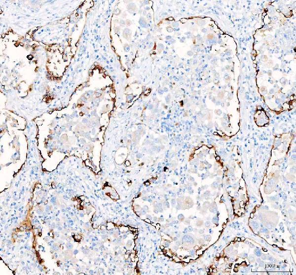
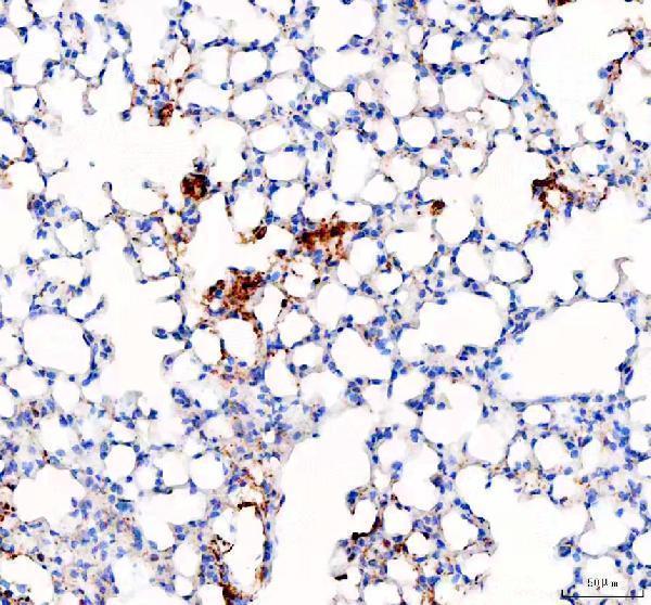
A03957-1 has IHC images from paraffin sections of human lung cancer and mouse and rat lung (catalog IHC image captions). No IF/ICC image is supplied (catalog IF images).

Real IHC data IHC analysis of SLC34A2 using anti-SLC34A2 antibody (A03957-1). SLC34A2 was detected in a paraffin-embedded section of human lung cancer tissue. Heat mediated antigen retrieval was performed in EDTA buffer (pH 8.0, epitope retrieval solution). The tissue section was blocked with 10% goat serum. The tissue section was then incubated with 2 μg/ml rabbit anti-SLC34A2 Antibody (A03957-1) overnight at 4°C. Peroxidase Conjugated Goat Anti-rabbit IgG was used as secondary antibody and incubated for 30 minutes at 37°C. The tissue section was developed using HRP Conjugated Rabbit IgG Super Vision Assay Kit (Catalog # SV0002) with DAB as the chromogen.
Anti-SLC34A2 Antibody ®
Cat # A03957-1

A03957-1 is listed for IHC in human, mouse, and rat (catalog applications; catalog reactivity). Its IHC images show human lung cancer, mouse lung, and rat lung paraffin sections (catalog IHC image captions).

Which to pick: Choose A03957-1 for tissue IHC because its own images show staining in paraffin sections from all three listed species (catalog IHC image captions; catalog reactivity). It is a rabbit antibody; clonality is unreported (catalog host; catalog clone). No IF/ICC antibody can be recommended from this payload because A03957-1 has no listed IF/ICC application or image; its paraffin-section captions do not report the fixative (catalog applications; catalog IF images; catalog IHC image captions).

Each figure is that product's own IHC / IF validation image from its datasheet.

References

  1. UniProt Consortium. UniProt entry O95436 (NPT2B_HUMAN, Sodium-dependent phosphate transport protein 2B).
  2. Human Protein Atlas. SLC34A2 tissue IHC expression (reliability: Enhanced).
  3. Human Protein Atlas. SLC34A2 subcellular location (ICC-IF): Mainly localized to the plasma membrane. In addition localized to the nucleoplasm and cytosol..
  4. Human Protein Atlas. SLC34A2 antibody validation summary (2 antibodies).
  5. Evaluation of the Prognostic Value of Solute Carrier Family 34 Member 2 "SLC34A2" in Papillary Thyroid Carcinoma: An Immunohistochemical Study. Analytical cellular pathology (Amsterdam) 2021 — PMC8298178.
  6. SLC34A2 promotes cell proliferation by activating STX17-mediated autophagy in esophageal squamous cell carcinoma. Thoracic cancer 2024 — PMC11168907.
  7. SLC34A2 Targets in Calcium/Phosphorus Homeostasis of Mammary Gland and Involvement in Development of Clinical Mastitis in Dairy Cows. Animals : an open access journal from MDPI 2024 — PMC11083581.
  8. Phosphate dysregulation via the XPR1-KIDINS220 protein complex is a therapeutic vulnerability in ovarian cancer. Nature cancer 2022 — PMC9246846.
  9. PubMed PMID:10329428 — UniProt-cited evidence.
  10. PubMed PMID:10610722 — UniProt-cited evidence.
  11. PubMed PMID:15815621 — UniProt-cited evidence.