This website uses cookies to ensure you get the best experience on our website.


- Table of Contents
Plan chromogenic IHC on paraffin sections using the catalog antibody’s documented protocol (datasheet A04761). Kidney distal tubules and colon glandular cells provide high-staining reference populations (HPA tissue IHC).

Expected localisation, validated protocols, controls and antibodies — the at-a-glance facts below, then the full design guide.
| Expected localisation | Membranous and cytoplasmic staining (HPA tissue IHC) | |
| Staining pattern | Glandular cells: membranous and cytoplasmic staining (HPA tissue IHC) | |
| Antigen retrieval | Citrate pH 6 HIER, heat-mediated (datasheet A04761) | |
| Positive control | Appendix+4 more · see all | |
| Negative control | Adipose tissue+4 more · see all |
| Fixation | Formaldehyde fixed in the kidney IHC example (selected-SKU IHC image A04761); keep fixation consistent (standard IHC practice; not target-specific) | |
| Caveat | Presumed off-target binding may complicate interpretation (HPA tissue IHC) | |
| Regulation | Glucose uptake enriches surface ZIP14 (UniProt) | |
| Isoform / epitope | 3 isoforms; check extracellular vs cytoplasmic epitope (UniProt) |
The catalog antibody has an IHC-P protocol (datasheet: A04761). Published SLC39A14 IHC protocols cover nasopharyngeal tissue (PMC11009181) and control liver and normal human brain (PMC4894980).
| Sample | Formaldehyde-fixed, paraffin-embedded human kidney tissue (datasheet A04761) |
| Fixation | Image formalin-fixed; duration unreported (datasheet A04761); verify before use. |
| Sectioning | 4–5 µm sections on charged slides (standard) |
| Deparaffinisation | Xylene, graded ethanol series to water (standard) |
| Antigen retrieval | Heat retrieval: Citrate pH 6 (datasheet A04761); 20 min, 95–100 °C (standard) |
| Peroxidase block | 3% H2O2, 10 min, room temperature (standard) |
| Blocking | 10% serum (datasheet A04761) |
| Primary antibody | Rabbit anti-SLC39A14, 1-2 μg/mL (datasheet A04761) |
| Primary incubation | Overnight at 4 °C (standard) |
| Detection | HRP-polymer secondary, DAB chromogen 5–10 min (standard) |
| Counterstain | Hematoxylin, blue, dehydrate and mount (standard) |
| Expected result | SLC39A14-positive staining in glandular cells of appendix (HPA tissue IHC: High). HPA tissue profile: Membranous and cytoplasmic expression in several tissues. No signal in the no-primary control. |
SLC39A14 is a six-pass membrane transporter found at the cell surface and on endosomal and lysosomal membranes (UniProt Q15043 topology and subcellular location). In paraffin-section IHC, expect membranous and cytoplasmic staining in selected epithelial cells, including kidney distal tubules and intestinal glandular cells (HPA: tissue IHC). HPA rates the tissue evidence Enhanced, while noting medium agreement with RNA data and presumed off-target staining that was disregarded (HPA: reliability description).
| Distinct membrane staining with some cytoplasmic signal in kidney distal tubules or intestinal glandular cells. | This fits the reported tissue pattern (HPA: High in kidney distal tubules and small-intestinal glandular cells; membranous and cytoplasmic profile). Score the named cell population and compartment together; intensity alone cannot establish specificity. Intracellular signal is plausible because endosome and lysosome membranes are reported locations (UniProt Q15043 subcellular location). |
| Predominantly nuclear staining, with little membrane or cytoplasmic signal. | Treat a nuclear-dominant pattern as discordant: the reported IHC pattern is membranous and cytoplasmic (HPA: tissue IHC profile), and the annotated locations are membranes (UniProt Q15043 subcellular location). Check the antibody and detection controls before interpreting it as SLC39A14. Neither source establishes a nuclear staining pattern. |
| Strong staining in a cell population listed as not detected, such as adipocytes or bone-marrow hematopoietic cells. | Investigate possible cross-reactivity or endogenous detection activity before scoring it as positive (HPA: Not detected in adipocytes and bone-marrow hematopoietic cells; general IHC practice). A not-detected reference is a comparator, not proof that every specimen or condition must be negative. Review staining in the expected cell populations on the same run. |
| Broad, diffuse color obscures cell borders and the tissue architecture. | Background prevents a compartment call even where SLC39A14 is expected (HPA: membranous and cytoplasmic tissue profile). Uneven reagent exposure, nonspecific binding, or detection-system background are general IHC possibilities. Compare a no-primary control and inspect whether color follows cells or spreads across the section (general IHC practice). |
| No detectable signal in kidney distal tubules or colon glandular cells. | A negative result in these high-staining reference populations warrants a run-level check before a biological conclusion (HPA: High in kidney distal tubules and colon glandular cells). Confirm that the positive-control section developed, then review antibody use, retrieval, and detection steps as general IHC checks. The supplied sources do not establish target-specific fixation sensitivity. |
| Membrane topology and compartment | SLC39A14 has 6 transmembrane segments, extracellular and cytoplasmic regions, and reported surface, endosome, and lysosome locations (UniProt Q15043 topology and subcellular location). These support a membrane-centered interpretation with possible intracellular staining. The sources do not identify this antibody’s epitope, so topology cannot predict its retrieval requirements. |
| Tissue and cell selection | The tissue pattern is cell-specific: kidney distal tubules and colon, rectum, appendix, and small-intestinal glandular cells are High, while adipocytes and bone-marrow hematopoietic cells are listed as not detected (HPA: tissue IHC). Use named cell populations when comparing sections; a whole-tissue label can hide a mixed result. |
| Evidence strength and antibody choice | HPA calls tissue IHC Enhanced for HPA056732 and reports presumed off-target binding that was disregarded in its summary (HPA: antibody validation and reliability description). HPA016508 is ICC Approved but has no listed IHC status (HPA: antibodies). These ratings support different applications; they do not validate every unexpected staining pattern. |
| IF/ICC Q&A: what location should I expect? | In cultured-cell IF, HPA approves plasma-membrane, endoplasmic-reticulum, and Golgi locations and lists images from A-431, U-251MG, and U2OS (HPA: subcellular ICC-IF). This is the IF/ICC interpretation answer; it does not turn those images or the ICC approval into an IHC protocol or establish the same organelle balance in tissue sections. |
| Situation | Likely cause | Next action |
|---|---|---|
| Positive-control tissue is blank. | The run may have failed, or the antibody and detection conditions may be unsuitable; high-staining reference cells are expected in kidney distal tubules or colon glands (HPA: tissue IHC). | Check the control section and reagent sequence, then review retrieval and detection against the IHC-validated antibody’s instructions (general IHC practice). Do not infer an SLC39A14-specific fixation effect from this result. |
| Color is strongest over nuclei. | Nuclear-dominant signal conflicts with the reported membrane and cytoplasmic tissue pattern (HPA: tissue IHC profile; UniProt Q15043 subcellular location). | Inspect a no-primary control and reassess detection background and antibody specificity (general IHC practice). Require convincing staining in expected cells and compartments before scoring the section. |
| Color appears in adipocytes or marrow hematopoietic cells. | Those cell populations are listed as not detected; unexpected color raises a specificity or detection-background question (HPA: tissue IHC). | Compare with an expected positive tissue and a no-primary control in the same run (general IHC practice). Record the cell type and compartment rather than calling the entire tissue positive. |
| A uniform brown haze obscures cell outlines. | Diffuse background cannot be matched confidently to HPA’s membranous and cytoplasmic pattern (HPA: tissue IHC profile). Nonspecific detection is a general IHC possibility. | Review blocking, washes, detection exposure, and the no-primary control (general IHC practice). Score only resolved cellular staining after background is addressed. |
| A granular cytoplasmic pattern appears without a clear surface rim. | Intracellular membrane signal is biologically plausible because endosomes and lysosomes are annotated locations (UniProt Q15043 subcellular location), but granular color alone is not proof of target identity. | Check whether granules occur in HPA-positive cell populations and whether controls show comparable color (HPA: tissue IHC; general IHC practice). Report the observed compartment without claiming organelle identity from chromogenic IHC alone. |
| IF images and tissue IHC seem to emphasize different compartments. | HPA’s cultured-cell ICC-IF summary includes ER and Golgi, whereas its tissue IHC summary reports membranous and cytoplasmic expression (HPA: subcellular ICC-IF and tissue IHC). | Interpret each result within its application and cell context. Use the IF/ICC guide for that assay; the ICC Approved status of HPA016508 does not supply an IHC validation claim (HPA: antibodies). |
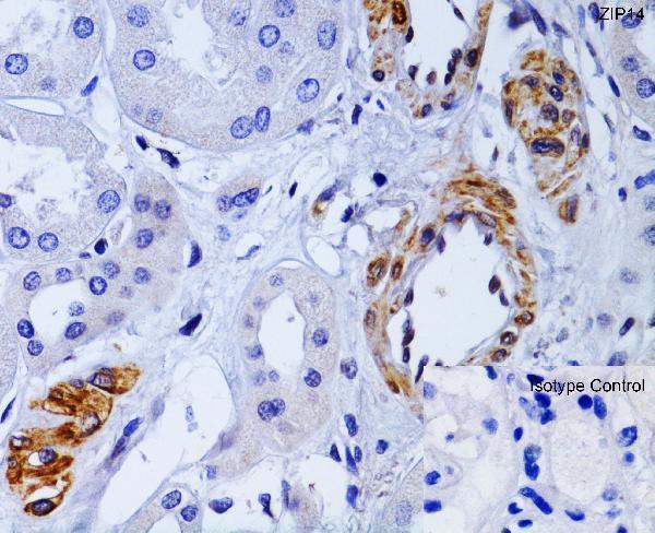
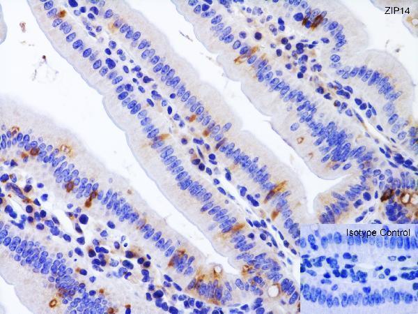


Comprehensive Human Protein Atlas IHC scoring per tissue (reliability: Enhanced — Medium consistency between antibody staining and RNA expression data. Presumed off target binding observed and disregarded.). Rows are taken directly from the HPA tissue chart — click any row's HPA link to view the source.
| Tissue | Cell type | Level | Evidence | Source |
|---|---|---|---|---|
| Appendix | Glandular cells | High | Protein (IHC) | HPA → |
| Colon | Glandular cells | High | Protein (IHC) | HPA → |
| Esophagus | Squamous epithelial cells | High | Protein (IHC) | HPA → |
| Kidney | Distal tubules | High | Protein (IHC) | HPA → |
| Rectum | Glandular cells | High | Protein (IHC) | HPA → |
| Tissue | Cell type | Level | Evidence | Source |
|---|---|---|---|---|
| Adipose tissue | Adipocytes | Not detected | Protein (IHC) | HPA → |
| Bone marrow | Hematopoietic cells | Not detected | Protein (IHC) | HPA → |
| Breast | Adipocytes | Not detected | Protein (IHC) | HPA → |
| Bronchus | Respiratory epithelial cells | Not detected | Protein (IHC) | HPA → |
| Caudate | Glial cells | Not detected | Protein (IHC) | HPA → |
Troubleshoot SLC39A14 staining by checking retrieval, cell type and membrane localisation before interpreting chromogenic signal.
The IHC-validated antibody A04761 has paraffin-section images from human kidney, mouse colon and rat kidney (A04761 image captions); no IF/ICC validation is listed (A04761 applications).
A04761 is listed for IHC-P in human, mouse and rat samples (A04761 applications and reactivity). Its images show paraffin sections of human kidney, mouse colon and rat kidney; the captions report formaldehyde fixation (A04761 image captions).
Which to pick: Choose A04761 for paraffin-section IHC: it is a rabbit polyclonal antibody listed for IHC-P, with 1–2 μg/mL as the catalog starting range (A04761 catalog entry and datasheet). For cross-species IHC, its captions show human kidney and mouse colon at 1 μg/mL and rat kidney at 2 μg/mL, all in paraffin sections fixed with formaldehyde (A04761 image captions). No IF/ICC application or figure is listed for A04761, so the payload does not support an IF/ICC pick (A04761 applications and image captions).