This website uses cookies to ensure you get the best experience on our website.


- Table of Contents
Plan paraffin-section IHC for SLC39A6 with the catalog antibody starting at 2.5 μg/mL (datasheet). Compare the observed cytoplasmic pattern with HPA tissue staining, while considering ZIP6’s membrane localization when interpreting signal (HPA tissue IHC; UniProt).

Expected localisation, validated protocols, controls and antibodies — the at-a-glance facts below, then the full design guide.
| Expected localisation | Cytoplasmic staining (HPA tissue IHC); membrane localization expected (UniProt) | |
| Staining pattern | General cytoplasmic staining; nuclear staining in some tissues (HPA tissue IHC) | |
| Antigen retrieval | Citrate pH 6.0 HIER, 95–98 °C, 20 min (rule: cytoplasmic / membrane antigen) | |
| Positive control | Adrenal gland+4 more · see all | |
| Negative control | Appendix+4 more · see all |
| Fixation | Keep formalin fixation consistent across sections (standard IHC practice; not target-specific); Matched tissue-IHC evidence does not establish the fixation claim. Validate the specimen-specific method before use. (selected-SKU IHC image A05693) | |
| Caveat | Placenta: high expression reported, but trophoblast IHC is absent (UniProt; HPA tissue IHC) | |
| Regulation | TCR activation recruits ZIP6 to the synapse (UniProt) | |
| Isoform / epitope | 2 isoforms; check whether the epitope is extracellular or cytoplasmic (UniProt) |
Compare the catalog antibody’s IHC-P protocol with published SLC39A6 tissue protocols (PMC8505289; PMC4595240; PMC8771636).
| Sample | Tissue sections; selected-image fixative not specified (standard IHC workflow) |
| Fixation | Image fixative and duration unreported (datasheet A05693); verify before use. |
| Sectioning | 4–5 µm sections on charged slides (standard) |
| Deparaffinisation | Xylene, graded ethanol series to water (standard) |
| Antigen retrieval | Heat-induced epitope retrieval in citrate buffer, pH 6.0, 20 min at 95–98 °C (standard rule: cytoplasmic / membrane antigen) |
| Peroxidase block | 3% H2O2, 10 min, room temperature (standard) |
| Blocking | 10% normal serum of the secondary host, 30 min, room temperature (standard) |
| Primary antibody | Rabbit anti-SLC39A6, 2.5 μg/mL (datasheet A05693) |
| Primary incubation | Overnight at 4 °C (standard) |
| Detection | HRP-polymer secondary, DAB chromogen 5–10 min (standard) |
| Counterstain | Hematoxylin, blue, dehydrate and mount (standard) |
| Expected result | SLC39A6-positive staining in glandular cells of adrenal gland (HPA tissue IHC: High). HPA tissue profile: General cytoplasmic expression. Additional nuclear expression in several tissues. No signal in the no-primary control. |
SLC39A6 is a six-pass membrane protein found at the cell membrane, including lamellipodia and membrane rafts (UniProt Q13433 topology/localization). In tissue IHC, expect predominantly cytoplasmic staining, with additional nuclear staining in some tissues (HPA: tissue IHC profile). Signal should follow the identified cell population: for example, adrenal glandular cells, fallopian tube non-ciliated cells and nasopharyngeal basal cells stain High (HPA: tissue IHC). HPA rates tissue staining Approved, with medium consistency against RNA data (HPA: reliability).
| Cytoplasmic staining in adrenal glandular cells, fallopian tube non-ciliated cells or nasopharyngeal basal cells. | This matches HPA's general cytoplasmic profile and its High calls for those cell populations (HPA: tissue IHC). A membrane component is biologically plausible for a six-pass transporter (UniProt Q13433 topology), but HPA's tissue profile does not require a crisp membrane outline in every positive cell. |
| A membrane rim or polarized membrane signal accompanies cytoplasmic staining. | This is compatible with cell membrane localization (UniProt Q13433 localization). Judge it alongside the tissue's named cell type and HPA pattern; membrane signal alone does not validate antibody specificity. UniProt describes apical localization in choroid plexus epithelial cells by similarity, so that detail is an inference, not a demonstrated tissue IHC benchmark (UniProt Q13433 localization). |
| Strong, exclusively nuclear staining dominates many cells, with little cytoplasmic signal. | Treat this as a pattern requiring investigation, since HPA reports general cytoplasmic staining and additional nuclear expression in several tissues (HPA: tissue IHC profile). Nuclear signal alone is therefore not proof of artefact. Compare the affected cell types and controls before assigning a cause; HPA's ICC-IF nucleoplasm call is uncertain (HPA: subcellular). |
| Staining appears in a cell population that HPA lists as Not detected, such as lung alveolar cells. | This conflicts with the stated cell-level IHC observation (HPA: lung alveolar cells, Not detected). Check morphology and detection controls: cross-reactivity or endogenous detection activity are possible general IHC explanations, not established causes for this specimen. A Not detected call is specific to the listed cells and assay, not every cell in the organ. |
| No signal appears in an expected High population, such as adrenal glandular cells. | The result conflicts with an HPA High tissue IHC call (HPA: adrenal glandular cells). First check that the expected cells are present and that the IHC run and counterstain are interpretable; then review antibody and detection conditions (general IHC practice). HPA's Approved rating has medium RNA concordance, so one negative slide cannot establish absent SLC39A6 (HPA: reliability). |
| Cell identity and tissue choice | HPA's calls are cell specific: adrenal glandular, fallopian tube non-ciliated and nasopharyngeal basal cells are High, while lung alveolar and placental trophoblastic cells are Not detected (HPA: tissue IHC). Confirm the scored cell population by morphology; do not generalize a cell-level call to the whole organ (general IHC practice). |
| Subcellular interpretation | Six transmembrane segments and extracellular and cytoplasmic regions support membrane localization (UniProt Q13433 topology). HPA tissue IHC nonetheless describes general cytoplasmic expression with nuclear staining in several tissues (HPA: tissue IHC profile). Record compartments separately when scoring; a membrane-only requirement would discard part of the observed IHC pattern. |
| Evidence and antibody validation | HPA rates tissue IHC Approved with medium consistency between staining and RNA expression (HPA: reliability). HPA042377 is IHC Approved but ICC Uncertain (HPA: antibody validation). These ratings support a cautious tissue IHC reference; they do not establish that every observed compartment or positive cell on a new slide is specific. |
| Epitope placement | SLC39A6 has extracellular and cytoplasmic regions (UniProt Q13433 topology), but the supplied record gives no epitope for the IHC-validated antibody. Antigen retrieval may be optimized as a general paraffin-IHC step (general IHC practice); these sources do not show a target-specific retrieval or fixation effect, so neither can explain a failed stain on its own. |
| Expression source and tissue discrepancy | UniProt reports high expression in placenta, while HPA lists placental trophoblastic cells as Not detected by tissue IHC (UniProt Q13433 tissue specificity; HPA: placenta trophoblastic cells). The statements cover different evidence and cell resolution. Do not make trophoblast a mandatory positive control or infer a fixation effect from the discrepancy. |
| Situation | Likely cause | Next action |
|---|---|---|
| Expected High cells show no chromogenic signal. | Possible assay failure or an unrecognized cell population; HPA lists those cells as High but reports only medium RNA concordance (HPA: tissue IHC; HPA: reliability). | Check section morphology and the expected cells, then review the IHC-validated antibody, retrieval, detection reagents and a working positive control (general IHC practice). Avoid calling the target absent from this slide alone. |
| Diffuse color covers cells and surrounding tissue. | Nonspecific binding, inadequate blocking or washout, or endogenous detection activity are general IHC possibilities (general IHC practice); HPA's cytoplasmic profile does not establish a cause (HPA: tissue IHC profile). | Inspect a no-primary control, blocking and wash steps, and the chromogen or secondary detection controls; score only cell-associated signal after background is resolved (general IHC practice). |
| Staining is predominantly nuclear across many cell types. | HPA allows nuclear expression in several tissues, but its overall tissue profile is cytoplasmic; the ICC-IF nucleoplasm assignment is uncertain (HPA: tissue IHC profile; HPA: subcellular). | Compare nuclear and cytoplasmic signal within the specified cell populations, inspect controls, and report the pattern rather than automatically marking all nuclear color positive or artefactual (general IHC practice). |
| A listed Not detected cell population stains strongly. | Possible cross-reactivity, endogenous detection activity or cell misidentification (general IHC practice); the result disagrees with HPA's cell-specific call (HPA: tissue IHC). | Recheck morphology and the exact HPA cell designation, then review no-primary and detection controls before assigning specificity (general IHC practice). |
| Placental trophoblastic cells are negative despite UniProt's high placental expression. | HPA reports trophoblastic cells Not detected by IHC, whereas UniProt describes expression at the tissue level (HPA: placenta trophoblastic cells; UniProt Q13433 tissue specificity). The supplied sources do not resolve that difference. | Keep the trophoblast result cell specific and select an HPA High cell population to assess assay performance; do not attribute the difference to fixation or retrieval without evidence (HPA: tissue IHC; general IHC practice). |
| Can the tissue IHC result determine the expected IF/ICC pattern? | HPA's ICC-IF summary places SLC39A6 mainly in cytosol (uncertain), additionally in nucleoplasm (uncertain) and plasma membrane (supported); HPA042377 is ICC Uncertain (HPA: subcellular; HPA: antibody validation). | Use the separate IF/ICC guide for assay design. Treat the membrane call as supported and the cytosol and nucleoplasm calls as uncertain; do not transfer tissue IHC validation or its chromogenic appearance directly to IF/ICC (HPA: subcellular; HPA: antibody validation). |
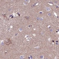
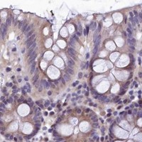


Comprehensive Human Protein Atlas IHC scoring per tissue (reliability: Approved — Medium consistency between antibody staining and RNA expression data.). Rows are taken directly from the HPA tissue chart — click any row's HPA link to view the source.
| Tissue | Cell type | Level | Evidence | Source |
|---|---|---|---|---|
| Adrenal gland | Glandular cells | High | Protein (IHC) | HPA → |
| Endometrium | Glandular cells | Medium | Protein (IHC) | HPA → |
| Fallopian tube | Non-ciliated cells | High | Protein (IHC) | HPA → |
| Nasopharynx | Basal cells | High | Protein (IHC) | HPA → |
| Bronchus | Ciliated cells (cell body) | Medium | Protein (IHC) | HPA → |
| Tissue | Cell type | Level | Evidence | Source |
|---|---|---|---|---|
| Appendix | Glandular cells | Not detected | Protein (IHC) | HPA → |
| Caudate | Glial cells | Not detected | Protein (IHC) | HPA → |
| Lung | Alveolar cells | Not detected | Protein (IHC) | HPA → |
| Lymph node | Germinal center cells | Not detected | Protein (IHC) | HPA → |
| Ovary | Follicle cells | Not detected | Protein (IHC) | HPA → |
Troubleshoot ZIP6 staining by checking retrieval, cell identity and compartment, then score signal against appropriate tissue controls (UniProt Q13433; HPA tissue IHC).
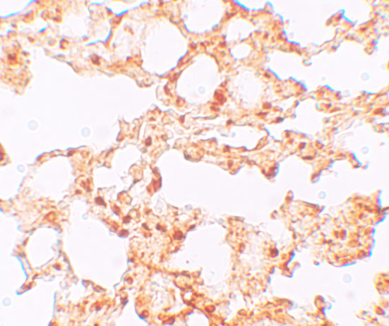
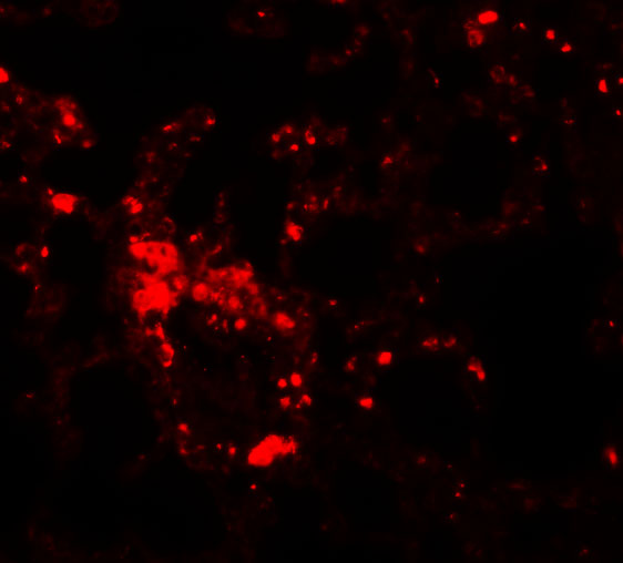
A05693 is listed for IHC-P and IF in human and mouse (catalog: applications and reactivity), with IHC and IF images from mouse lung tissue (image captions).
A05693 will render with a mouse lung IHC image at 5 μg/mL (IHC image caption). The catalog also lists IF and shows a mouse lung IF image at 20 μg/mL (catalog: applications; IF image caption).
Which to pick: For paraffin-section tissue IHC, choose A05693 (catalog: IHC-P); its mouse lung IHC image reports 5 μg/mL, while the fixative is unreported (IHC image caption). For IF, choose A05693 (catalog: IF; IF image caption: mouse lung); ICC validation is unreported (catalog: applications). For work across human and mouse samples, A05693 lists both species (catalog: reactivity), though the supplied images show mouse tissue only (IHC and IF image captions); clonality is unreported (catalog: clone).