This website uses cookies to ensure you get the best experience on our website.


- Table of Contents
Plan SLC3A2 paraffin IHC around membranous staining in renal proximal tubules and placental trophoblasts (HPA tissue IHC). The catalog antibody has a documented chromogenic IHC workflow (datasheet A01794-4).

Expected localisation, validated protocols, controls and antibodies — the at-a-glance facts below, then the full design guide.
| Expected localisation | Membranous staining in several tissue cell types (HPA tissue IHC) | |
| Staining pattern | Membranous in proximal tubules, islets and squamous cells (HPA tissue IHC) | |
| Antigen retrieval | EDTA pH 8.0 HIER, heat-mediated (datasheet A01794-4) | |
| Positive control | Kidney+4 more · see all | |
| Negative control | Adipose tissue+4 more · see all |
| Fixation | Keep fixation consistent across sections (standard IHC practice; not target-specific) | |
| Caveat | Plasma membrane localization depends on SLC7A5/8 pairing (UniProt) | |
| Regulation | Placental expression is stronger at full term (UniProt) | |
| Isoform / epitope | Four isoforms; check whether the epitope is extracellular or cytoplasmic (UniProt) |
The catalog antibody’s IHC-P protocol is followed by 3 published SLC3A2 protocols for xenograft and clinical paraffin sections (PMC9043103; PMC3749780; PMC5428444).
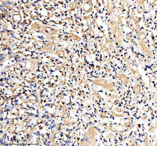
| Sample | Paraffin-embedded human glioma tissue; fixative not specified (datasheet A01794-4) |
| Fixation | Image fixative and duration unreported (datasheet A01794-4); verify before use. |
| Sectioning | 4–5 µm sections on charged slides (standard) |
| Deparaffinisation | Xylene, graded ethanol series to water (standard) |
| Antigen retrieval | Heat retrieval: EDTA pH 8.0 (datasheet A01794-4); 20 min, 95–100 °C (standard) |
| Peroxidase block | 3% H2O2, 10 min, room temperature (standard) |
| Blocking | 10% goat serum (datasheet A01794-4) |
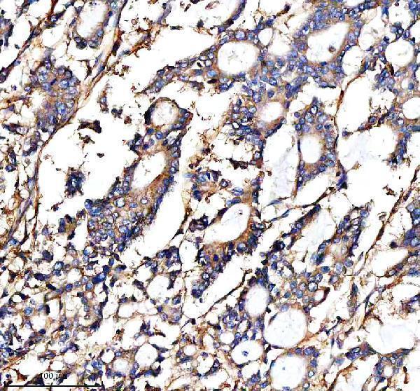
| Primary antibody | Rabbit anti-SLC3A2, 1:50 recommended; image 1:100 (datasheet A01794-4) |
| Primary incubation | Overnight at 4 °C (datasheet A01794-4) |
| Detection | HRP-conjugated secondary, DAB chromogen (datasheet A01794-4) |
| Counterstain | Hematoxylin, blue, dehydrate and mount (standard) |
| Expected result | SLC3A2-positive staining in proximal tubules (cell body) of kidney (HPA tissue IHC: High). HPA tissue profile: Membranous expression in several cell types, mainly in seminiferous ducts, islets of Langerhans, squamous epithelial cells and renal proximal tubules. No signal in the no-primary control. |
SLC3A2 is a single-pass membrane protein with an extracellular region spanning residues 105–529 (UniProt P08195 topology). In paraffin-section IHC, expect mainly membranous staining in renal proximal tubules, pancreatic endocrine cells, placental trophoblasts and testicular Sertoli cells (HPA: tissue IHC). HPA rates the tissue staining profile Enhanced, reflecting high consistency between antibody staining and RNA expression (HPA: reliability).
| Distinct cell borders stain in renal proximal tubules, pancreatic endocrine cells, placental trophoblasts or Sertoli cells (HPA: High in each cell type). | This matches the reported membranous IHC profile (HPA: tissue IHC) and plasma-membrane localization (UniProt P08195 subcellular location). Assess the identified cells and their boundaries; a uniformly stained section does not establish the expected cell-specific pattern (general IHC interpretation). |
| Nuclei dominate the chromogenic signal, with little staining at cell borders. | Predominantly nuclear IHC is discordant with the membranous tissue profile (HPA: tissue IHC). Investigate nonspecific staining or detection artefact (general IHC practice). HPA also reports uncertain nucleoplasmic localization in ICC-IF, so that observation alone does not establish a nuclear IHC pattern (HPA: subcellular ICC-IF). |
| Strong signal appears in adipocytes or another listed cell type scored Not detected (HPA: adipose tissue and breast adipocytes). | Check cell identification and compare with a positive tissue on the same run (general IHC practice). Unexpected signal may reflect cross-reactivity or endogenous detection activity (general IHC practice). An HPA Not detected score is an observation in that cell type, not proof that every specimen must be negative (HPA: tissue IHC). |
| Color spreads across extracellular space or appears similarly in unrelated cells and the tissue background. | Diffuse signal lacks the expected cell-border pattern (HPA: tissue IHC). Examine the no-primary control, blocking and detection reagents for background or endogenous activity (general IHC practice); background alone cannot be assigned to SLC3A2 (general IHC interpretation). |
| A known-positive section has no convincing signal in renal proximal tubules or placental trophoblasts (HPA: High in both cell types). | First assess tissue preservation, retrieval, antibody and detection controls (general IHC practice). An absent result conflicts with the reported positive-cell profile (HPA: tissue IHC), but it does not by itself show that SLC3A2 is absent from the specimen or identify which workflow step failed (general IHC interpretation). |
| Membrane topology and compartment | SLC3A2 has one transmembrane segment at residues 84–104, a cytoplasmic region at 2–83 and an extracellular region at 105–529 (UniProt P08195 topology). Its plasma-membrane location depends in part on association with SLC7A5 or SLC7A8 (UniProt P08195 subcellular location). Read border staining in the context of the cell type and tissue architecture (general IHC interpretation). |
| Tissue and cell-type variation | HPA scores proximal tubules, pancreatic endocrine cells, trophoblasts and Sertoli cells High, but cervical and esophageal squamous epithelial cells Medium (HPA: tissue IHC). Adipocytes in adipose tissue and breast are scored Not detected (HPA: tissue IHC). Compare like cell types when judging intensity; a whole-section average can obscure the reported distribution (general IHC interpretation). |
| Antibody validation | HPA lists Enhanced IHC validation for HPA017980 and CAB010455 (HPA: antibodies). The tissue profile has Enhanced reliability because staining is highly consistent with RNA expression (HPA: reliability). These assessments support use of the reported pattern as a reference; they do not validate every antibody, specimen or staining run (general IHC interpretation). |
| Antigen retrieval and epitope access | Retrieval conditions and antibody dilution should be optimized against a known-positive section and appropriate controls (general IHC practice). The supplied UniProt and HPA records do not report an SLC3A2-specific retrieval requirement or fixation sensitivity (UniProt P08195; HPA: tissue IHC). Topology and four annotated glycosylation sites do not establish how fixation changes staining (UniProt P08195 topology and glycosylation). |
| IF/ICC Q: What localization should be expected? | A: HPA reports supported plasma-membrane localization and uncertain nucleoplasmic localization in ICC-IF (HPA: subcellular ICC-IF). Interpret IF/ICC on its own guide page; this paraffin-section IHC pattern does not establish an IF/ICC protocol or make nuclear fluorescence a confirmed IHC finding (HPA: tissue IHC; HPA: subcellular ICC-IF). |
| Situation | Likely cause | Next action |
|---|---|---|
| Positive-control cell borders are faint or absent. | The run may have insufficient detectable signal; the HPA High designation identifies an expected positive cell population but cannot diagnose the workflow (HPA: tissue IHC; general IHC practice). | Confirm the expected cells are present, then check retrieval, antibody dilution, detection reagents and counterstain against run controls (general IHC practice). Avoid scoring the study tissue negative until the positive control performs (general IHC interpretation). |
| Most cells show broad brown staining without distinct borders. | This differs from the reported membranous distribution (HPA: tissue IHC). Background from blocking, detection chemistry or overly concentrated antibody is possible (general IHC practice). | Review the no-primary control and reagent-only controls where appropriate; optimize blocking, washes and antibody dilution using a positive tissue (general IHC practice). Reassess whether cell borders remain distinguishable after background is reduced (general IHC interpretation). |
| Nuclear staining is stronger than membrane staining in IHC. | The nuclear-dominant pattern does not match HPA tissue IHC (HPA: tissue IHC). HPA labels nucleoplasmic ICC-IF localization uncertain, so it does not confirm this chromogenic pattern (HPA: subcellular ICC-IF). | Check the no-primary control, counterstain and detection background; compare with membranous staining in a known-positive cell population (general IHC practice; HPA: tissue IHC). Do not score nuclear color alone as the expected IHC result (general IHC interpretation). |
| An HPA Not detected cell type stains strongly. | Misidentified cells, cross-reactivity or endogenous detection activity could explain a discordant result (general IHC practice; HPA: tissue IHC). | Verify the cell type on the counterstained section and inspect no-primary and detection controls (general IHC practice). Compare the same run with an HPA High population before assigning the unexpected color to SLC3A2 (HPA: tissue IHC; general IHC interpretation). |
| A whole tissue looks weak despite some clearly positive cells. | HPA scores specific cells within tissues, and its reported levels vary by cell type (HPA: tissue IHC). Tissue-wide visual averages can therefore misrepresent a localized pattern (general IHC interpretation). | Score the relevant cell population and its membrane staining separately from surrounding cells (general IHC interpretation). Use the HPA cell-type entries as the comparison, including Medium squamous epithelium and High proximal tubules (HPA: tissue IHC). |


Comprehensive Human Protein Atlas IHC scoring per tissue (reliability: Enhanced — High consistency between antibody staining and RNA expression data.). Rows are taken directly from the HPA tissue chart — click any row's HPA link to view the source.
| Tissue | Cell type | Level | Evidence | Source |
|---|---|---|---|---|
| Kidney | Proximal tubules (cell body) | High | Protein (IHC) | HPA → |
| Pancreas | Pancreatic endocrine cells | High | Protein (IHC) | HPA → |
| Parathyroid gland | Glandular cells | High | Protein (IHC) | HPA → |
| Placenta | Trophoblastic cells | High | Protein (IHC) | HPA → |
| Testis | Sertoli cells | High | Protein (IHC) | HPA → |
| Tissue | Cell type | Level | Evidence | Source |
|---|---|---|---|---|
| Adipose tissue | Adipocytes | Not detected | Protein (IHC) | HPA → |
| Adrenal gland | Glandular cells | Not detected | Protein (IHC) | HPA → |
| Appendix | Glandular cells | Not detected | Protein (IHC) | HPA → |
| Bone marrow | Hematopoietic cells | Not detected | Protein (IHC) | HPA → |
| Breast | Adipocytes | Not detected | Protein (IHC) | HPA → |
Troubleshoot SLC3A2 staining in paraffin sections by checking retrieval, membrane localisation and cell-specific controls (datasheet A01794-4; HPA tissue IHC; UniProt P08195).
A01794-4 has IHC images of human glioma, colon cancer, placenta, and liver cancer paraffin sections (catalog IHC captions); IF/ICC and rat reactivity are listed (catalog applications/reactivity).
A01794-4 shows chromogenic IHC in human glioma, colon cancer, placenta, and liver cancer paraffin sections (catalog IHC captions). IF/ICC are listed applications for this human- and rat-reactive antibody, but no IF image is supplied (catalog applications/reactivity; IF image alts).
Which to pick: For tissue IHC, choose A01794-4: its images document paraffin sections with EDTA retrieval at pH 8.0, a 1:100 primary dilution, and DAB detection; the fixative is unreported (catalog IHC captions). For IF/ICC or rat samples, A01794-4 is the listed option because it is polyclonal, includes IF/ICC among its applications, and lists human and rat reactivity; the supplied images document human IHC only (catalog dilution_raw/applications/reactivity; IHC and IF image alts).