SLC45A3 / Solute carrier family 45 member 3 · IHC design guide

Design Immunohistochemistry for SLC45A3

Plan chromogenic IHC for SLC45A3 in paraffin sections using prostate glandular cells as a positive reference (HPA tissue IHC). The catalog antibody has an IHC dilution range of 1:100–1:300 (datasheet M11232-1); adipose tissue offers a negative reference (HPA tissue IHC).

Evidence assembled Oct 2026 · For research use; verify linked source records and product datasheet before use
Immunohistochemistry protocol sheet for SLC45A3 (IHC for SLC45A3): expected localisation Membrane protein; tissue compartment unreported (UniProt; HPA tissue IHC), antibody M11232-1, validated IHC image, and IHC protocol steps
Printable SLC45A3 IHC protocol sheet — expected localisation Membrane protein; tissue compartment unreported (UniProt; HPA tissue IHC), antibody M11232-1, controls and protocol steps. Open the full SLC45A3 IHC guide →

SLC45A3 Immunohistochemistry Experimental Design Guide

Expected localisation, validated protocols, controls and antibodies — the at-a-glance facts below, then the full design guide.

Must know before staining
Expected localisation Membrane protein; tissue compartment unreported (UniProt; HPA tissue IHC)
Staining pattern High staining in prostate glandular cells; compartment unreported (HPA tissue IHC)
Antigen retrieval EDTA pH 9.0 HIER, heat-mediated (datasheet M11232-1)
Positive control ⓘ Prostate
Negative control ⓘ Adipose tissue+4 more · see all
Important caveats
Reasons your staining may differ from the expected pattern.
Fixation Keep fixation consistent across sections (standard IHC practice; not target-specific)
Caveat Negative staining outside prostate may reflect tissue specificity (HPA tissue IHC)
Regulation Prostate-enriched expression (HPA tissue IHC)
Isoform / epitope No annotated isoforms; extracellular versus cytoplasmic epitope access matters (UniProt)
Section 1

Recommended SLC45A3 IHC & IF Protocols

The catalog antibody’s IHC-P protocol (datasheet M11232-1) is accompanied by one published prostate cancer IHC protocol (PMC5650326 methods).

Recommended immunohistochemistry (IHC-P) protocol parameters
SampleParaffin-embedded human Prostate tissue; fixative not specified (datasheet M11232-1)
FixationImage fixative and duration unreported (datasheet M11232-1); verify before use.
Sectioning4–5 µm sections on charged slides (standard)
DeparaffinisationXylene, graded ethanol series to water (standard)
Antigen retrievalHeat retrieval: EDTA pH 9.0, 20min (datasheet M11232-1)
Peroxidase block3% H2O2, 10 min, room temperature (standard)
Blocking10% normal serum of the secondary host, 30 min, room temperature (standard)
Primary antibodyRabbit anti-SLC45A3, 1:100-1:300 (datasheet M11232-1)
Primary incubationOvernight at 4 °C (standard)
DetectionHRP-polymer secondary, DAB chromogen 5–10 min (standard)
CounterstainHematoxylin, blue, dehydrate and mount (standard)
Expected resultSLC45A3-positive staining in glandular cells of prostate (HPA tissue IHC: High). HPA tissue profile: Selective expression in prostate. No signal in the no-primary control.
💡Decision noteStart with heat-mediated EDTA retrieval at pH 9.0 for the catalog antibody (datasheet M11232-1).
Section 2

What Is the Expected SLC45A3 Staining Pattern?

SLC45A3 is an 11-pass membrane protein (UniProt Q96JT2 topology). In prostate IHC, expect high staining in glandular cells (HPA: High in prostate glandular cells). HPA rates the tissue pattern Enhanced because antibody staining is consistent with RNA expression (HPA: Enhanced reliability). ICC-IF places the protein mainly in vesicles, with additional nucleoplasmic localization; that observation does not define its appearance in paraffin sections (HPA: approved subcellular locations).

What am I looking at on my slide?
Strong staining in prostate glandular cells, with a membrane-associated or punctate appearance.The cell type and intensity fit the tissue result (HPA: High in prostate glandular cells). Vesicular staining is plausible from ICC-IF (HPA: Vesicles, approved), but the supplied tissue IHC record does not specify a subcellular pattern.
Predominantly nuclear staining in prostate glands, without a convincing glandular membrane-associated or punctate pattern.Treat this as an uncertain IHC result and check controls. Nucleoplasmic localization is reported in ICC-IF (HPA: Nucleoplasm, approved), so nuclear signal alone cannot establish an artefact or a correct paraffin-section pattern.
Staining in cells from an HPA-negative comparison tissue, such as adipocytes in adipose tissue.This disagrees with the reported tissue result (HPA: Not detected in adipose-tissue adipocytes). Consider antibody cross-reactivity or endogenous chromogenic activity; inspect the no-primary control before assigning a cause (general IHC practice).
Diffuse color across glands and surrounding tissue, obscuring cell boundaries.The slide cannot be scored confidently for the selective glandular pattern (HPA: selective expression in prostate). Diffuse background can arise from nonspecific binding or detection chemistry; controls are needed to distinguish them (general IHC practice).
No staining in an adequately preserved prostate section expected to be positive.A negative result conflicts with the reported high glandular signal (HPA: High in prostate glandular cells). Check the positive control and staining run before interpreting the specimen as lacking SLC45A3 (general IHC practice).
💡Expected SLC45A3 appearanceCall a prostate section positive when glandular cells show clear, relatively high staining (HPA: High in prostate glandular cells); widespread staining in an HPA-negative cell population is suspect (HPA: Not detected in adipose-tissue adipocytes), while the precise IHC compartment remains unreported.
How each factor affects the staining
Tissue and cell selectionProstate glandular cells provide the documented positive comparator; adipose-tissue adipocytes provide a documented negative comparator (HPA: High in prostate glandular cells; Not detected in adipose-tissue adipocytes). These ratings describe the listed cell populations, not every cell in either tissue.
Membrane topology and epitope accessSLC45A3 has 11 transmembrane segments (UniProt Q96JT2 topology). The payload gives no antibody epitope or target-specific retrieval result, so topology alone cannot prescribe retrieval, permeabilisation, or an expected chromogenic subcellular pattern.
Antibody evidence for tissue IHCBoth listed antibodies, HPA019073 and HPA019075, have Enhanced IHC status (HPA: antibody validation). Enhanced tissue evidence supports the reported distribution; it does not validate every staining compartment or every laboratory's detection conditions.
IF/ICC interpretation — does it establish an IHC pattern?No. ICC-IF reports mainly vesicles and additional nucleoplasm, with Approved localization for each (HPA: subcellular ICC-IF). Use those observations as context when reviewing compartments, while scoring paraffin IHC against its tissue and cell evidence (HPA: tissue IHC).
Endogenous chromogenic activityA colored deposit may arise from the detection system even without specific primary-antibody binding (general IHC practice). A no-primary control helps assess this possibility; HPA tissue staining levels do not identify the cause of background in a new run.
Why is my staining missing, weak or wrong?
SituationLikely causeNext action
Prostate glandular cells are negative.The run may have insufficient detectable signal; the expected tissue result is high (HPA: High in prostate glandular cells).Confirm the positive control stained and review antibody dilution, retrieval, detection reagents, and counterstain against the laboratory's established IHC procedure (general IHC practice). No SLC45A3-specific retrieval condition is supplied.
The whole section has weak, diffuse color.Nonspecific binding or detection background can obscure the selective prostate pattern (general IHC practice; HPA: selective expression in prostate).Compare with a no-primary control, review blocking and washes, and adjust antibody concentration within the validated workflow (general IHC practice). Do not score diffuse color as glandular positivity.
An HPA-negative comparison cell population stains.Cross-reactivity or endogenous chromogenic activity is possible when a reported negative population is colored (HPA: Not detected in adipose-tissue adipocytes; general IHC practice).Inspect no-primary and positive controls, then reassess the antibody and detection conditions (general IHC practice). Judge the comparison by its named cell population, not the whole tissue.
Staining is mainly nuclear.Compartment assignment is uncertain in paraffin IHC: additional nucleoplasm was observed by ICC-IF, but no matching tissue IHC compartment is specified (HPA: Nucleoplasm, approved; HPA: tissue IHC).Check controls and whether prostate glandular cells retain the expected selective signal (HPA: High in prostate glandular cells). Avoid classifying nuclear staining alone as proven specific or artefactual.
Punctate staining is present, but cell identity is unclear.Vesicles are the main reported ICC-IF location, yet puncta alone do not identify the stained cell or validate an IHC result (HPA: Vesicles, approved).Use morphology and the counterstain to identify glandular cells, then compare the distribution with the reported prostate pattern (HPA: High in prostate glandular cells; general IHC practice).
A prostate section stains unevenly across the slide.Uneven reagent coverage or section handling can produce regional variation (general IHC practice); the supplied sources do not establish a target-specific fixation effect.Review section integrity, reagent coverage, and run controls before scoring weak areas (general IHC practice). Record the uneven result rather than inferring loss of SLC45A3 expression.

Sample controls for SLC45A3 IHC & IF

🧪Run prostate first and require staining in glandular cells (HPA: High in prostate glandular cells). Use adipose tissue adipocytes as the negative comparator (HPA: Not detected in adipocytes); use nonglandular cells on the prostate slide to assess background, but do not assume they are target-negative because HPA provides no negative cell-type call for prostate.
Positive control tissue: Prostate (Glandular cells, HPA High)
Negative control tissue: Adipose tissue (HPA Not detected)
ICC-IF cell lines (HPA subcellular resource): HPA ICC-IF images show SLC45A3 in A-431, U2OS, U-251MG, with annotated localisation: Vesicles (approved) (HPA subcellular).
Technical controls: Include a no-primary, secondary-only control; a host-species- and isotype-matched control for a monoclonal primary or normal host IgG for a polyclonal primary; and, if available, a verified SLC45A3 knockout or immunizing-peptide block (standard IHC controls). For chromogenic prostate IHC, quench endogenous peroxidase and check for endogenous biotin if using biotin-based detection (standard IHC practice).
⚠️Feasibility: A target-specific fixation window or fixation effect is unreported, and the selected M11232-1 tissue-IHC caption does not state the fixative; its paraffin section therefore does not establish fixation (M11232-1 caption). EDTA retrieval at pH 9.0 and >98°C for 20 minutes is a documented starting condition, but a retrieval dependency has not been established (M11232-1 caption). The evidence does not establish whether frozen sections or IF are easier, and it reports no prostate-specific artefact; score staining in glandular cells rather than acellular luminal material (HPA: High in prostate glandular cells; M11232-1 caption). The selected M11232-1 tissue-IHC caption documents paraffin sections, but does not specify the fixative (selected-SKU IHC image M11232-1).

HPA tissue IHC evidence for SLC45A3

Comprehensive Human Protein Atlas IHC scoring per tissue (reliability: Enhanced — High consistency between antibody staining and RNA expression data.). Rows are taken directly from the HPA tissue chart — click any row's HPA link to view the source.

Positive expression · recommended positive controls

TissueCell typeLevelEvidenceSource
Prostate Glandular cells High Protein (IHC) HPA →

Undetected expression · recommended negative controls

TissueCell typeLevelEvidenceSource
Adipose tissue Adipocytes Not detected Protein (IHC) HPA →
Adrenal gland Glandular cells Not detected Protein (IHC) HPA →
Appendix Endocrine cells Not detected Protein (IHC) HPA →
Bone marrow Hematopoietic cells Not detected Protein (IHC) HPA →
Breast Adipocytes Not detected Protein (IHC) HPA →
Section 3

Advanced SLC45A3 IHC Tips

Troubleshoot SLC45A3 staining in paraffin sections by checking retrieval, compartment, glandular cell specificity, controls and scoring before interpreting signal.

What retrieval conditions should I start with for SLC45A3 paraffin sections?
Start with heat-mediated retrieval in EDTA at pH 9.0 (datasheet M11232-1). The catalog antibody’s paraffin-section image used heating above 98°C for 20 minutes, followed by primary antibody at 1:200 overnight at 4°C (datasheet M11232-1). Keep section thickness, heating and cooling consistent across the run so differences in retrieval do not masquerade as differences in expression. If glandular staining is weak, check tissue adhesion and heating uniformity before trying another buffer as a fallback. Compare any adjustment against a prostate control, where glandular-cell staining is reported as high (HPA tissue IHC).
Could fixation explain weak or uneven SLC45A3 staining?
The selected tissue image identifies paraffin-embedded prostate but does not state its fixative, so target-specific fixation sensitivity is unknown (datasheet M11232-1). Record each specimen’s fixative and fixation duration, then compare sections processed and stained together using the same retrieval conditions. In routine IHC, prolonged fixation can reduce epitope access, while poor preservation can produce uneven morphology; neither effect has been established specifically for SLC45A3 here. Check whether signal loss follows processing batches or damaged areas before changing antibody concentration. Use intact prostatic glandular cells as the within-run reference because they are reported to express SLC45A3 (HPA tissue IHC; UniProt Q96JT2 tissue specificity).
Which staining pattern and cells should count as plausible SLC45A3 signal?
Prioritize staining in prostatic glandular cells, where expression is reported as high, and review the pattern at cell level (HPA tissue IHC). SLC45A3 is an 11-pass membrane protein (UniProt Q96JT2 topology), while cell imaging places it mainly in vesicles with an additional nucleoplasmic location (HPA subcellular). A punctate or membrane-associated glandular pattern is therefore plausible, but a compartment alone cannot establish specificity. Compare staining with tissue morphology, the negative control and similarly processed prostate sections. Treat staining confined to unrelated cell populations or damaged tissue as suspect until it is reproduced with appropriate controls.
How do I troubleshoot staining if the antibody epitope is unspecified?
Check whether the antibody documentation identifies the immunogen or epitope before interpreting a negative paraffin section. SLC45A3 has 11 annotated transmembrane segments and no annotated isoforms, domains or glycosylation sites in the supplied record (UniProt Q96JT2). Those annotations do not reveal which side of the membrane this antibody recognizes or whether its epitope survives processing. Keep retrieval and detection settings fixed while comparing a known positive prostate section with the test section, then change one condition at a time. An absent signal should remain unresolved if the epitope and its accessibility have not been established.
How should I assess SLC45A3 by multiplex IF alongside the IHC result?
For a separate IF assay, multiplex SLC45A3 with a validated prostatic glandular-cell marker, the cell population reported positive in tissue (HPA tissue IHC). Choose fluorophores after measuring tissue autofluorescence, favoring channels with clear separation from that background, and include single-stain controls. Because SLC45A3 spans the membrane 11 times (UniProt Q96JT2 topology), choose permeabilisation only after establishing whether the antibody epitope is accessible from the cell exterior or requires intracellular access. Cell imaging reports mainly vesicular and additional nucleoplasmic localisation (HPA subcellular). Treat IF fixation, permeabilisation and staining conditions as assay-specific; the paraffin-section caption does not establish them (datasheet M11232-1).
How can I distinguish diffuse background from specific glandular staining?
First compare the specimen with a no-primary control and inspect whether color deposits follow glandular cells or spread across stroma, folds and section edges. Prostatic glandular cells are the expected positive population (HPA tissue IHC; UniProt Q96JT2 tissue specificity). In a chromogenic workflow, check peroxidase blocking, secondary-reagent background and wash quality before increasing antibody dilution; these are general IHC controls. The selected paraffin image used primary and secondary antibodies at 1:200, with secondary incubation for 30 minutes at room temperature (datasheet M11232-1). Reassess suspicious diffuse color against the same-run controls before counting any cells as positive.
What should I score when comparing SLC45A3 staining across sections? ⚠ ANSWER MARKED FOR VERIFICATION
Define the region of interest before scoring and count intact prostatic glandular cells, the reported positive population (HPA tissue IHC). Record the percentage of positive glandular cells and staining intensity, then calculate an H-score if intensity grades are reproducible across the run. Normalise cell counts to the number of evaluable glandular cells, or report positive-cell density per mm² of evaluable glandular area when area is the denominator. Keep retrieval, chromogen development and image thresholds consistent between sections. Exclude folds, necrosis and edge staining from the denominator, and report those exclusions alongside the score so low values remain interpretable.
When is a strong SLC45A3 IHC signal likely to be artefactual?
A credible result should involve prostatic glandular cells, the tissue population with high reported staining (HPA tissue IHC). SLC45A3 is a membrane protein with 11 transmembrane segments (UniProt Q96JT2 topology), and cell imaging reports mainly vesicular plus additional nucleoplasmic localisation (HPA subcellular). Signal limited to unrelated cells, section edges or necrotic regions deserves review even when its color is intense. Check a no-primary control for nonspecific detection and the peroxidase block for endogenous enzyme activity; both are standard chromogenic IHC checks. Interpret an isolated nuclear pattern cautiously in tissue and seek agreement with morphology and controls before calling it specific.
Boster reagents

Best SLC45A3 / Solute carrier family 45 member 3 IHC Antibodies

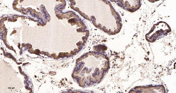
The IHC-validated antibody has an IHC image from paraffin-embedded human prostate tissue; no IF image is supplied (catalog: M11232-1 image caption and applications).

Real IHC data Immunohistochemical analysis of paraffin-embedded human Prostate tissue. 1,primary Antibody was diluted at 1:200(4°C,overnight). 2, EDTA pH 9.0 was used for antibody retrieval(>98°C,20min). 3,Secondary antibody was diluted at 1:200(room tempeRature, 30min).
Anti-Prostein Rabbit Monoclonal Antibody
Cat # M11232-1

M11232-1 will render with an IHC figure from paraffin-embedded human prostate tissue (catalog: M11232-1 image caption). Its listed reactivity is Human, and its listed application is IHC (catalog: M11232-1 reactivity and applications).

Which to pick: Choose M11232-1 for human prostate paraffin-section IHC; its own caption documents EDTA pH 9.0 retrieval and a 1:200 primary antibody dilution, but does not report the fixative (catalog: M11232-1 image caption). Neither catalog SKU lists IF/ICC validation or non-human reactivity, so neither is a supported choice here for IF/ICC or cross-species work (catalog: M11232 and M11232-1 applications and reactivity).

Each figure is that product's own IHC / IF validation image from its datasheet.

References

  1. UniProt Consortium. UniProt entry Q96JT2 (S45A3_HUMAN, Solute carrier family 45 member 3).
  2. Human Protein Atlas. SLC45A3 tissue IHC expression (reliability: Enhanced).
  3. Human Protein Atlas. SLC45A3 subcellular location (ICC-IF): Mainly localized to vesicles. In addition localized to the nucleoplasm..
  4. Human Protein Atlas. SLC45A3 antibody validation summary (2 antibodies).
  5. ERG overexpression plus SLC45A3 (prostein) and PTEN expression loss: Strong association of the triple hit phenotype with an aggressive pathway of prostate cancer progression. Oncotarget 2017 — PMC5650326.
  6. Immunohistochemical markers as predictors of prognosis in multifocal prostate cancer. Virchows Archiv : an international journal of pathology 2024 — PMC11329545.
  7. In vivo miR-138-5p inhibition alleviates monocrotaline-induced pulmonary hypertension and normalizes pulmonary KCNK3 and SLC45A3 expression. Respiratory research 2020 — PMC7364627.
  8. PubMed PMID:11245466 — UniProt-cited evidence.
  9. PubMed PMID:14702039 — UniProt-cited evidence.
  10. PubMed PMID:15489334 — UniProt-cited evidence.