This website uses cookies to ensure you get the best experience on our website.


- Table of Contents
Plan chromogenic IHC for SLC5A5 using the IHC-validated antibody A04465-1 (datasheet). Compare membranous and cytoplasmic staining in glandular cells, with thyroid gland and stomach as high-staining reference tissues (HPA tissue IHC).

Expected localisation, validated protocols, controls and antibodies — the at-a-glance facts below, then the full design guide.
| Expected localisation | Membranous and cytoplasmic staining (HPA tissue IHC) | |
| Staining pattern | Glandular cells: membranous and cytoplasmic staining (HPA tissue IHC) | |
| Antigen retrieval | EDTA pH 8.0 HIER, heat-mediated (datasheet A04465-1) | |
| Positive control | Choroid plexus+3 more · see all | |
| Negative control | Adipose tissue+4 more · see all |
| Fixation | Matched tissue-IHC evidence does not establish the fixation claim. Validate the specimen-specific method before use. (selected-SKU IHC image A04465-1) | |
| Caveat | Presumed off-target staining requires careful interpretation (HPA tissue IHC) | |
| Regulation | Expression is reduced in tumors (UniProt) | |
| Isoform / epitope | No annotated isoforms; epitope side matters across 13 spans (UniProt) |
The catalog antibody has an IHC-P protocol (datasheet: A04465-1). The published protocols below report SLC5A5 staining in paraffin sections and frozen sections (PMC5754505; PMC12155144; PMC6735285).
| Sample | Paraffin-embedded rat stomach tissue; fixative not specified (datasheet A04465-1) |
| Fixation | Image fixative and duration unreported (datasheet A04465-1); verify before use. |
| Sectioning | 4–5 µm sections on charged slides (standard) |
| Deparaffinisation | Xylene, graded ethanol series to water (standard) |
| Antigen retrieval | Heat retrieval: EDTA pH 8.0 (datasheet A04465-1); 20 min, 95–100 °C (standard) |
| Peroxidase block | 3% H2O2, 10 min, room temperature (standard) |
| Blocking | 10% goat serum (datasheet A04465-1) |
| Primary antibody | Rabbit anti-SLC5A5, 1:50 recommended; image 1:100 (datasheet A04465-1) |
| Primary incubation | Overnight at 4 °C (datasheet A04465-1) |
| Detection | HRP-conjugated secondary, DAB chromogen (datasheet A04465-1) |
| Counterstain | Hematoxylin, blue, dehydrate and mount (standard) |
| Expected result | SLC5A5-positive staining in ependymal cells of choroid plexus (HPA tissue IHC: High). HPA tissue profile: Selective cytoplasmic and membranous expression, most abundant in choroid plexus, salivary ducts, thyroid gland and stomach. No signal in the no-primary control. |
SLC5A5 is a 13-transmembrane protein assigned to the cell membrane and cytoplasm (UniProt Q92911 topology and subcellular location). In paraffin-section IHC, expect selective membranous and cytoplasmic staining in glandular cells of thyroid and stomach, ependymal cells of choroid plexus, and glandular cells of salivary gland (HPA tissue IHC). HPA rates its tissue staining reliability “Enhanced” and notes that presumed off-target staining was disregarded (HPA tissue IHC).
| Distinct cell-boundary staining, with some cytoplasmic signal, in the expected cells. | This matches the reported membrane and cytoplasm locations (UniProt Q92911 subcellular location) and HPA’s selective membranous and cytoplasmic tissue pattern (HPA tissue IHC). Judge the result by both compartment and cell type; chromogen colour alone does not establish specificity (general IHC practice). |
| Predominantly nuclear staining, or staining detached from recognisable cell boundaries and cytoplasm. | A predominantly nuclear pattern is unsupported by the supplied localisation records (UniProt Q92911 subcellular location; HPA tissue IHC). Treat it as suspect and compare with controls and tissue morphology before interpreting it as SLC5A5 (general IHC practice). |
| Strong staining in an unexpected cell population, including cells listed as not detected. | HPA reports no detection in adipocytes of adipose tissue or hematopoietic cells of bone marrow, among other listed populations (HPA tissue IHC). Unexpected signal can reflect cross-reactivity or detection-system activity; check a reagent-only control and confirm which cells carry the stain (general IHC practice). |
| Diffuse colour across tissue or in areas without intact cells. | This does not resemble HPA’s selective cell-associated pattern (HPA tissue IHC). Background from the staining workflow can obscure genuine membrane signal; inspect the negative control and review blocking, washes and detection reagents (general IHC practice). |
| No staining in an otherwise interpretable thyroid, stomach or choroid plexus section. | HPA reports High staining in thyroid and stomach glandular cells and choroid plexus ependymal cells (HPA tissue IHC). Absence in the expected cells calls for a control and workflow check before concluding that the specimen lacks SLC5A5 (general IHC practice). |
| Cell compartment and topology | UniProt assigns SLC5A5 to membrane and cytoplasm and annotates 13 transmembrane segments (UniProt Q92911 topology and subcellular location). Use cell-associated membrane signal as the localisation anchor; topology alone does not specify which membrane face an IHC antibody recognises (general IHC practice). |
| Choice of positive and comparison tissue | HPA records High staining in choroid plexus ependymal cells and thyroid and stomach glandular cells, versus Medium in salivary gland glandular cells (HPA tissue IHC). Compare the named cells, since an overall tissue score can hide an incorrect cell pattern (general IHC practice). |
| Strength of pattern evidence | HPA rates tissue staining “Enhanced” for consistency with RNA expression and records IHC “Enhanced” for 2 antibodies, HPA049055 and CAB022364 (HPA tissue IHC; HPA antibodies). This supports the reported pattern, while HPA also notes that presumed off-target binding was disregarded (HPA tissue IHC). |
| Scope of IF/ICC evidence | HPA’s subcellular summary says “Membrane,” but provides no main-location assignment or ICC-IF image-bearing cell lines; the listed antibodies have no ICC status (HPA subcellular; HPA antibodies). That summary can inform an IF/ICC localisation check, but it does not supply an IF/ICC staining result for this guide (HPA subcellular; HPA antibodies). |
| Situation | Likely cause | Next action |
|---|---|---|
| The positive-control section has no visible staining. | Thyroid and stomach glandular cells and choroid plexus ependymal cells are reported High by HPA, so absent signal may reflect a staining-run problem or the cells present in the section (HPA tissue IHC; general IHC practice). | Confirm that the named cells are present, then review the antibody, antigen-retrieval and detection steps against the validated IHC workflow; use a same-run positive control (general IHC practice). |
| The slide is brown throughout, including areas without the expected cells. | Diffuse colour is inconsistent with HPA’s selective cell-associated pattern; incomplete blocking, washing or control of endogenous detection activity can contribute to IHC background (HPA tissue IHC; general IHC practice). | Inspect the primary-antibody-omission control, review blocking and wash steps, and check the detection system’s endogenous-activity control before scoring cells (general IHC practice). |
| Nuclei stain more strongly than cell borders. | The supplied sources place SLC5A5 at the membrane and in cytoplasm, and HPA describes membranous and cytoplasmic tissue staining (UniProt Q92911 subcellular location; HPA tissue IHC). A nuclear-dominant result is therefore unsupported by this evidence. | Check the counterstain and detection controls, then reassess localisation in morphologically intact cells; do not score nuclear-only colour as a supported SLC5A5 pattern (general IHC practice; UniProt Q92911 subcellular location). |
| A listed negative cell type stains strongly. | HPA lists adipose-tissue adipocytes and bone-marrow hematopoietic cells as not detected (HPA tissue IHC). Signal there may reflect nonspecific binding or detection-system activity, though a single unexpected slide does not identify its cause (general IHC practice). | Verify cell identity and compare with a reagent-only control; repeat the interpretation against a same-run expected-positive tissue and the HPA cell-specific pattern (general IHC practice; HPA tissue IHC). |
| Cytoplasmic staining appears without a clear cell-border signal. | Cytoplasm is a reported location, but HPA describes a selective cytoplasmic and membranous tissue pattern (UniProt Q92911 subcellular location; HPA tissue IHC). Cytoplasmic colour alone gives weaker localisation support than a matching cell and membrane pattern (general IHC practice). | Confirm that staining is confined to an HPA-reported positive cell population, inspect a negative control, and score the cytoplasmic and membrane components separately (HPA tissue IHC; general IHC practice). |
| Q: Can the IHC result be used as an IF/ICC protocol or validation result? | HPA lists a membrane subcellular summary but no ICC-IF image-bearing cell lines or ICC validation status for the listed antibodies (HPA subcellular; HPA antibodies). | A: Use the separate IF/ICC guide for that application; describe this section’s cell and tissue expectations as IHC evidence, and do not transfer IHC antibody validation to IF/ICC (HPA tissue IHC; HPA antibodies; general IHC practice). |
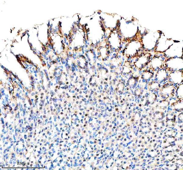
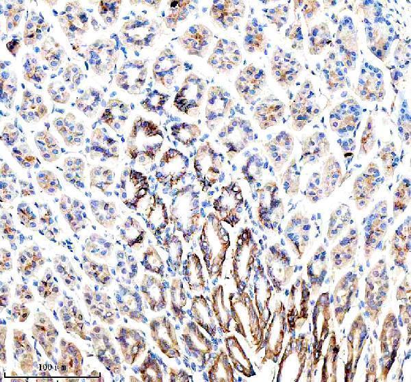


Comprehensive Human Protein Atlas IHC scoring per tissue (reliability: Enhanced — High consistency between antibody staining and RNA expression data. Presumed off target binding observed and disregarded.). Rows are taken directly from the HPA tissue chart — click any row's HPA link to view the source.
| Tissue | Cell type | Level | Evidence | Source |
|---|---|---|---|---|
| Choroid plexus | Ependymal cells | High | Protein (IHC) | HPA → |
| Stomach | Glandular cells | High | Protein (IHC) | HPA → |
| Thyroid gland | Glandular cells | High | Protein (IHC) | HPA → |
| Salivary gland | Glandular cells | Medium | Protein (IHC) | HPA → |
| Tissue | Cell type | Level | Evidence | Source |
|---|---|---|---|---|
| Adipose tissue | Adipocytes | Not detected | Protein (IHC) | HPA → |
| Adrenal gland | Glandular cells | Not detected | Protein (IHC) | HPA → |
| Appendix | Glandular cells | Not detected | Protein (IHC) | HPA → |
| Bone marrow | Hematopoietic cells | Not detected | Protein (IHC) | HPA → |
| Breast | Adipocytes | Not detected | Protein (IHC) | HPA → |
Troubleshoot SLC5A5 staining in paraffin sections by checking retrieval, cell type, membrane localisation and assay controls before interpreting chromogenic signal.
A04465-1 has real IHC images of paraffin-embedded human, mouse, and rat stomach (catalog image captions); no IF image is provided (catalog IF images).
A04465-1 is listed for IHC and human, mouse, and rat reactivity (catalog applications and reactivity). Its own images show paraffin-embedded stomach IHC in all three species with EDTA retrieval at pH 8.0 (catalog image captions).
Which to pick: For tissue IHC, choose A04465-1: this rabbit polyclonal antibody is listed for IHC, and its own images show staining in paraffin-embedded stomach sections (catalog host, dilution data, applications, and image captions). For IF/ICC, no SKU in this payload has a listed IF/ICC application or IF image, so there is no supported pick (catalog applications and IF images). For cross-species IHC, A04465-1 lists human, mouse, and rat reactivity and shows stomach IHC in each; its image captions do not report the fixative (catalog reactivity and image captions).