SLC6A6 / Sodium- and chloride-dependent taurine transporter · IHC design guide

Design Immunohistochemistry for SLC6A6

Plan SLC6A6 paraffin IHC with the catalog antibody’s 1:100–1:300 dilution range (datasheet: A06936/A06936-1). Use retinal nerve fibers and stomach glandular cells as positive tissue references, and assess membranous and cytoplasmic staining (HPA tissue IHC).

Evidence assembled Oct 2026 · For research use; verify linked source records and product datasheet before use
Immunohistochemistry protocol sheet for SLC6A6 (IHC for SLC6A6): expected localisation Membranous and cytoplasmic in most tissues (HPA tissue IHC), antibody A06936, validated IHC image, and IHC protocol steps
Printable SLC6A6 IHC protocol sheet — expected localisation Membranous and cytoplasmic in most tissues (HPA tissue IHC), antibody A06936, controls and protocol steps. Open the full SLC6A6 IHC guide →

SLC6A6 Immunohistochemistry Experimental Design Guide

Expected localisation, validated protocols, controls and antibodies — the at-a-glance facts below, then the full design guide.

Must know before staining
Expected localisation Membranous and cytoplasmic in most tissues (HPA tissue IHC)
Staining pattern Membranous and cytoplasmic staining in most tissues (HPA tissue IHC)
Antigen retrieval Citrate pH 6.0 HIER, 95–98 °C, 20 min (rule: cytoplasmic / membrane antigen)
Positive control ⓘ Retina+4 more · see all
Negative control ⓘ Adipose tissue+4 more · see all
Important caveats
Reasons your staining may differ from the expected pattern.
Fixation Matched tissue-IHC evidence does not establish the fixation claim. Validate the specimen-specific method before use. (selected-SKU IHC image A06936)
Caveat Skeletal myocytes lack staining despite reported abundance (HPA tissue IHC; UniProt)
Regulation PKA favors plasma membrane localisation (UniProt)
Isoform / epitope 2 isoforms; check whether the epitope faces inward or outward (UniProt)
Section 1

Recommended SLC6A6 IHC & IF Protocols

Compare the catalog antibody’s IHC-P protocol (datasheet) with published SLC6A6 chromogenic IHC methods (PMC9917619; PMC5291972; PMC12130737; PMC4052839).

Recommended immunohistochemistry (IHC-P) protocol parameters
SampleParaffin-embedded human lung carcinoma tissue; fixative not specified (datasheet A06936)
FixationImage fixative and duration unreported (datasheet A06936); verify before use.
Sectioning4–5 µm sections on charged slides (standard)
DeparaffinisationXylene, graded ethanol series to water (standard)
Antigen retrievalHeat-induced epitope retrieval in citrate buffer, pH 6.0, 20 min at 95–98 °C (standard rule: cytoplasmic / membrane antigen)
Peroxidase block3% H2O2, 10 min, room temperature (standard)
Blocking10% normal serum of the secondary host, 30 min, room temperature (standard)
Primary antibodyRabbit anti-SLC6A6, 1:100-1:300 (datasheet A06936)
Primary incubationOvernight at 4 °C (standard)
DetectionHRP-polymer secondary, DAB chromogen 5–10 min (standard)
CounterstainHematoxylin, blue, dehydrate and mount (standard)
Expected resultSLC6A6-positive staining in nerve fiber layer of retina (HPA tissue IHC: High). HPA tissue profile: Membranous and cytoplasmic expression in most tissues. No signal in the no-primary control.
💡Decision noteFor paraffin sections, start with citrate pH 6.0 retrieval at 95–98 °C for 20 min (page retrieval rule); a pancreatic study used microwave citrate retrieval for 10 min (PMC12130737).
Section 2

What Is the Expected SLC6A6 Staining Pattern?

SLC6A6 is a 12-pass membrane transporter with cell membrane and mitochondrial inner membrane annotations (UniProt P31641 topology and localization). In paraffin IHC, expect membranous and cytoplasmic staining in selected cells (HPA: tissue IHC profile); HPA rates the tissue pattern Enhanced, with medium consistency between staining and RNA data (HPA: tissue IHC reliability). Retina nerve fiber layer and stomach glandular cells are strong reference sites (HPA: High in both).

What am I looking at on my slide?
Distinct membranous and cytoplasmic stain in stomach glandular cells or the retinal nerve fiber layer.This fits the reported tissue pattern and High staining in those sites (HPA: tissue IHC). Assess the identified cells and compartments together; a dark deposit elsewhere in the section does not establish the same result.
Strong, predominantly nuclear stain with little convincing membranous or cytoplasmic signal.A nuclear-only pattern conflicts with HPA's membranous and cytoplasmic tissue profile and UniProt's membrane localization (HPA: tissue IHC; UniProt P31641 localization). Treat it as suspect and review controls and detection conditions.
Prominent staining of adipocytes or skeletal myocytes.Those cell types are Not detected in HPA tissue IHC; consider cross-reactivity or endogenous chromogenic activity (HPA: adipose tissue and skeletal muscle). UniProt reports abundant skeletal muscle expression, so the myocyte discrepancy needs independent resolution (UniProt P31641 tissue specificity).
Diffuse color across tissue and background, without a clear cell boundary or compartment.The result cannot be scored confidently against HPA's membranous and cytoplasmic profile (HPA: tissue IHC). Background can arise from nonspecific reagent binding or detection activity; compare a no-primary control and inspect the same cell types in a positive reference section.
No signal in stomach glandular cells or the retinal nerve fiber layer.This misses two High HPA reference patterns (HPA: tissue IHC). Check section quality, primary antibody conditions and detection controls before calling the specimen negative; HPA's Enhanced rating still carries only medium staining–RNA consistency (HPA: reliability description).
💡Expected SLC6A6 appearanceCall a positive when clear membranous and cytoplasmic staining marks stomach glandular cells or the retinal nerve fiber layer at the reported High level (HPA: tissue IHC); isolated nuclear color or widespread featureless deposit is suspect against that pattern (HPA: tissue IHC; UniProt P31641 localization).
How each factor affects the staining
Topology and epitope positionSLC6A6 has 12 transmembrane segments and cytoplasmic and extracellular loops (UniProt P31641 topology). An antibody's epitope position could affect accessibility, but no epitope or retrieval comparison is supplied; do not infer an SLC6A6-specific retrieval requirement.
Tissue and cell-type choiceUse stomach glandular cells or retinal nerve fiber layer as observed high-staining references (HPA: tissue IHC). Adipocytes and skeletal myocytes are reported Not detected by HPA; UniProt's abundant skeletal muscle expression makes the latter an evidence discrepancy, not a definitive negative.
Antibody evidenceHPA015028 is Enhanced for IHC, while HPA016488 is Supported (HPA: antibody validation). These ratings help interpret concordance; neither rating supplies an antibody dilution, antigen retrieval setting or target-specific fixation effect.
IF/ICC Q&A: should fluorescence reproduce the paraffin IHC pattern?HPA reports cell junctions and cytosol in ICC-IF images from A-549 and U2OS (HPA: subcellular ICC-IF). This is a localization comparison, not an IHC protocol option; HPA015028 has Uncertain ICC validation (HPA: antibody validation).
Why is my staining missing, weak or wrong?
SituationLikely causeNext action
The positive reference section has no stain.The assay may have failed; both stomach glandular cells and retinal nerve fiber layer have High HPA tissue staining (HPA: tissue IHC).Confirm the reference cells are present, then check primary antibody application and chromogenic detection with appropriate process controls. Reassess the run before interpreting test sections as negative.
Only nuclei are strongly colored.The dominant compartment conflicts with the reported membranous and cytoplasmic pattern (HPA: tissue IHC) and membrane localization (UniProt P31641).Compare a no-primary control, inspect staining at cell borders and cytoplasm, and avoid scoring nuclear-only color as SLC6A6.
Adipocytes or skeletal myocytes stain strongly.HPA reports both cell types as Not detected in tissue IHC (HPA: adipose tissue; skeletal muscle). Cross-reactivity or endogenous detection activity is possible.Check a no-primary control and the cell identity. For myocytes, document the conflict with abundant skeletal muscle expression reported by UniProt (UniProt P31641 tissue specificity).
Color appears throughout the section without cell-specific detail.Diffuse background obscures the membranous and cytoplasmic pattern needed for interpretation (HPA: tissue IHC); nonspecific binding or endogenous chromogenic activity may contribute.Inspect a no-primary control, verify blocking and washing steps, and optimize detection conditions using a reference section with HPA-reported High staining.
A retrieval change alters staining intensity.No supplied source establishes SLC6A6-specific retrieval behavior or fixation sensitivity; intensity alone cannot identify the cause.Compare retrieval conditions with matched positive and no-primary controls, recording whether the reported cells and compartments remain recognizable (HPA: tissue IHC pattern).
ICC-IF looks junctional or cytosolic while paraffin IHC looks membranous and cytoplasmic.Those are the respective reported observations (HPA: subcellular ICC-IF; tissue IHC). HPA015028 is Enhanced for IHC but Uncertain for ICC (HPA: antibody validation).Evaluate each application against its own HPA evidence; do not use the ICC-IF image as an IHC retrieval, dilution or scoring standard.

Sample controls for SLC6A6 IHC & IF

🧪Run stomach first: glandular cells should stain (HPA: High in stomach glandular cells). Use oral mucosa squamous epithelial cells as the negative tissue (HPA: Not detected in oral mucosa squamous epithelial cells); on the stomach slide, use adjacent nonglandular cells to assess background without assuming they lack SLC6A6.
Positive control tissue: Retina (Nerve fiber layer, HPA High)
Negative control tissue: Adipose tissue (HPA Not detected)
ICC-IF cell lines (HPA subcellular resource): HPA ICC-IF images show SLC6A6 in A-549, U2OS, with annotated localisation: Cell Junctions (approved), Cytosol (approved) (HPA subcellular).
Technical controls: Include no-primary (secondary-only) and host- and isotype-matched controls, plus peptide competition with the matching immunogen as a biological specificity control (A06936 IHC caption: peptide blocked). For chromogenic stomach IHC, quench endogenous peroxidase and check glandular background before scoring (standard IHC practice).
⚠️Feasibility: No supplied source reports an SLC6A6-specific fixation window or fixation effect; the selected A06936 paraffin-section caption leaves the fixative unreported (A06936 IHC caption). Retrieval dependence and whether frozen sections or IF are easier are unreported, so optimize retrieval empirically for the IHC-validated antibody (standard IHC practice). In stomach glands, distinguish cellular staining from luminal mucus and background (HPA: High in stomach glandular cells; standard IHC practice).

HPA tissue IHC evidence for SLC6A6

Comprehensive Human Protein Atlas IHC scoring per tissue (reliability: Enhanced — Medium consistency between antibody staining and RNA expression data.). Rows are taken directly from the HPA tissue chart — click any row's HPA link to view the source.

Positive expression · recommended positive controls

TissueCell typeLevelEvidenceSource
Retina Nerve fiber layer High Protein (IHC) HPA →
Stomach Glandular cells High Protein (IHC) HPA →
Adrenal gland Glandular cells Medium Protein (IHC) HPA →
Appendix Glandular cells Medium Protein (IHC) HPA →
Breast Glandular cells Medium Protein (IHC) HPA →

Undetected expression · recommended negative controls

TissueCell typeLevelEvidenceSource
Adipose tissue Adipocytes Not detected Protein (IHC) HPA →
Caudate Glial cells Not detected Protein (IHC) HPA →
Hippocampus Glial cells Not detected Protein (IHC) HPA →
Oral mucosa Squamous epithelial cells Not detected Protein (IHC) HPA →
Ovary Ovarian stroma cells Not detected Protein (IHC) HPA →
Section 3

Advanced SLC6A6 IHC Tips

Troubleshoot SLC6A6 staining in paraffin sections by checking retrieval, compartment, cell identity and controls before comparing chromogenic signal across samples.

How should I retrieve SLC6A6 in paraffin sections when staining is weak?
Start with citrate buffer at pH 6.0, heated to 95–98 °C for 20 minutes (page retrieval rule: cytoplasmic and membrane antigen). Keep section thickness, heating time and cooling conditions consistent across test and control slides so a change in signal can be attributed to retrieval (standard IHC practice). Because SLC6A6 has 12 transmembrane segments, accessibility may depend on the antibody’s epitope, which is not specified here (UniProt P31641 topology). If signal remains weak, compare a carefully optimized alternative retrieval condition on matched sections, watching for tissue damage and increased background (standard IHC practice).
Can I adjust fixation to recover weak SLC6A6 staining?
Target-specific fixation sensitivity is unknown from the supplied evidence; the A06936 image describes paraffin-embedded human lung carcinoma but does not state its fixative (caption A06936). Document the actual fixative, fixation duration and processing history for each specimen before attributing weak staining to fixation (standard IHC practice). Compare sections processed together with the same retrieval and chromogenic detection conditions, using a known positive section in each run (standard IHC practice). If fixation history varies, interpret differences cautiously and optimize retrieval on matched material first; neither tissue staining patterns nor SLC6A6 topology establish a fixation effect (standard IHC practice; UniProt P31641 topology).
Which staining compartments should count as plausible SLC6A6 signal?
Assess membrane-associated and cytoplasmic staining separately, since both occur in tissue IHC observations (HPA: membranous and cytoplasmic expression in most tissues). Cell junction and cytosol localisation also appear in subcellular imaging, while UniProt lists the cell membrane and mitochondrial inner membrane (HPA: subcellular; UniProt P31641 localisation). A diffuse nuclear-only chromogenic signal lacks support from those localisation records and warrants review against a negative control and tissue morphology (HPA: tissue IHC and subcellular; UniProt P31641 localisation; standard IHC practice). Do not call fine cytoplasmic puncta mitochondrial solely from brightfield staining; that assignment needs an appropriate compartment marker or independent evidence (standard IHC practice).
Could the antibody epitope explain inconsistent staining between sections?
SLC6A6 has 2 annotated isoforms, but the supplied evidence does not map this antibody’s epitope to either one (UniProt P31641 isoforms; caption A06936). Its 12 membrane-spanning segments separate cytoplasmic and extracellular regions, so retrieval and processing may expose candidate epitopes differently (UniProt P31641 topology; standard IHC practice). Extracellular residues 163, 179 and 190 are annotated glycosylation sites, while phosphorylation is annotated at 21, 25 and 322; no effect on this antibody’s staining is established (UniProt P31641 modifications). Obtain the immunogen or epitope specification before assigning an isoform-specific or modification-specific interpretation to chromogenic signal (standard IHC practice).
How can I investigate an uncertain IHC pattern by immunofluorescence?
Use IF as an orthogonal localisation check and multiplex SLC6A6 with a validated marker for the cell type being assessed (standard IF practice). For example, HPA reports high signal in retina nerve fiber layer and stomach glandular cells, so select a corresponding cell marker only when examining those tissues (HPA: tissue IHC). Choose spectrally separated fluorophores after checking the specimen’s autofluorescence, and include single-stain and secondary-only controls (standard IF practice). If the antibody recognises a cytoplasmic epitope, permeabilise appropriately; an accessible extracellular epitope may need less permeabilisation, but this antibody’s epitope is unspecified (UniProt P31641 topology; standard IF practice).
What should I check when SLC6A6 chromogenic staining looks widespread?
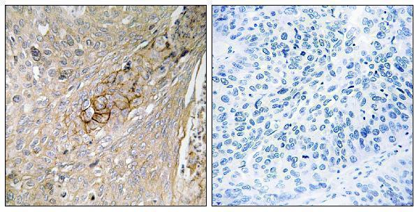
Inspect a no-primary control alongside the stained section to identify chromogen deposition caused by the detection system (standard IHC practice). Block endogenous peroxidase before peroxidase-based detection, and adjust blocking and washes if nonspecific staining persists (standard IHC practice). The selected A06936 image includes a synthesized-peptide-blocked comparison in paraffin-embedded human lung carcinoma, which can help assess antibody-dependent signal in that displayed specimen (caption A06936). Compare suspected background with tissue morphology: HPA describes membranous and cytoplasmic expression in most tissues, but reports no detectable signal in several specific cell populations, including adipocytes (HPA: tissue IHC).
How should I quantify SLC6A6 IHC across samples? ⚠ ANSWER MARKED FOR VERIFICATION
Define the cell population and whether membrane, cytoplasmic or combined signal is scored before examining group labels (HPA: tissue IHC; standard IHC practice). Record the percentage of positive target cells and staining intensity, or calculate an H-score from intensity categories 0–3 weighted by their cell percentages, yielding 0–300 (standard IHC practice). Normalize counts to the number of evaluable target cells or stained area to tissue area in mm², rather than comparing raw positive counts across unequal sections (standard IHC practice). Hold retrieval, imaging, thresholds and exclusion rules constant, and report membrane and cytoplasmic results separately when their proportions differ (standard IHC practice).
How do I distinguish true SLC6A6 staining from artefact?
Favor reproducible signal in morphologically intact cells with membrane or cytoplasmic distribution, matching the reported tissue pattern (HPA: tissue IHC; standard IHC practice). Check cell identity: HPA reports high staining in retina nerve fiber layer and stomach glandular cells, but no detection in adipocytes or skeletal muscle myocytes (HPA: tissue IHC). Exclude section edges, necrotic regions and pigment-like deposits before scoring, and compare no-primary controls for endogenous enzyme or detection-related color (standard IHC practice). The A06936 peptide-blocked image supports assessment of that displayed paraffin-embedded lung carcinoma specimen, but does not establish specificity in every tissue or compartment (caption A06936; standard IHC practice).
Boster reagents

Best SLC6A6 / Sodium- and chloride-dependent taurine transporter IHC Antibodies

Both antibodies have IHC images from paraffin-embedded human lung carcinoma (catalog image captions); IF is listed for both, while ICC is listed only for A06936-1 (catalog applications).

Real IHC data Immunohistochemistry analysis of paraffin-embedded human lung carcinoma tissue, using SLC6A6 Antibody. The picture on the right is blocked with the synthesized peptide.
Anti-TAUT SLC6A6 Antibody
Cat # A06936
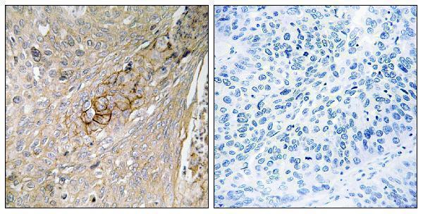
Real IHC data Immunohistochemistry analysis of paraffin-embedded human lung carcinoma tissue, using SLC6A6 Antibody. The picture on the right is blocked with the synthesized peptide.
Anti-SLC6A6/Taut Antibody
Cat # A06936-1

A06936 lists IHC and IF for human, mouse, and rat; its IHC image shows paraffin-embedded human lung carcinoma with a peptide-blocked comparison (catalog applications, reactivity, and image caption). A06936-1 also lists ICC and shows the same sample type and comparison in its own IHC image (catalog applications and image caption).

Which to pick: For tissue IHC, either A06936 or A06936-1 has a paraffin-section image and a listed 1:100–1:300 IHC dilution (each SKU’s catalog image caption and dilution). For IF/ICC, choose A06936-1 if ICC is required; both list IF and both are rabbit polyclonals (catalog applications, host, and dilution data). Both list human, mouse, and rat reactivity, but each IHC image shows only human lung carcinoma, and neither caption reports the fixative (catalog reactivity and each SKU’s image caption).

Each figure is that product's own IHC / IF validation image from its datasheet.

References

  1. UniProt Consortium. UniProt entry P31641 (SC6A6_HUMAN, Sodium- and chloride-dependent taurine transporter).
  2. Human Protein Atlas. SLC6A6 tissue IHC expression (reliability: Enhanced).
  3. Human Protein Atlas. SLC6A6 subcellular location (ICC-IF): Localized to the cell junctions and cytosol..
  4. Human Protein Atlas. SLC6A6 antibody validation summary (2 antibodies).
  5. Ant-Neointimal Formation Effects of SLC6A6 in Preventing Vascular Smooth Muscle Cell Proliferation and Migration via Wnt/β-Catenin Signaling. International journal of molecular sciences 2023 — PMC9917619.
  6. miR-3156-3p is downregulated in HPV-positive cervical cancer and performs as a tumor-suppressive miRNA. Virology journal 2017 — PMC5291972.
  7. Taurine Alleviates Pancreatic β-Cell Senescence by Inhibition of p53 Pathway. Journal of diabetes 2025 — PMC12130737.
  8. Widespread microRNA dysregulation in multiple system atrophy - disease-related alteration in miR-96. The European journal of neuroscience 2014 — PMC4052839.
  9. PubMed PMID:8382624 — UniProt-cited evidence.
  10. PubMed PMID:8010975 — UniProt-cited evidence.
  11. PubMed PMID:8654117 — UniProt-cited evidence.