SLC7A7 / Y+L amino acid transporter 1 · IHC design guide

Design Immunohistochemistry for SLC7A7

Plan chromogenic paraffin-section IHC using kidney tubule cells as a positive tissue reference (HPA tissue IHC). This guide helps you score cytoplasmic and membranous staining (HPA tissue IHC) against the basolateral membrane expectation (UniProt).

Evidence assembled Oct 2026 · For research use; verify linked source records and product datasheet before use
Immunohistochemistry protocol sheet for SLC7A7 (IHC for SLC7A7): expected localisation Kidney cytoplasm and membrane (HPA tissue IHC); basolateral membrane expected (UniProt), antibody A03602-2, validated IHC image, and IHC protocol steps
Printable SLC7A7 IHC protocol sheet — expected localisation Kidney cytoplasm and membrane (HPA tissue IHC); basolateral membrane expected (UniProt), antibody A03602-2, controls and protocol steps. Open the full SLC7A7 IHC guide →

SLC7A7 Immunohistochemistry Experimental Design Guide

Expected localisation, validated protocols, controls and antibodies — the at-a-glance facts below, then the full design guide.

Must know before staining
Expected localisation Kidney cytoplasm and membrane (HPA tissue IHC); basolateral membrane expected (UniProt)
Staining pattern Kidney tubule cells: high cytoplasmic and membranous staining (HPA tissue IHC)
Antigen retrieval EDTA pH 8.0 HIER, heat-mediated (datasheet A03602-2)
Positive control ⓘ Kidney+1 more · see all
Negative control ⓘ Adipose tissue+4 more · see all
Important caveats
Reasons your staining may differ from the expected pattern.
Fixation Keep fixation consistent across paraffin sections. (standard IHC practice; not target-specific)
Caveat Cytoplasmic staining can complicate membrane scoring (HPA tissue IHC)
Regulation No specific expression regulator reported (UniProt)
Isoform / epitope No annotated isoforms or cleavage; epitope side affects access (UniProt)
Section 1

Recommended SLC7A7 IHC & IF Protocols

Compare the catalog antibody’s IHC-P protocol (datasheet: A03602-2) with four published SLC7A7 chromogenic IHC protocols (PMC7905560; PMC11903784; PMC7380045; PMC12374663).

Recommended immunohistochemistry (IHC-P) protocol parameters
SampleParaffin-embedded human ovarian cancer tissue; fixative not specified (datasheet A03602-2)
FixationImage fixative and duration unreported (datasheet A03602-2); verify before use.
Sectioning4–5 µm sections on charged slides (standard)
DeparaffinisationXylene, graded ethanol series to water (standard)
Antigen retrievalHeat retrieval: EDTA pH 8.0 (datasheet A03602-2); 20 min, 95–100 °C (standard)
Peroxidase block3% H2O2, 10 min, room temperature (standard)
Blocking10% goat serum (datasheet A03602-2)
Primary antibodyRabbit anti-SLC7A7, 2-5 μg/ml (datasheet A03602-2)
Primary incubationOvernight at 4 °C (datasheet A03602-2)
DetectionHRP-conjugated secondary, DAB chromogen (datasheet A03602-2)
CounterstainHematoxylin, blue, dehydrate and mount (standard)
Expected resultSLC7A7-positive staining in cells in tubules of kidney (HPA tissue IHC: High). HPA tissue profile: Cytoplasmic and membranous expression in kidney. No signal in the no-primary control.
💡Decision noteStart with heat-mediated EDTA retrieval at pH 8.0 (datasheet: A03602-2); the lung study provides a low-pH citrate alternative (PMC7905560).
Section 2

What Is the Expected SLC7A7 Staining Pattern?

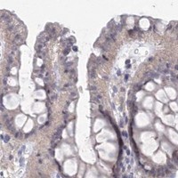
SLC7A7 is a 12-pass transporter at the basolateral cell membrane (UniProt Q9UM01 topology and subcellular location). In IHC, expect the strongest staining in kidney tubule cells, with both membranous and cytoplasmic signal reported; spermatogonia show medium staining (HPA tissue IHC). HPA rates the tissue pattern Enhanced, while reporting medium consistency between antibody staining and RNA expression (HPA tissue IHC).

What am I looking at on my slide?
Kidney tubule cells show strong membrane-associated staining with some cytoplasmic signal.This matches the reported high tubular staining and cytoplasmic and membranous kidney profile (HPA tissue IHC). Basolateral membrane enrichment is consistent with UniProt localisation (UniProt Q9UM01 subcellular location). Interpret cytoplasmic signal in context; it is part of HPA’s kidney description (HPA tissue IHC).
Nuclei stain strongly while tubule cell membranes show little or no signal.A predominantly nuclear pattern conflicts with the reported kidney pattern and membrane localisation (HPA tissue IHC; UniProt Q9UM01 subcellular location). Treat it as suspect staining and compare the antibody control and detection-only control before assigning it to SLC7A7 (general IHC practice).
Adipocytes or adrenal glandular cells stain as strongly as kidney tubules.HPA reports SLC7A7 as not detected in those cell types, versus high staining in kidney tubules (HPA tissue IHC). Such a reversal raises concern about cross-reactivity or endogenous chromogen activity; verify the cell identity and inspect detection controls (general IHC practice).
Chromogen appears broadly across tissue structures, with little cell-bound contrast.Diffuse staining cannot be scored as the reported tubular pattern (HPA tissue IHC). Review background on the detection-only control, then assess blocking, washes and primary-antibody concentration as general IHC variables (general IHC practice).
Kidney tubules have no visible signal despite acceptable tissue morphology.This conflicts with HPA’s high tubular staining (HPA tissue IHC). First confirm that the positive-control section and detection reagents worked; then review the antibody’s validated IHC conditions. One negative slide does not establish absence of SLC7A7 (general IHC practice).
💡Expected SLC7A7 appearanceCall a result positive when kidney tubule cells show high membranous staining, allowing the cytoplasmic component reported by HPA (HPA tissue IHC); predominantly nuclear staining or equally strong staining in HPA-negative adipocytes is suspect (UniProt Q9UM01 subcellular location; HPA tissue IHC).
How each factor affects the staining
Membrane topology and compartmentSLC7A7 has 12 transmembrane segments and a basolateral membrane location (UniProt Q9UM01 topology and subcellular location). The record gives no antibody epitope, so it does not establish which side of the membrane the antibody recognises or predict a target-specific antigen-retrieval response.
Choice of comparison tissue and cellsKidney tubule cells are high, spermatogonia are medium, and lung macrophages are low by HPA IHC (HPA tissue IHC). Compare the same cell types when judging intensity; weak staining in a low-level population cannot substitute for a kidney tubule positive control (general IHC practice).
Strength and limits of IHC evidenceHPA calls the tissue staining Enhanced but describes only medium consistency with RNA expression; antibody HPA036227 also has Enhanced IHC validation (HPA tissue IHC; HPA antibody record). Use the observed cell pattern as the reference, while treating unexpected staining as a finding to verify.
Processing and antibody accessibilityUniProt lists one SLC7A7 chain, residues 1–511, with no signal peptide or propeptide (UniProt Q9UM01 processing). It also lists glycosylation at residue 325 (UniProt Q9UM01 glycosylation). Without an antibody epitope or target-specific fixation data, these annotations cannot justify a cleavage-pattern or fixation-sensitivity claim.
IF/ICC: what localisation is expected?A membrane pattern is the available HPA subcellular summary, consistent with UniProt’s basolateral membrane annotation (HPA subcellular; UniProt Q9UM01 subcellular location). HPA provides no ICC/IF image cell line or main-location entry, so this record does not support a cell-line-specific IF pattern (HPA subcellular).
Why is my staining missing, weak or wrong?
SituationLikely causeNext action
Kidney positive control is blank.The result conflicts with HPA’s high kidney tubule signal; a failed detection step or unsuitable assay conditions are possibilities (HPA tissue IHC; general IHC practice).Check the section and detection controls, then repeat using the antibody’s validated IHC conditions. Review retrieval only as a general IHC variable; target-specific retrieval or fixation sensitivity is unreported.
Only nuclei stain in the kidney section.Nuclear dominance is inconsistent with the membrane location and reported kidney pattern (UniProt Q9UM01 subcellular location; HPA tissue IHC).Check the detection-only control and antibody specificity, then rescore membrane and cytoplasmic staining in identified tubule cells (general IHC practice; HPA tissue IHC).
Adipocytes or adrenal glandular cells appear strongly positive.Both are listed as not detected by HPA tissue IHC; cross-reactivity or endogenous detection activity is possible (HPA tissue IHC; general IHC practice).Confirm the cell type and compare a detection-only control with the kidney tubule positive control. If chromogen appears without primary antibody, address the detection background (general IHC practice).
Staining covers the section with poor cell contrast.Broad background obscures the cell pattern HPA reports in kidney (HPA tissue IHC). Excess primary antibody, insufficient washing or detection background are general IHC possibilities (general IHC practice).Compare controls, optimise antibody concentration and washes, and score only cell-associated staining above background (general IHC practice).
Testis staining is weaker than kidney staining.That difference is expected: HPA scores spermatogonia medium and kidney tubule cells high (HPA tissue IHC).Identify spermatogonia before comparing sections; use kidney tubules to check that the assay can reveal the stronger reference signal (HPA tissue IHC; general IHC practice).
Cytoplasmic kidney staining appears alongside membrane staining.HPA explicitly describes kidney expression as cytoplasmic and membranous, while UniProt assigns SLC7A7 to the basolateral membrane (HPA tissue IHC; UniProt Q9UM01 subcellular location).Record both compartments and judge whether the signal remains concentrated in tubule cells. Investigate only staining that loses cellular specificity or fails controls (HPA tissue IHC; general IHC practice).

Sample controls for SLC7A7 IHC & IF

🧪Run kidney first; cells in tubules should stain (HPA: High in kidney cells in tubules). Run adipose tissue as a negative, where adipocytes should lack specific staining (HPA: Not detected in adipocytes); on the kidney slide, use cells without specific staining as an internal background reference, without assigning an unlisted renal cell type a negative status.
Positive control tissue: Kidney (Cells in tubules, HPA High)
Negative control tissue: Adipose tissue (HPA Not detected)
ICC-IF cell lines (HPA subcellular resource): HPA carries no ICC-IF cell line for SLC7A7; derive a cell-line control from the positive tissue's cell type (Cells in tubules) and confirm it by RNA or western blot first.
Technical controls: Include a no-primary, secondary-only control and a concentration-matched rabbit IgG isotype control matched to the primary antibody’s clonality (caption: rabbit anti-SLC7A7 antibody). Use SLC7A7 knockout tissue or cells as a biological negative where available; for kidney chromogenic sections, quench and check endogenous peroxidase before interpreting DAB signal (caption: HRP/DAB detection).
⚠️Feasibility: A target-specific fixation window or fixation effect is unreported, and the selected A03602-2 paraffin-section caption does not state the fixative (caption: fixative unreported). The caption reports heat retrieval in EDTA at pH 8.0; optimize retrieval on matched sections, since the supplied evidence does not establish whether frozen sections or IF are easier (caption: EDTA retrieval; HPA: no ICC-IF images). In kidney, assess tubular background when scoring the expected basolateral membrane pattern (UniProt Q9UM01: basolateral cell membrane).

HPA tissue IHC evidence for SLC7A7

Comprehensive Human Protein Atlas IHC scoring per tissue (reliability: Enhanced — Medium consistency between antibody staining and RNA expression data.). Rows are taken directly from the HPA tissue chart — click any row's HPA link to view the source.

Positive expression · recommended positive controls

TissueCell typeLevelEvidenceSource
Kidney Cells in tubules High Protein (IHC) HPA →
Testis Spermatogonia cells Medium Protein (IHC) HPA →

Undetected expression · recommended negative controls

TissueCell typeLevelEvidenceSource
Adipose tissue Adipocytes Not detected Protein (IHC) HPA →
Adrenal gland Glandular cells Not detected Protein (IHC) HPA →
Breast Adipocytes Not detected Protein (IHC) HPA →
Cerebellum Cells in granular layer Not detected Protein (IHC) HPA →
Cervix Glandular cells Not detected Protein (IHC) HPA →
Section 3

Advanced SLC7A7 IHC Tips

Troubleshoot SLC7A7 staining in paraffin sections by checking retrieval, membrane-associated localisation, cell identity and controls (UniProt Q9UM01; HPA tissue IHC).

What retrieval should I try first when SLC7A7 staining is weak?
Start with heat-mediated retrieval in EDTA, pH 8.0, for paraffin-section IHC (datasheet A03602-2). The selected antibody image used that retrieval before 2 μg/mL primary antibody overnight at 4°C, so keep those conditions together for the first comparison (datasheet A03602-2). If staining remains weak, compare a different retrieval buffer on serial sections while holding detection and exposure to chromogen constant (standard IHC practice). Excessive heating can damage tissue morphology or increase background, so assess staining alongside section integrity (standard IHC practice). Include kidney tubules as a positive comparator when available (HPA: High in kidney tubule cells).
Could fixation explain weak or patchy SLC7A7 staining?
Target-specific sensitivity to fixation is unknown: the selected tissue-IHC caption says the section was paraffin embedded but does not state its fixative (datasheet A03602-2). Record the fixative and fixation duration for each specimen, then compare sections processed under matched conditions (standard IHC practice). If signal varies between blocks, first repeat EDTA, pH 8.0 retrieval and the 2 μg/mL overnight primary incubation before changing detection conditions (datasheet A03602-2). Examine morphology and a control section processed with the same fixative, because poor preservation or uneven reagent access can complicate interpretation (standard IHC practice). Do not assign a SLC7A7-specific fixation effect without direct comparative evidence.
Should SLC7A7 stain the membrane, cytoplasm, or both?
Expect membrane-associated staining, particularly at the basolateral surface of polarized cells (UniProt Q9UM01: basolateral cell membrane). Kidney tissue IHC also reports cytoplasmic and membranous staining, so a cytoplasmic component alone is not sufficient to reject a section (HPA: kidney profile). Compare the signal with cell boundaries and tissue architecture at the same magnification, and score membrane and cytoplasmic patterns separately (standard IHC practice). SLC7A7 has 12 annotated transmembrane segments and forms a heterodimer with SLC3A2, which supports careful assessment of membrane localisation (UniProt Q9UM01 topology and subunit). Diffuse staining without recognisable cells warrants a background-control check (standard IHC practice).
How should an unknown antibody epitope affect retrieval decisions?
The record lists no annotated isoforms, but the supplied evidence does not identify this antibody’s epitope (UniProt Q9UM01 isoforms; datasheet A03602-2). SLC7A7 contains 12 transmembrane segments, a glycosylation site at residue 325, and phosphoserines at residues 18 and 25 (UniProt Q9UM01 topology and modified residues). Without an epitope sequence, do not attribute weak staining to any one modification or membrane-facing region (standard IHC interpretation). Begin with the documented EDTA, pH 8.0 retrieval, then compare serial sections only if signal remains inadequate (datasheet A03602-2; standard IHC practice). Use an independent antibody with a documented, distinct epitope if epitope-dependent staining remains a concern (standard IHC practice).
How can IF help resolve ambiguous SLC7A7 tissue-IHC localisation?
Use IF as a separate localisation check, since the supplied HPA subcellular record lists no ICC/IF image cell lines (HPA subcellular). Multiplex SLC7A7 with a marker for the cell population under examination; kidney tubule cells are a supported positive population, while lung macrophages have low reported staining (HPA tissue IHC). Choose fluorophores after inspecting unstained tissue for autofluorescence, and use single-stain controls to check channel bleed-through (standard IF practice). Plan permeabilisation only after establishing whether the antibody epitope faces an accessible surface or requires intracellular access; its location is unspecified here (datasheet A03602-2; standard IF practice). Do not transfer the paraffin-section retrieval conditions directly to an IF specimen (datasheet A03602-2; standard IF practice).
What should I check when DAB appears outside expected SLC7A7-positive cells?
Compare the stained section with a no-primary control to identify signal from secondary reagents, endogenous enzymes, or chromogen deposition (standard IHC practice). The selected tissue example used peroxidase-conjugated secondary detection and DAB; those reagents make a peroxidase block and timed chromogen development relevant workflow checks (datasheet A03602-2; standard IHC practice). It also used 10% goat serum blocking and 2 μg/mL primary antibody overnight at 4°C, which provide documented starting conditions (datasheet A03602-2). Review any diffuse staining against kidney tubule cell boundaries and the reported membranous and cytoplasmic pattern (HPA tissue IHC). If background persists, compare a shorter DAB development on adjacent sections (standard IHC practice).
How should I score SLC7A7 staining across paraffin sections? ⚠ ANSWER MARKED FOR VERIFICATION
Define the cell population and compartment before scoring; kidney tubule cells have high reported staining, with cytoplasmic and membranous expression described (HPA tissue IHC). For chromogenic sections, report the percentage of positive target cells and an H-score that combines staining intensity with that percentage (standard IHC practice). Record membrane and cytoplasmic scores separately, and normalise positive-cell counts to the total eligible cells or a density per mm² of evaluable tissue (standard IHC practice). Exclude necrotic or poorly preserved regions using the same rule across specimens, and compare sections processed with matched retrieval and DAB development (standard IHC practice). State the threshold and analysis region alongside each score (standard IHC practice).
How can I distinguish true SLC7A7 staining from artefact?
A convincing positive should follow recognisable cells and a plausible membrane-associated pattern; SLC7A7 is annotated at the basolateral membrane, while kidney IHC reports cytoplasmic and membranous expression (UniProt Q9UM01 subcellular location; HPA tissue IHC). Check cell identity before calling a field positive: kidney tubule cells stain highly, whereas adipocytes in adipose tissue were reported as undetected (HPA tissue IHC). Staining confined to section edges, necrotic areas, or the no-primary control suggests a processing or detection artefact (standard IHC practice). Check endogenous peroxidase activity when interpreting DAB deposits, and verify a disputed pattern on an adjacent section (standard IHC practice). Treat an unexpected compartment or cell population as requiring independent confirmation (standard IHC practice).
Boster reagents

Best SLC7A7 / Y+L amino acid transporter 1 IHC Antibodies

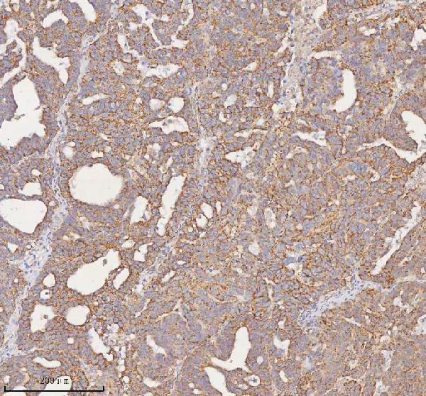
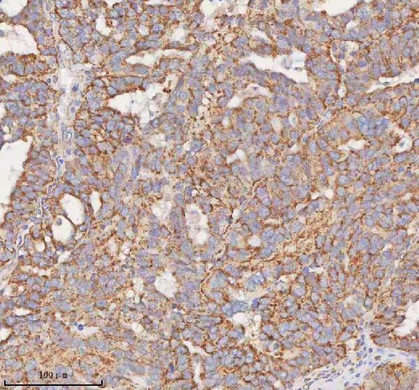
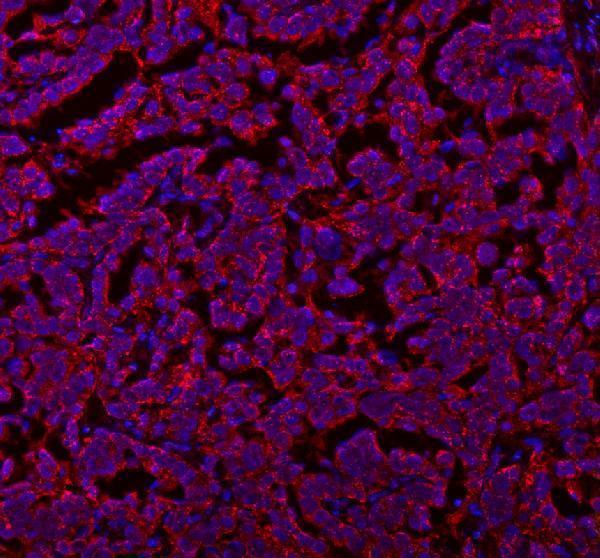
A03602-2 has IHC and IF images from paraffin sections of human ovarian cancer tissue (catalog image captions); its listed reactivity is human, mouse, and rat (catalog reactivity).

Real IHC data IHC analysis of SLC7A7 using anti-SLC7A7 antibody (A03602-2). SLC7A7 was detected in a paraffin-embedded section of human ovarian cancer tissue. Heat mediated antigen retrieval was performed in EDTA buffer (pH 8.0, epitope retrieval solution). The tissue section was blocked with 10% goat serum. The tissue section was then incubated with 2 μg/ml rabbit anti-SLC7A7 Antibody (A03602-2) overnight at 4°C. Peroxidase Conjugated Goat Anti-rabbit IgG was used as secondary antibody and incubated for 30 minutes at 37°C. The tissue section was developed using HRP Conjugated Rabbit IgG Super Vision Assay Kit (Catalog # SV0002) with DAB as the chromogen.
Anti-SLC7A7 Antibody ®
Cat # A03602-2

A03602-2 is the only SKU shown, with IHC and IF listed as applications (catalog applications). Its IHC and IF images each show a paraffin section of human ovarian cancer tissue (catalog image captions).

Which to pick: For tissue IHC, choose A03602-2: its IHC image documents a paraffin section, while the fixative is unreported (catalog IHC caption). For IF, choose the same SKU for paraffin sections; ICC validation is unreported (catalog IF caption; catalog applications). For work across species, A03602-2 lists human, mouse, and rat reactivity, but its supplied IHC and IF images show human tissue only; the antibody host is rabbit and clonality is unreported (catalog reactivity; catalog image captions; catalog host and clone fields).

Each figure is that product's own IHC / IF validation image from its datasheet.

References

  1. UniProt Consortium. UniProt entry Q9UM01 (YLAT1_HUMAN, Y+L amino acid transporter 1).
  2. Human Protein Atlas. SLC7A7 tissue IHC expression (reliability: Enhanced).
  3. Human Protein Atlas. SLC7A7 subcellular location (ICC-IF): Membrane.
  4. Human Protein Atlas. SLC7A7 antibody validation summary (1 antibodies).
  5. SLC7A7 is a prognostic biomarker correlated with immune infiltrates in non-small cell lung cancer. Cancer cell international 2021 — PMC7905560.
  6. Exosomes containing miR-152-3p targeting FGFR3 mediate SLC7A7-induced angiogenesis in bladder cancer. NPJ precision oncology 2025 — PMC11903784.
  7. Expression profile of the amino acid transporters SLC7A5, SLC7A7, SLC7A8 and the enzyme TDO2 in basal cell carcinoma. The British journal of dermatology 2019 — PMC7380045.
  8. Multiomics integration analysis identifies tumor cell-derived MIF as a therapeutic target and potentiates anti-PD-1 therapy in osteosarcoma. Journal for immunotherapy of cancer 2025 — PMC12374663.
  9. PubMed PMID:9829974 — UniProt-cited evidence.
  10. PubMed PMID:9878049 — UniProt-cited evidence.
  11. PubMed PMID:10080183 — UniProt-cited evidence.