This website uses cookies to ensure you get the best experience on our website.


- Table of Contents
Use this guide to plan SLPI IHC-P and assess cytoplasmic epithelial staining in cervix and respiratory tissue (HPA tissue IHC). The catalog antibody starts at 10 μg/mL; its stated IHC validation is in mouse tissue, so verify staining in human sections (datasheet).

Expected localisation, validated protocols, controls and antibodies — the at-a-glance facts below, then the full design guide.
| Expected localisation | Cytoplasmic staining in select epithelia (HPA tissue IHC) | |
| Staining pattern | Cytoplasmic glandular and respiratory epithelial staining (HPA tissue IHC) | |
| Antigen retrieval | Citrate pH 6.0 HIER, 95–98 °C, 20 min (rule: cytoplasmic / membrane antigen) | |
| Positive control | Cervix+4 more · see all | |
| Negative control | Adipose tissue+4 more · see all |
| Fixation | Matched tissue-IHC evidence does not establish the fixation claim. Validate the specimen-specific method before use. (selected-SKU IHC image A01682) | |
| Caveat | Secreted SLPI may appear beyond producing cells (UniProt) | |
| Regulation | No expression regulator established in the record (UniProt) | |
| Isoform / epitope | No isoforms reported; signal peptide 1–25 is cleaved (UniProt) |
The catalog antibody protocol is paired with published SLPI chromogenic IHC workflows for eye sections (PMC2234354) and head and neck tissue (PMC4164221).
| Sample | Tissue sections; selected-image fixative not specified (standard IHC workflow) |
| Fixation | Image fixative and duration unreported (datasheet A01682); verify before use. |
| Sectioning | 4–5 µm sections on charged slides (standard) |
| Deparaffinisation | Xylene, graded ethanol series to water (standard) |
| Antigen retrieval | Heat-induced epitope retrieval in citrate buffer, pH 6.0, 20 min at 95–98 °C (standard rule: cytoplasmic / membrane antigen) |
| Peroxidase block | 3% H2O2, 10 min, room temperature (standard) |
| Blocking | 10% normal serum of the secondary host, 30 min, room temperature (standard) |
| Primary antibody | Rabbit anti-SLPI, 10 μg/mL (datasheet A01682) |
| Primary incubation | Overnight at 4 °C (standard) |
| Detection | HRP-polymer secondary, DAB chromogen 5–10 min (standard) |
| Counterstain | Hematoxylin, blue, dehydrate and mount (standard) |
| Expected result | SLPI-positive staining in glandular cells of cervix (HPA tissue IHC: High). HPA tissue profile: Cytoplasmic expression in cervix, respiratory epithelium, fallopian tube and seminal vesicle. No signal in the no-primary control. |
In paraffin-section IHC, expect SLPI mainly in the cytoplasm of cervix glandular cells and nasopharyngeal goblet cells, with staining in other listed epithelia (HPA: tissue IHC). SLPI is secreted and has no transmembrane segment (UniProt P03973: topology). HPA rates tissue staining Enhanced, while reporting medium agreement between staining and RNA because a secreted protein can appear away from its site of production (HPA: tissue IHC).
| Distinct cytoplasmic staining in cervix glandular cells or nasopharyngeal goblet cells; nearby cells stain less (HPA: tissue IHC). | This matches the strongest reported tissue patterns: both cell populations are High (HPA: tissue IHC). Compare the distribution among cells as well as intensity; secretion can separate the protein's location from its RNA source (HPA: reliability description; UniProt P03973: secreted). |
| A dominant nuclear or crisp cell-membrane pattern replaces epithelial cytoplasmic staining (HPA: tissue IHC pattern). | Treat that compartment mismatch as a possible artefact and check the control slide and detection reagents (HPA: tissue IHC; standard IHC practice). SLPI has no transmembrane segment, although its secreted status alone cannot identify the source of an unexpected stain (UniProt P03973: topology). |
| Cells outside the expected epithelial populations stain strongly, including adipocytes in an adipose-tissue control (HPA: tissue IHC). | Consider antibody cross-reactivity or endogenous detection activity, then compare controls and staining conditions (standard IHC practice). HPA reports adipose-tissue adipocytes as Not detected; that finding is cell-specific, not a claim that every cell in the tissue must be unstained (HPA: tissue IHC). |
| A broad, even haze obscures cell boundaries across the section, including areas without an expected positive population (standard IHC practice). | This is difficult to score as cell-associated SLPI. Review blocking, antibody concentration, washing and the detection control for background signal (standard IHC practice). HPA describes a selective cytoplasmic epithelial pattern, so diffuse haze alone does not reproduce its reported result (HPA: tissue IHC). |
| No convincing stain appears in cervix glandular cells or nasopharyngeal goblet cells (HPA: tissue IHC). | A blank known-positive region makes a negative test section inconclusive (standard IHC practice). Confirm that the positive-control cells are present, then review retrieval and detection conditions as general workflow variables; the supplied sources do not establish SLPI-specific retrieval sensitivity (HPA: tissue IHC; standard IHC practice). |
| Secretion and processing (UniProt P03973). | SLPI has a signal peptide at residues 1–25 and a mature chain at 26–132, with no transmembrane segment (UniProt P03973). Interpret tissue staining in light of secretion; protein location need not match the cells with highest RNA (HPA: reliability description). |
| Cell and tissue context (HPA: tissue IHC). | HPA reports High staining in cervix glandular cells and nasopharyngeal goblet cells, Medium in bronchial respiratory epithelium, and Medium at fallopian-tube cilia tips (HPA: tissue IHC). Score the named cells and compartment rather than assigning one expected intensity to every tissue (standard IHC practice). |
| Antibody validation (HPA: antibody records). | HPA027774 and CAB002303 each have Enhanced IHC status (HPA: antibodies). That supports comparison with their reported tissue distribution but does not guarantee that an unexpected signal with a different antibody is SLPI (HPA: antibody records; standard IHC practice). |
| RNA and protein comparison (HPA: tissue IHC; UniProt P03973). | HPA calls SLPI RNA group enriched in cervix and salivary gland, yet lists salivary glandular-cell IHC as Low (HPA: tissue IHC). UniProt reports SLPI in parotid secretions (UniProt P03973: tissue specificity). Do not use RNA enrichment alone to demand strong glandular-cell staining (HPA: reliability description). |
| Situation | Likely cause | Next action |
|---|---|---|
| The positive-control cervix section is blank in glandular cells (HPA: tissue IHC). | Possible general workflow failure in retrieval, antibody incubation or detection; the supplied sources give no SLPI-specific retrieval setting (standard IHC practice). | Verify that glandular cells are present, inspect the staining run's controls, and optimize retrieval and antibody conditions for the IHC-validated antibody before interpreting test negatives (standard IHC practice). |
| The whole section has diffuse chromogen, including tissue expected to be negative (standard IHC practice). | Possible excess antibody, inadequate washing or endogenous detection activity (standard IHC practice). | Review antibody concentration, wash steps and the detection control; suppress endogenous enzyme activity when the chosen chromogen system requires it (standard IHC practice). Then reassess the selective epithelial pattern (HPA: tissue IHC). |
| Adipocytes stain as strongly as the expected epithelial cells (HPA: tissue IHC). | Possible nonspecific antibody binding or endogenous detection activity; HPA lists adipose-tissue adipocytes as Not detected (HPA: tissue IHC; standard IHC practice). | Compare a control lacking primary antibody and an independent IHC-validated antibody, if available; interpret any persistent signal in its cell-specific context (standard IHC practice; HPA: antibody records). |
| The main signal looks nuclear or sharply membrane bound (HPA: tissue IHC pattern). | Possible artefact or off-target staining; HPA's tissue summary describes cytoplasmic expression, and UniProt lists no transmembrane segment (HPA: tissue IHC; UniProt P03973: topology). | Recheck the compartment at higher magnification and compare an IHC-validated antibody and detection control before calling those cells positive (HPA: antibody records; standard IHC practice). |
| Salivary glandular cells stain weakly despite RNA enrichment (HPA: tissue IHC). | That combination is compatible with HPA's Low glandular-cell stain and its warning that secreted protein and RNA locations may differ (HPA: tissue IHC). | Score the observed glandular-cell signal against HPA's Low category, and use a high-staining epithelial control to judge run performance (HPA: tissue IHC; standard IHC practice). |
| Q: Does a Golgi-pattern IF/ICC image contradict cytoplasmic tissue IHC? (HPA: subcellular; tissue IHC) | A: HPA approves Golgi localization in ICC-IF and describes cytoplasmic expression in tissue IHC; these are observations from different assay contexts (HPA: subcellular; tissue IHC). | Use the Golgi result to interpret an IF/ICC image on its own guide page; judge paraffin-section IHC against the reported epithelial tissue pattern (HPA: subcellular; tissue IHC). |


Comprehensive Human Protein Atlas IHC scoring per tissue (reliability: Enhanced — Medium consistency between antibody staining and RNA expression data. Secreted protein, tissue location of RNA and protein is expected to differ.). Rows are taken directly from the HPA tissue chart — click any row's HPA link to view the source.
| Tissue | Cell type | Level | Evidence | Source |
|---|---|---|---|---|
| Cervix | Glandular cells | High | Protein (IHC) | HPA → |
| Nasopharynx | Goblet cells | High | Protein (IHC) | HPA → |
| Bronchus | Respiratory epithelial cells | Medium | Protein (IHC) | HPA → |
| Fallopian tube | Ciliated cells (tip of cilia) | Medium | Protein (IHC) | HPA → |
| Kidney | Cells in tubules | Medium | Protein (IHC) | HPA → |
| Tissue | Cell type | Level | Evidence | Source |
|---|---|---|---|---|
| Adipose tissue | Adipocytes | Not detected | Protein (IHC) | HPA → |
| Adrenal gland | Glandular cells | Not detected | Protein (IHC) | HPA → |
| Appendix | Endocrine cells | Not detected | Protein (IHC) | HPA → |
| Bone marrow | Hematopoietic cells | Not detected | Protein (IHC) | HPA → |
| Breast | Adipocytes | Not detected | Protein (IHC) | HPA → |
Troubleshoot SLPI staining in paraffin section IHC by checking retrieval, compartment, controls, and cell specific scoring before interpreting signal.
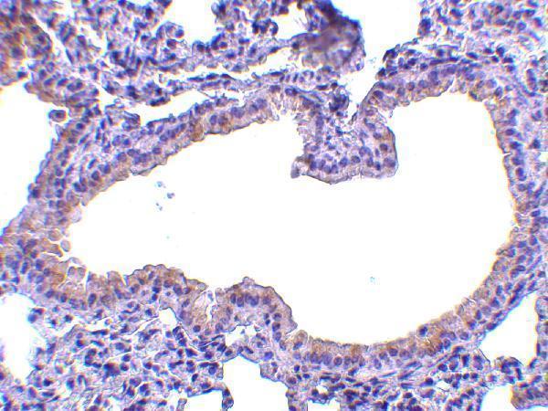
A01682 has a real SLPI IHC image from mouse lung tissue (catalog: IHC image caption). Human and Mouse are listed as reactive species (catalog: reactivity); no IF image is supplied (catalog: IF image alts).
A01682 will render with an IHC image of mouse lung tissue stained at 10 μg/mL (catalog: IHC image caption). It lists IHC-P and Human/Mouse reactivity (catalog: applications/reactivity), while the supplied IHC image documents mouse tissue only (catalog: IHC image caption).
Which to pick: Choose A01682 for tissue IHC-P: its application list includes IHC-P, and its image shows mouse lung tissue at 10 μg/mL; the fixative is unreported (catalog: applications/IHC image caption). No listed SKU has IF/ICC validation or an IF image, so there is no supported IF/ICC pick (catalog: applications/IF image alts). A01682 is the only cross-species candidate because Human and Mouse are listed as reactive, although the IHC image documents mouse tissue only; its host is Rabbit and its clone is unreported (catalog: reactivity/IHC image caption/host/clone).