This website uses cookies to ensure you get the best experience on our website.


- Table of Contents
Plan chromogenic SLU7 IHC in paraffin sections using the nuclear and cytoplasmic tissue pattern (HPA tissue IHC). This guide covers the catalog antibody’s 2–5 μg/ml range (datasheet A07557-2) and the uncertain consistency of tissue staining with RNA expression (HPA tissue IHC).

Expected localisation, validated protocols, controls and antibodies — the at-a-glance facts below, then the full design guide.
| Expected localisation | Nuclear and cytoplasmic (HPA tissue IHC) | |
| Staining pattern | Most tissues show nuclear and cytoplasmic staining (HPA tissue IHC) | |
| Antigen retrieval | EDTA pH 8.0 HIER, heat-mediated (datasheet A07557-2) | |
| Positive control | Adrenal gland+4 more · see all | |
| Negative control | Spleen |
| Fixation | Keep fixation conditions consistent across sections. (standard IHC practice; not target-specific) | |
| Caveat | Heat shock can shift SLU7 toward the cytoplasm (UniProt) | |
| Regulation | UV-C reduces nuclear SLU7 (UniProt) | |
| Isoform / epitope | No isoforms annotated; chain spans aa 2–586 (UniProt) |
Compare the catalog antibody’s IHC-P protocol (datasheet A07557-2) with a published SLU7 protocol for paraffin-embedded bladder cancer tissue (PMC10870071).
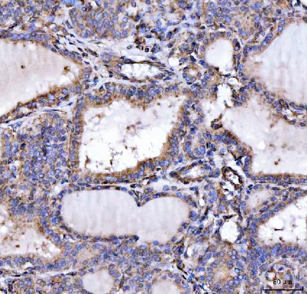
| Sample | Paraffin-embedded human thyroid cancer tissue; fixative not specified (datasheet A07557-2) |
| Fixation | Image fixative and duration unreported (datasheet A07557-2); verify before use. |
| Sectioning | 4–5 µm sections on charged slides (standard) |
| Deparaffinisation | Xylene, graded ethanol series to water (standard) |
| Antigen retrieval | Heat retrieval: EDTA pH 8.0 (datasheet A07557-2); 20 min, 95–100 °C (standard) |
| Peroxidase block | 3% H2O2, 10 min, room temperature (standard) |
| Blocking | 10% goat serum (datasheet A07557-2) |
| Primary antibody | Rabbit anti-SLU7, 2-5 μg/ml (datasheet A07557-2) |
| Primary incubation | Overnight at 4 °C (datasheet A07557-2) |
| Detection | HRP-conjugated secondary, DAB chromogen (datasheet A07557-2) |
| Counterstain | Hematoxylin, blue, dehydrate and mount (standard) |
| Expected result | SLU7-positive staining in glandular cells of adrenal gland (HPA tissue IHC: High). HPA tissue profile: Cytoplasmic and nuclear expression in most tissues. No signal in the no-primary control. |
SLU7 is predominantly nuclear, with nucleoplasmic and nuclear-speckle localization; cytoplasmic staining is also possible (UniProt O95391 localization; HPA: supported ICC-IF locations). In tissue IHC, expect nuclear and cytoplasmic signal in many cell types, including glandular cells and hepatocytes (HPA: tissue IHC). Treat intensity as provisional because HPA rates tissue staining Uncertain owing to low agreement with RNA expression (HPA: tissue IHC reliability). SLU7 has no transmembrane segment (UniProt O95391 topology).
| Nuclear staining, with possible cytoplasmic signal, in glandular cells or hepatocytes. | This fits the reported tissue pattern and predominantly nuclear localization (HPA: tissue IHC; UniProt O95391 localization). Judge staining within the relevant cell type, since a whole-section impression can hide which cells carry the signal (general IHC interpretation). |
| Strong staining confined to a membrane-like outline, with little nuclear signal. | Treat the compartment mismatch as possible artefact: SLU7 has no transmembrane segment and is predominantly nuclear (UniProt O95391 topology and localization). Cytoplasmic signal alone does not prove an artefact, because it is reported in tissues (HPA: tissue IHC). |
| Strong staining mainly in cells expected to be low or unstained. | Check for cross-reactivity or endogenous detection activity (general IHC interpretation). HPA reports low staining in bronchial respiratory epithelium and no detection in splenic red-pulp cells; its tissue IHC reliability is Uncertain (HPA: tissue IHC). |
| Diffuse color covers cells and spaces without a discernible cellular pattern. | Background may obscure nuclear localization; compare with a no-primary control and inspect whether signal follows tissue structures (general chromogenic IHC practice). A uniform haze does not independently establish SLU7 expression (general IHC interpretation). |
| No signal in a tissue-cell pair reported as high. | First check the staining run and cell identity (general IHC practice). HPA reports high signal in renal tubular cells and hepatocytes, but those observations are provisional because tissue IHC reliability is Uncertain (HPA: tissue IHC). |
| Cell type within the section | HPA reports High staining in adrenal, cervical, epididymal and stomach glandular cells, renal tubular cells, hepatocytes and placental decidual cells (HPA: tissue IHC). Score the specified cells rather than treating every cell in those organs as a positive reference (general IHC interpretation). |
| Lower-staining comparators | Appendiceal glandular cells are Medium; bronchial respiratory epithelial cells, prostatic glandular cells and adipocytes are low; splenic red-pulp cells are not detected (HPA: tissue IHC). These are observed staining levels, not guaranteed controls (HPA: tissue IHC reliability: Uncertain). |
| Expected compartment | SLU7 is predominantly nuclear but can occur in cytoplasm (UniProt O95391 localization). HPA describes cytoplasmic and nuclear expression in most tissues (HPA: tissue IHC). Assess both compartments while giving a purely membrane-like pattern extra scrutiny (UniProt O95391 topology; general IHC interpretation). |
| Cellular stress | UV-C reduces nuclear SLU7 concentration; heat shock can move it into cytoplasm and vesicle-like structures (UniProt O95391 localization). These reported responses do not establish what an unstressed paraffin section must show (UniProt O95391 localization; general interpretation). |
| Strength of tissue evidence | HPA rates tissue IHC Uncertain because antibody staining and RNA expression have low consistency (HPA: tissue IHC reliability). HPA035907 is IHC Uncertain; HPA058120 has no listed IHC rating (HPA: antibodies). Interpret a single unexpected tissue result cautiously. |
| IF/ICC Q: Should speckles appear? | A: Nucleoplasm is the supported main location; nuclear speckles are a supported additional location in ICC-IF (HPA: subcellular). This guides compartment interpretation but does not require resolvable speckles in chromogenic tissue IHC (HPA: subcellular; general IHC interpretation). |
| Situation | Likely cause | Next action |
|---|---|---|
| Reported high-staining cells show no signal. | The run or cell identification may need checking (general IHC practice); HPA's high calls are provisional (HPA: tissue IHC reliability: Uncertain). | Confirm the expected cell type, review controls, then check the antibody's IHC-P instructions and routine retrieval and detection steps (general IHC practice). No SLU7-specific fixation sensitivity is established by the supplied evidence. |
| Signal is mainly at cell borders. | A border-only pattern conflicts with SLU7's lack of a transmembrane segment and predominant nuclear location (UniProt O95391 topology and localization). | Compare with a no-primary control, inspect nuclear counterstain and reassess the stained structures before calling the result positive (general chromogenic IHC practice). |
| Cytoplasm stains more strongly than nuclei. | Cytoplasmic SLU7 is possible, including after reported stress responses (UniProt O95391 localization), but tissue IHC also shows mixed compartments with uncertain reliability (HPA: tissue IHC). | Check the cellular pattern against a reported high-staining tissue-cell pair and the run controls; record the compartment balance rather than rejecting cytoplasmic signal automatically (HPA: tissue IHC; general IHC practice). |
| Diffuse staining obscures cellular boundaries. | Nonspecific background or endogenous detection activity may contribute (general chromogenic IHC practice). | Review no-primary and detection controls; adjust blocking, washing or detection conditions according to the staining system before interpreting localization (general chromogenic IHC practice). |
| Splenic red-pulp cells stain strongly. | HPA reports them as Not detected, though tissue IHC reliability is Uncertain (HPA: tissue IHC). Cross-reactivity or endogenous activity merits consideration (general IHC interpretation). | Verify the cells being scored, inspect controls and compare staining with a reported high-staining cell type before assigning SLU7 positivity (HPA: tissue IHC; general IHC practice). |
| Two antibodies give different tissue patterns. | Agreement is not assured: HPA035907 is IHC Uncertain, and HPA058120 has no listed IHC rating (HPA: antibodies). | Document antibody identity, controls, stained cell types and compartments for each run; weigh disagreement against HPA's Uncertain tissue assessment rather than treating either pattern as definitive (HPA: antibodies and tissue IHC; general IHC practice). |
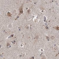


Comprehensive Human Protein Atlas IHC scoring per tissue (reliability: Uncertain — Low consistency between antibody staining and RNA expression data.). Rows are taken directly from the HPA tissue chart — click any row's HPA link to view the source.
| Tissue | Cell type | Level | Evidence | Source |
|---|---|---|---|---|
| Adrenal gland | Glandular cells | High | Protein (IHC) | HPA → |
| Cervix | Glandular cells | High | Protein (IHC) | HPA → |
| Epididymis | Glandular cells | High | Protein (IHC) | HPA → |
| Kidney | Cells in tubules | High | Protein (IHC) | HPA → |
| Liver | Hepatocytes | High | Protein (IHC) | HPA → |
| Tissue | Cell type | Level | Evidence | Source |
|---|---|---|---|---|
| Spleen | Cells in red pulp | Not detected | Protein (IHC) | HPA → |
Use compartment controls and the documented paraffin-section conditions to troubleshoot SLU7 staining, while treating tissue staining patterns cautiously (HPA: IHC reliability uncertain).
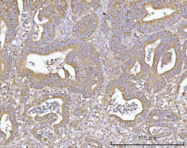
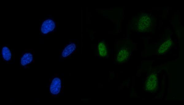
A07557-2 has IHC images from human paraffin sections and IF images from human liver cancer paraffin sections and U87 cells (catalog image captions; datasheet: Human reactivity).
A07557-2 is listed for human IHC, ICC and IF (catalog applications; datasheet: Human reactivity). Its IHC captions show human thyroid cancer, colorectal adenocarcinoma and liver cancer paraffin sections; its IF captions show U87 cells and human liver cancer paraffin sections (A07557-2 image captions).
Which to pick: For tissue IHC, choose A07557-2: its paraffin-section captions document EDTA retrieval at pH 8.0 and primary antibody at 2 μg/ml; the fixative is unreported (A07557-2 IHC image captions). For IF/ICC, the same rabbit antibody has IF images of U87 cells and human liver cancer paraffin sections, with a listed IF dilution of 5 μg/ml (catalog host and IF image captions; datasheet: 5 μg/ml). Cross-species use is unvalidated in this payload because A07557-2 lists Human reactivity only; no clone is specified (catalog reactivity and clone fields).