This website uses cookies to ensure you get the best experience on our website.


- Table of Contents
Plan chromogenic SMAD9 IHC in paraffin sections using the catalog antibody (datasheet A04932). Choose tissue controls and interpret cytoplasmic and nuclear staining using the tissue profile, while accounting for its low consistency with RNA expression (HPA tissue IHC).

Expected localisation, validated protocols, controls and antibodies — the at-a-glance facts below, then the full design guide.
| Expected localisation | Cytoplasmic and nuclear staining (HPA tissue IHC) | |
| Staining pattern | Glandular and epithelial cells stain; cytoplasm and nuclei (HPA tissue IHC) | |
| Antigen retrieval | Citrate pH 6.0 HIER, heat-mediated (datasheet A04932) | |
| Positive control | Appendix+4 more · see all | |
| Negative control | Adipose tissue+1 more · see all |
| Fixation | Keep fixation conditions consistent across sections. (standard IHC practice; not target-specific) | |
| Caveat | Antibody staining has low consistency with RNA expression (HPA tissue IHC) | |
| Regulation | SMAD4 binding may shift nuclear signal (UniProt; by similarity) | |
| Isoform / epitope | 2 isoforms (A and B); epitope differences unspecified (UniProt) |
The catalog antibody’s IHC-P protocol is supplemented by two published SMAD9 chromogenic IHC protocols (PMC7923649; PMC10685465).
| Sample | Paraffin-embedded human liver cancer tissue; fixative not specified (datasheet A04932) |
| Fixation | Image fixative and duration unreported (datasheet A04932); verify before use. |
| Sectioning | 4–5 µm sections on charged slides (standard) |
| Deparaffinisation | Xylene, graded ethanol series to water (standard) |
| Antigen retrieval | Heat retrieval: Citrate pH 6.0 (datasheet A04932); 20 min, 95–100 °C (standard) |
| Peroxidase block | 3% H2O2, 10 min, room temperature (standard) |
| Blocking | 10% normal serum of the secondary host, 30 min, room temperature (standard) |
| Primary antibody | Rabbit anti-SMAD9, 1:50-1:200 (datasheet A04932) |
| Primary incubation | Overnight at 4 °C (standard) |
| Detection | HRP-polymer secondary, DAB chromogen 5–10 min (standard) |
| Counterstain | Hematoxylin, blue, dehydrate and mount (standard) |
| Expected result | SMAD9-positive staining in glandular cells of appendix (HPA tissue IHC: High). HPA tissue profile: Cytoplasmic and nuclear expression in several tissues. No signal in the no-primary control. |
SMAD9 staining may appear in the cytoplasm and nucleus of epithelial and glandular cells in paraffin tissue sections (HPA: tissue IHC profile). Several such cell populations show High staining, including colonic glandular and bronchial respiratory epithelial cells (HPA: tissue IHC). SMAD9 has no transmembrane segment (UniProt O15198: topology). Interpret the pattern cautiously: HPA rates its IHC data Approved but reports low consistency between antibody staining and RNA expression (HPA: reliability description).
| Cytoplasmic and nuclear staining in colonic glandular cells or bronchial respiratory epithelial cells. | This fits the reported tissue pattern and High staining in those cells (HPA: tissue IHC). Score each compartment and cell population separately; nuclear staining alone does not establish pathway activation (UniProt O15198: ligand-dependent localization, by similarity). |
| A predominantly plasma-membrane outline, with little cellular staining. | This conflicts with the reported cytoplasmic and nuclear distribution and absence of a transmembrane segment (HPA: tissue IHC; UniProt O15198: topology). Review morphology and controls before assigning the outline to SMAD9 (general IHC practice). |
| Strong signal in adipocytes or cells of the cerebellar granular layer. | Both populations are reported as Not detected (HPA: tissue IHC). Unexpected signal could reflect cross-reactivity or endogenous detection activity; check controls before treating either population as positive (general IHC practice). |
| Chromogen spread across tissue, including spaces between cells. | A widespread deposit lacks the compartment and cell-type pattern reported for SMAD9 (HPA: tissue IHC). Uneven reagent coverage, inadequate washing, or endogenous detection activity are general IHC possibilities (general IHC practice). |
| No cellular signal in an otherwise interpretable colon or bronchus section. | These contain cell populations reported as High (HPA: tissue IHC). Check section quality, retrieval, antibody application, and detection controls before concluding absence of SMAD9 (general IHC practice). |
| Cell population and tissue context | HPA reports High staining in several glandular and epithelial populations, but Not detected in adipocytes and cerebellar granular-layer cells (HPA: tissue IHC). Compare like cell populations rather than scoring an entire section uniformly (general IHC practice). |
| Conditional compartment distribution | UniProt places SMAD9 in the cytoplasm without ligand and describes migration to the nucleus with SMAD4 by similarity (UniProt O15198: subcellular location). Compartment balance can vary; a static stain cannot establish why SMAD9 is nuclear (general IHC interpretation). |
| Antibody evidence | Two listed antibodies have Approved IHC status, while HPA notes low consistency between staining and RNA expression (HPA: antibody validation; tissue reliability). Treat an unexpected cell pattern as needing control-based review rather than assuming it represents SMAD9 (general IHC practice). |
| Isoforms and epitope coverage | UniProt lists 2 isoforms, A and B (UniProt O15198: isoforms). The supplied evidence does not locate the catalog antibody's epitope or establish isoform-specific IHC staining, so do not assign an observed compartment or cell pattern to one isoform. |
| IF evidence and transfer to sections | HPA reports mainly nucleoplasmic, with additional cytosolic, localization in ICC-IF (HPA: subcellular). That observation helps assess compartments but does not set an IHC intensity threshold in paraffin sections (HPA: tissue IHC; general IHC interpretation). |
| Situation | Likely cause | Next action |
|---|---|---|
| Expected glandular or respiratory epithelial staining is absent. | The section may have a detection or preparation problem; these populations are reported as High (HPA: tissue IHC; general IHC practice). | Confirm tissue morphology and run the catalog antibody's IHC-P procedure with its specified retrieval and detection controls; compare a known-positive section in the same run (general IHC practice). |
| Adipocytes or cerebellar granular-layer cells stain strongly. | That conflicts with their Not detected HPA observations; cross-reactivity or endogenous activity is possible (HPA: tissue IHC; general IHC practice). | Review a reagent control and the cell morphology; for chromogenic detection, check the relevant endogenous-enzyme blocking control before scoring the cells (general IHC practice). |
| The slide has diffuse chromogen or poor cellular contrast. | Background can arise from detection reagents, inadequate washing, or uneven reagent handling (general IHC practice). | Check a control lacking primary antibody, washing, and reagent coverage; interpret only localized cellular signal against the expected tissue pattern (general IHC practice; HPA: tissue IHC). |
| A membrane rim dominates while cytoplasm and nuclei are faint. | The rim is discordant with SMAD9's cytoplasmic and nuclear locations and lack of a transmembrane segment (HPA: tissue IHC; UniProt O15198: topology). | Check whether the rim follows tissue edges or nonspecific deposits, then assess controls and a morphologically intact area before assigning localization (general IHC practice). |
| Nuclear staining varies among cells in one section. | SMAD9 can occur in both compartments, with nuclear migration described in a SMAD4 complex by similarity (UniProt O15198: subcellular location). Variation alone does not identify the underlying signaling state. | Record nuclear and cytoplasmic staining separately by cell type and compare with controls; avoid calling BMP activation from a single static section (general IHC interpretation; UniProt O15198: function). |
| Why does ICC-IF look more nuclear than the paraffin-section IHC? | HPA describes mainly nucleoplasmic ICC-IF staining, while its tissue IHC profile includes cytoplasmic and nuclear expression (HPA: subcellular; tissue IHC). The supplied evidence does not establish why the preparations differ. | Interpret each assay against its own reported pattern and controls; do not use ICC-IF intensity as an IHC scoring cutoff (HPA: subcellular; tissue IHC; general IHC interpretation). |
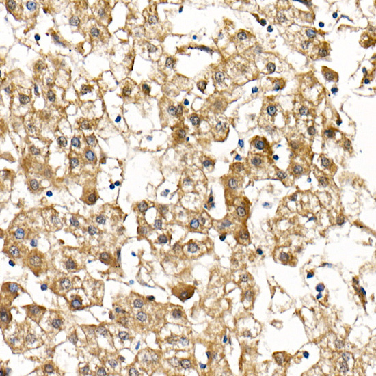
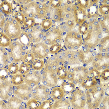
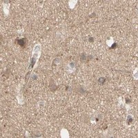


Comprehensive Human Protein Atlas IHC scoring per tissue (reliability: Approved — Low consistency between antibody staining and RNA expression data.). Rows are taken directly from the HPA tissue chart — click any row's HPA link to view the source.
| Tissue | Cell type | Level | Evidence | Source |
|---|---|---|---|---|
| Appendix | Glandular cells | High | Protein (IHC) | HPA → |
| Breast | Glandular cells | High | Protein (IHC) | HPA → |
| Bronchus | Respiratory epithelial cells | High | Protein (IHC) | HPA → |
| Cervix | Squamous epithelial cells | High | Protein (IHC) | HPA → |
| Colon | Glandular cells | High | Protein (IHC) | HPA → |
Use compartment-aware scoring and matched controls when troubleshooting SMAD9 staining in paraffin sections (UniProt O15198 localisation; standard IHC practice).
The catalog antibody has real IHC images from paraffin-embedded human liver cancer and placenta; IF/ICC is listed for human, mouse and rat samples (A04932 catalog; A04932 IHC captions).
A04932 is the sole card and shows IHC staining of paraffin-embedded human liver cancer and placenta at 1:100 after citrate retrieval (A04932 IHC captions). IF/ICC is listed, with human, mouse and rat reactivity, but no IF image is supplied (A04932 catalog).
Which to pick: Choose A04932 for paraffin-section IHC: its own captions document human liver cancer and placenta staining, while the fixative is unreported (A04932 IHC captions). A04932 is also the listed IF/ICC and cross-species option because its applications include IF/ICC and its stated reactivity covers human, mouse and rat; the supplied images demonstrate IHC in human tissue only (A04932 catalog; A04932 IHC captions). It is a rabbit polyclonal antibody (A04932 IHC captions).