This website uses cookies to ensure you get the best experience on our website.


- Table of Contents
Plan chromogenic IHC for SMARCA1 in paraffin sections with the IHC-validated antibody at 1:50–1:200 (datasheet A05676). Compare nuclear and cytoplasmic staining with tissue examples, bearing in mind that staining has low consistency with RNA expression (HPA tissue IHC).

Expected localisation, validated protocols, controls and antibodies — the at-a-glance facts below, then the full design guide.
| Expected localisation | Nuclear and cytoplasmic staining in several tissues (HPA tissue IHC) | |
| Staining pattern | Nuclear and cytoplasmic staining across several cell types (HPA tissue IHC) | |
| Antigen retrieval | Citrate pH 6.0 HIER, heat-mediated (datasheet A05676) | |
| Positive control | Breast+4 more · see all | |
| Negative control | Adipose tissue+4 more · see all |
| Fixation | Keep fixation consistent (standard IHC practice; not target-specific); target-specific effects are unknown. | |
| Caveat | Staining has low consistency with RNA expression (HPA tissue IHC) | |
| Regulation | Expression regulation not specified (UniProt) | |
| Isoform / epitope | 2 isoforms; epitope coverage is unspecified (UniProt) |
The catalog antibody uses citrate pH 6.0 retrieval (datasheet A05676). One published protocol stains paraffin-embedded soft tissue sarcoma sections (PMC6513346).
| Sample | Paraffin-embedded human lung cancer tissue; fixative not specified (datasheet A05676) |
| Fixation | Image fixative and duration unreported (datasheet A05676); verify before use. |
| Sectioning | 4–5 µm sections on charged slides (standard) |
| Deparaffinisation | Xylene, graded ethanol series to water (standard) |
| Antigen retrieval | Heat retrieval: Citrate pH 6.0 (datasheet A05676); 20 min, 95–100 °C (standard) |
| Peroxidase block | 3% H2O2, 10 min, room temperature (standard) |
| Blocking | 10% normal serum of the secondary host, 30 min, room temperature (standard) |
| Primary antibody | Rabbit anti-SMARCA1, 1:50-1:200 (datasheet A05676) |
| Primary incubation | Overnight at 4 °C (standard) |
| Detection | HRP-polymer secondary, DAB chromogen 5–10 min (standard) |
| Counterstain | Hematoxylin, blue, dehydrate and mount (standard) |
| Expected result | SMARCA1-positive staining in glandular cells of breast (HPA tissue IHC: Medium). HPA tissue profile: Nuclear and cytoplasmic expression in several tissues. No signal in the no-primary control. |
SMARCA1 is a nuclear and chromosome-associated protein with no transmembrane segment (UniProt P28370: subcellular location; topology). In paraffin-section IHC, look for staining in breast glandular cells, lung alveolar cells, brain glia, and cells in testicular seminiferous ducts (HPA tissue IHC: medium staining). HPA reports nuclear and cytoplasmic staining across several tissues, but rates its tissue IHC profile Uncertain because antibody staining has low consistency with RNA expression (HPA tissue IHC: profile; reliability).
| Distinct staining in nuclei of the listed cell populations, with possible cytoplasmic staining. | This fits SMARCA1's nuclear and chromosome location (UniProt P28370: subcellular location) and the reported IHC profile (HPA tissue IHC: nuclear and cytoplasmic expression). Judge intensity against the relevant cell population: the listed positive populations are reported at medium level, not uniformly across every cell in those tissues (HPA tissue IHC: positive cells and levels). |
| Signal is confined to cell borders, extracellular material, or another unexpected compartment, with little convincing nuclear staining. | Investigate background or an antibody-dependent pattern before assigning it to SMARCA1: UniProt places the protein in the nucleus and on chromosomes and reports no transmembrane segment (UniProt P28370: subcellular location; topology). Cytoplasmic staining alone deserves caution rather than automatic rejection, because HPA also reports cytoplasmic tissue IHC staining (HPA tissue IHC: profile). |
| Strong staining appears chiefly in a cell population reported as unstained, while expected positive cells remain faint. | Cross-reactivity or endogenous chromogenic activity is possible; compare cell identity, compartment, and control slides. For example, HPA reports breast glandular cells at medium level and adipocytes as not detected (HPA tissue IHC: breast; adipose tissue). These entries are comparison points, not definitive proof of target absence, because HPA rates the IHC profile Uncertain (HPA tissue IHC: reliability). |
| Brown signal spreads across tissue or blank areas without recognizable cell boundaries or nuclear contrast. | Treat the diffuse deposit as background until controls show otherwise. Check whether it persists when the primary antibody is omitted, and review blocking, washing, and chromogen development as general chromogenic IHC practice. The broad deposit cannot establish a SMARCA1 distribution; HPA's reported staining is described in cells and tissues (HPA tissue IHC: profile). |
| No visible signal appears in a section containing an expected positive population. | First confirm that the expected cells are present: HPA reports medium staining in lung alveolar cells, breast glandular cells, brain glia, and seminiferous-duct cells (HPA tissue IHC: positive cells and levels). Then check the assay controls and general IHC steps. A single blank section does not establish biological absence, particularly with HPA's Uncertain IHC reliability (HPA tissue IHC: reliability). |
| Tissue and cell selection | Use the named HPA-positive populations as practical comparison sites; staining is reported at medium level in breast glandular cells, lung alveolar cells, brain glia, and testicular seminiferous-duct cells (HPA tissue IHC: positive cells and levels). HPA reports low tissue RNA specificity and an Uncertain IHC profile, so a tissue label alone does not settle antibody specificity (HPA tissue IHC: RNA specificity; reliability). |
| Antibody validation | The HPA tissue IHC antibody HPA003335 is rated Uncertain for IHC, while HPA064712 is rated Supported for ICC (HPA antibodies: validation status). These are application-specific ratings; use tissue IHC controls to evaluate an IHC result rather than treating the ICC rating as tissue IHC validation (HPA antibodies: validation status; general IHC practice). |
| Sequence and processing | UniProt lists 2 isoforms, no signal peptide or propeptide, and no transmembrane segment (UniProt P28370: isoforms; processing; topology). These facts help frame antibody-epitope questions but do not identify which isoforms an unspecified antibody detects. Target-specific fixation sensitivity and antigen-retrieval requirements are unreported in the supplied UniProt and HPA records. |
| IF/ICC Q&A | Q: What location should an IF/ICC image support? A: Predominantly nucleoplasmic signal; HPA also reports vesicles as an additional location (HPA subcellular: supported nucleoplasm; approved vesicles). This ICC observation is useful for localization comparison, while HPA's tissue IHC profile separately reports nuclear and cytoplasmic staining with Uncertain reliability (HPA tissue IHC: profile; reliability). |
| Situation | Likely cause | Next action |
|---|---|---|
| Expected positive cells are blank. | The target population may be absent from the section, or a general IHC step may have failed; the chosen HPA-positive populations are reported at medium level (HPA tissue IHC: positive cells and levels). | Verify the cell population on the counterstained section, run a suitable positive-control section, then review retrieval, primary incubation, and detection against the antibody's documented IHC procedure (general IHC practice). Do not infer a SMARCA1-specific retrieval requirement from HPA staining levels. |
| Signal is widespread and lacks cellular structure. | Nonspecific binding, insufficient washing, or excessive chromogen development can produce diffuse background (general chromogenic IHC practice). | Compare a primary-omission control, inspect washes and blocking, and shorten chromogen development if the control also stains (general chromogenic IHC practice). Reassess nuclear and cell-specific staining only after background is controlled (UniProt P28370: nuclear location; HPA tissue IHC: positive cells). |
| Primary-omission control still develops color. | Endogenous detection activity or another reagent-dependent deposit is plausible when color forms without primary antibody (general chromogenic IHC practice). | Check the detection system's endogenous-activity block and reagent-only controls before interpreting cellular color (general chromogenic IHC practice). Do not attribute control-slide color to SMARCA1. |
| Only cytoplasm stains in the expected cells. | HPA reports cytoplasmic as well as nuclear tissue staining, but UniProt places SMARCA1 in the nucleus and on chromosomes (HPA tissue IHC: profile; UniProt P28370: subcellular location). | Compare the nuclear counterstain and a second well-controlled section. Record the cytoplasmic-only result as ambiguous if nuclear signal remains absent, given HPA's Uncertain tissue IHC reliability (HPA tissue IHC: reliability; general IHC practice). |
| Reported negative cells stain more strongly than expected positive cells. | Cell misidentification, cross-reactivity, or endogenous detection activity may explain the mismatch (general IHC practice); HPA reports adipocytes as not detected and breast glandular cells at medium level (HPA tissue IHC: adipose tissue; breast). | Confirm cell identity and compare primary-omission and positive-control sections. Treat the HPA negative entry as a reference observation rather than a universal exclusion rule, because the tissue IHC profile is Uncertain (HPA tissue IHC: reliability). |
| Signal appears as isolated membrane-like outlines. | A membrane-confined pattern conflicts with the reported nuclear and chromosome location and lack of transmembrane segment (UniProt P28370: subcellular location; topology). | Inspect the same cells for convincing nuclear staining and compare control sections; investigate edge staining or detection background before scoring the outlines as positive (general chromogenic IHC practice). |
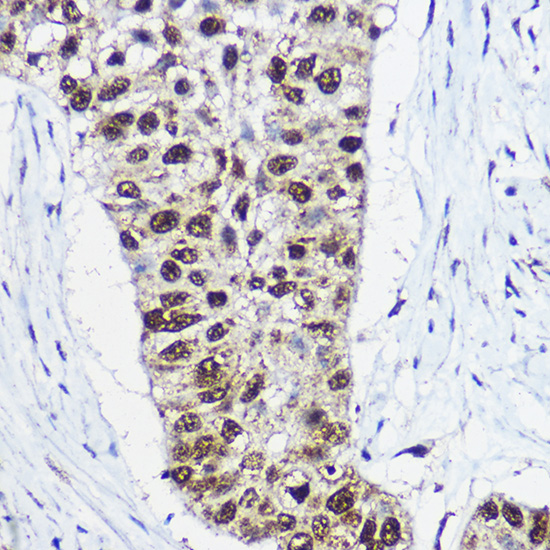
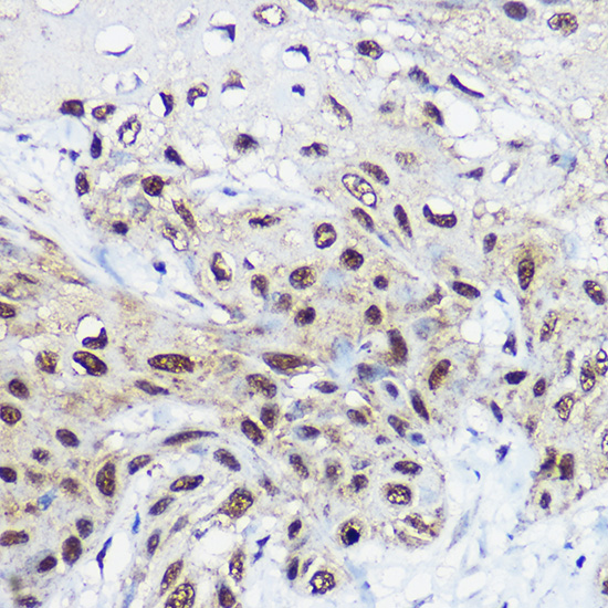
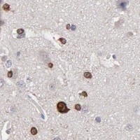


Comprehensive Human Protein Atlas IHC scoring per tissue (reliability: Uncertain — Low consistency between antibody staining and RNA expression data.). Rows are taken directly from the HPA tissue chart — click any row's HPA link to view the source.
| Tissue | Cell type | Level | Evidence | Source |
|---|---|---|---|---|
| Breast | Glandular cells | Medium | Protein (IHC) | HPA → |
| Caudate | Glial cells | Medium | Protein (IHC) | HPA → |
| Cerebellum | Cells in molecular layer | Medium | Protein (IHC) | HPA → |
| Cerebral cortex | Glial cells | Medium | Protein (IHC) | HPA → |
| Hippocampus | Glial cells | Medium | Protein (IHC) | HPA → |
| Tissue | Cell type | Level | Evidence | Source |
|---|---|---|---|---|
| Adipose tissue | Adipocytes | Not detected | Protein (IHC) | HPA → |
| Adrenal gland | Glandular cells | Not detected | Protein (IHC) | HPA → |
| Appendix | Glandular cells | Not detected | Protein (IHC) | HPA → |
| Bronchus | Respiratory epithelial cells | Not detected | Protein (IHC) | HPA → |
| Cervix | Glandular cells | Not detected | Protein (IHC) | HPA → |
Use nuclear localization, the selected antibody’s paraffin-section evidence, and tissue controls to troubleshoot SMARCA1 staining (UniProt P28370; datasheet A05676; HPA tissue IHC).
A05676 has real IHC images from paraffin-embedded human lung and esophageal cancer (catalog image captions). Human, mouse, and rat reactivity is listed; IF/ICC data are absent (catalog: reactivity and applications).
A05676 is listed for IHC and has images of paraffin-embedded human lung and esophageal cancer sections (catalog: applications and IHC image captions). Human, mouse, and rat reactivity is listed, but the supplied images show human tissue only (catalog: reactivity and IHC image captions).
Which to pick: For tissue IHC, choose A05676: its paraffin-section images use 1:100 antibody dilution and high-pressure retrieval in 10 mM citrate buffer at pH 6.0; the fixative is unreported (catalog: IHC image captions). No SKU here is listed for IF/ICC, so there is no IF/ICC pick (catalog: applications). For mouse or rat IHC, A05676 is the only listed option based on stated reactivity, although its supplied images show human tissue; it is a rabbit polyclonal antibody (catalog: reactivity, IHC image captions, and host/clonality).