This website uses cookies to ensure you get the best experience on our website.


- Table of Contents
Plan SMARCA5 chromogenic IHC on human paraffin sections using the catalog antibody at 2–5 μg/mL (datasheet A02687-1). Assess nuclear staining against tissue controls, such as colon glandular cells with high staining and adipocytes with no detected staining (HPA tissue IHC).

Expected localisation, validated protocols, controls and antibodies — the at-a-glance facts below, then the full design guide.
| Expected localisation | General nuclear staining (HPA tissue IHC) | |
| Staining pattern | Nuclear staining across diverse tissue cell types (HPA tissue IHC) | |
| Antigen retrieval | EDTA pH 8.0 HIER, heat-mediated (datasheet A02687-1) | |
| Positive control | Appendix+4 more · see all | |
| Negative control | Adipose tissue+1 more · see all |
| Fixation | Keep fixation consistent across paraffin sections (standard IHC practice; not target-specific) | |
| Caveat | Adipocytes and cholangiocytes may show no staining (HPA tissue IHC) | |
| Regulation | Ubiquitously expressed (UniProt) | |
| Isoform / epitope | No isoforms annotated; no signal peptide or propeptide (UniProt) |
The catalog antibody’s IHC-P protocol is presented alongside three published SMARCA5 IHC protocols with usable methods details (PMC12274346; PMC13378543; PMC11538284).
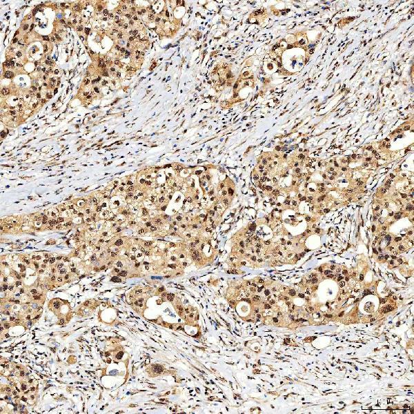
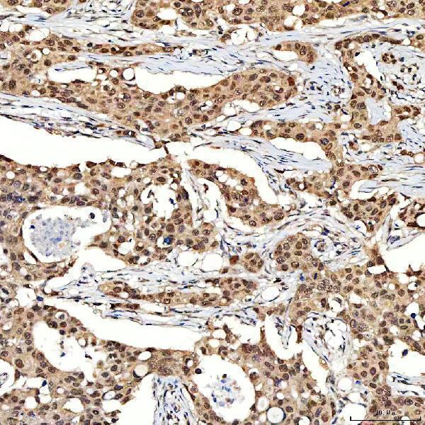
| Sample | Paraffin-embedded human appendix adenocarcinoma tissue; fixative not specified (datasheet A02687-1) |
| Fixation | Image fixative and duration unreported (datasheet A02687-1); verify before use. |
| Sectioning | 4–5 µm sections on charged slides (standard) |
| Deparaffinisation | Xylene, graded ethanol series to water (standard) |
| Antigen retrieval | Heat retrieval: EDTA pH 8.0 (datasheet A02687-1); 20 min, 95–100 °C (standard) |
| Peroxidase block | 3% H2O2, 10 min, room temperature (standard) |
| Blocking | 10% goat serum (datasheet A02687-1) |
| Primary antibody | Rabbit anti-SMARCA5, 2-5 μg/ml (datasheet A02687-1) |
| Primary incubation | Overnight at 4 °C (datasheet A02687-1) |
| Detection | HRP-conjugated secondary, DAB chromogen (datasheet A02687-1) |
| Counterstain | Hematoxylin, blue, dehydrate and mount (standard) |
| Expected result | SMARCA5-positive staining in glandular cells of appendix (HPA tissue IHC: High). HPA tissue profile: General nuclear expression. No signal in the no-primary control. |
SMARCA5 is a nuclear chromatin-remodeling protein with no transmembrane segment (UniProt O60264). In paraffin-section IHC, expect predominantly nuclear staining across many cell types, including colon glandular cells and lymph-node germinal-center cells with high reported staining (HPA tissue IHC). HPA describes general nuclear expression, but rates its tissue IHC evidence “Supported,” with only medium consistency between antibody staining and RNA expression (HPA tissue IHC).
| Clear nuclear staining in colon glandular cells or lymph-node germinal-center cells, with recognizable tissue architecture. | This matches HPA’s high staining in those specific cell populations and its general nuclear profile (HPA tissue IHC). Score the relevant cells and their nuclei; a positive field alone does not show that every cell in the tissue should stain (HPA tissue IHC; general IHC practice). |
| Predominantly cytoplasmic, membranous, or extracellular color, with little nuclear signal. | That distribution conflicts with the nuclear and chromosome localization of SMARCA5 and its lack of a transmembrane segment (UniProt O60264). Treat it as suspect staining and check the detection controls and morphology before assigning a biological explanation (general IHC practice). |
| Strong staining in adipocytes or liver cholangiocytes, especially when the signal is outside nuclei. | HPA reports SMARCA5 as not detected in these named cell populations (HPA tissue IHC). Unexpected staining may reflect antibody cross-reactivity or endogenous detection activity; assess each possibility with controls (general IHC practice). HPA’s observation does not label the entire adipose or liver section negative (HPA tissue IHC). |
| A diffuse haze covers nuclei, cytoplasm, and surrounding tissue, obscuring cell boundaries. | The haze cannot establish the reported nuclear pattern (HPA tissue IHC; general IHC practice). Possible general IHC causes include excess primary or detection reagent, inadequate washing, and endogenous detection activity; interpret only after controls distinguish background from cell-associated signal (general IHC practice). |
| No nuclear signal in a colon glandular-cell or lymph-node germinal-center-cell positive-control region. | This disagrees with HPA’s high staining reports for those cells (HPA tissue IHC). Check that the expected cells are present, then review primary-antibody, retrieval, and detection steps as general IHC troubleshooting (general IHC practice). A single failed section cannot establish SMARCA5 absence (general IHC practice). |
| Cell population and scoring | Expression is broadly distributed but varies by cell type: HPA lists high staining in several glandular, squamous, hematopoietic, and germinal-center populations, low staining in breast and salivary glandular cells, and no detected staining in adipocytes or cholangiocytes (HPA tissue IHC). Compare like cells, not whole-organ averages (general IHC practice). |
| Evidence strength and antibody choice | HPA calls tissue IHC “Supported” and notes medium antibody-staining/RNA consistency (HPA tissue IHC). HPA008751 and CAB005227 each have “Supported” IHC status; HPA008751 also has “Supported” ICC status (HPA antibodies). These labels support a starting expectation, while controls remain necessary for a new assay (general IHC practice). |
| Nuclear distribution and cell state | UniProt places SMARCA5 in the nucleus and on mitotic chromosomes, and describes associations with replication foci during S phase and DNA-damage sites (UniProt O60264). Those annotations provide biological context; the supplied tissue IHC record supports a general nuclear pattern, not a required punctate pattern in paraffin sections (HPA tissue IHC). |
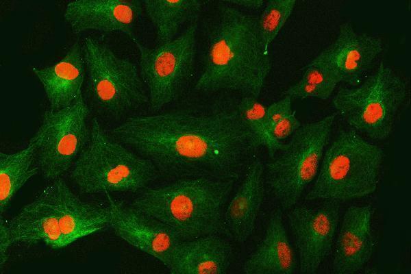
| IF/ICC Q&A: where should signal appear? | Mainly in the nucleoplasm, with additional localization to nucleoli fibrillar centers in HPA’s ICC-IF data (HPA subcellular ICC-IF). Use that localization to interpret an IF/ICC image; it is a separate application from the chromogenic paraffin-section IHC pattern described here (HPA subcellular ICC-IF; HPA tissue IHC). |
| Situation | Likely cause | Next action |
|---|---|---|
| Positive-control nuclei are blank. | The result conflicts with high staining reported for the chosen positive cell population (HPA tissue IHC); a missed or ineffective assay step is possible (general IHC practice). | Confirm the expected cells on the section, then review primary-antibody application, detection, and retrieval using the assay’s controls (general IHC practice). No SMARCA5-specific retrieval requirement is supplied by UniProt or HPA. |
| Color is chiefly cytoplasmic or membranous. | The compartment differs from SMARCA5’s nuclear localization (UniProt O60264); nonspecific staining or detection background is possible (general IHC practice). | Inspect a no-primary control, reassess the IHC-validated antibody’s staining pattern, and score only clearly localized nuclear signal (general IHC practice; HPA antibodies). |
| Adipocytes or cholangiocytes stain strongly. | HPA reports these particular cell types as not detected (HPA tissue IHC); cross-reactivity or endogenous detection activity may account for unexpected color (general IHC practice). | Compare the same cell type with a no-primary control and a high-staining positive-control population; keep cell identity and nuclear localization in the comparison (HPA tissue IHC; general IHC practice). |
| Background obscures nuclei throughout the section. | Excess reagent, insufficient washing, or endogenous activity can produce diffuse color in a chromogenic assay (general IHC practice). | Use a no-primary control to locate detection background, then review blocking, reagent concentration, and washes within the chosen assay workflow (general IHC practice). |
| Breast or salivary glandular cells show faint nuclear color. | HPA lists low staining in those cell populations, so a weak result can fit the reported tissue pattern (HPA tissue IHC). | Compare matched cell types and a high-staining control before judging the assay unsuccessful; preserve the distinction between faint nuclear signal and diffuse background (HPA tissue IHC; general IHC practice). |
| A dark field appears positive, but nuclei cannot be distinguished. | Overall color alone does not demonstrate the general nuclear expression reported by HPA (HPA tissue IHC; general IHC practice). | Assess tissue morphology and nuclear counterstain, compare the no-primary control, and record a positive interpretation only where signal can be assigned to nuclei in identifiable cells (general IHC practice). |


Comprehensive Human Protein Atlas IHC scoring per tissue (reliability: Supported — Medium consistency between antibody staining and RNA expression data.). Rows are taken directly from the HPA tissue chart — click any row's HPA link to view the source.
| Tissue | Cell type | Level | Evidence | Source |
|---|---|---|---|---|
| Appendix | Glandular cells | High | Protein (IHC) | HPA → |
| Bone marrow | Hematopoietic cells | High | Protein (IHC) | HPA → |
| Cervix | Glandular cells | High | Protein (IHC) | HPA → |
| Colon | Glandular cells | High | Protein (IHC) | HPA → |
| Duodenum | Glandular cells | High | Protein (IHC) | HPA → |
Troubleshoot chromogenic SMARCA5 IHC by checking nuclear staining, section processing and controls; use the IF guidance for multiplex imaging.
A02687-1 has human paraffin-section IHC images and an A549 IF image (catalog image captions); M02687 lists ICC/IF and Human, Mouse and Rat reactivity (catalog applications and reactivity).
A02687-1 will render with an IHC image from human paraffin-embedded appendix adenocarcinoma; its catalog also shows IF in A549 cells (A02687-1 image captions). M02687 will render with listed ICC/IF applications and Human, Mouse and Rat reactivity, but no supplied IHC or IF image (M02687 catalog).
Which to pick: Choose A02687-1 for tissue IHC: its image documents staining of a human paraffin section at 2 μg/ml (A02687-1 IHC caption). For IF/ICC, A02687-1 has an A549 IF image at 5 μg/ml (A02687-1 IF caption); M02687 is a monoclonal option with listed ICC/IF use and Human, Mouse and Rat reactivity, though no IF image is supplied (M02687 catalog). The A02687-1 IHC caption supports paraffin sections; it does not report the fixative (A02687-1 IHC caption).