This website uses cookies to ensure you get the best experience on our website.
- Table of Contents
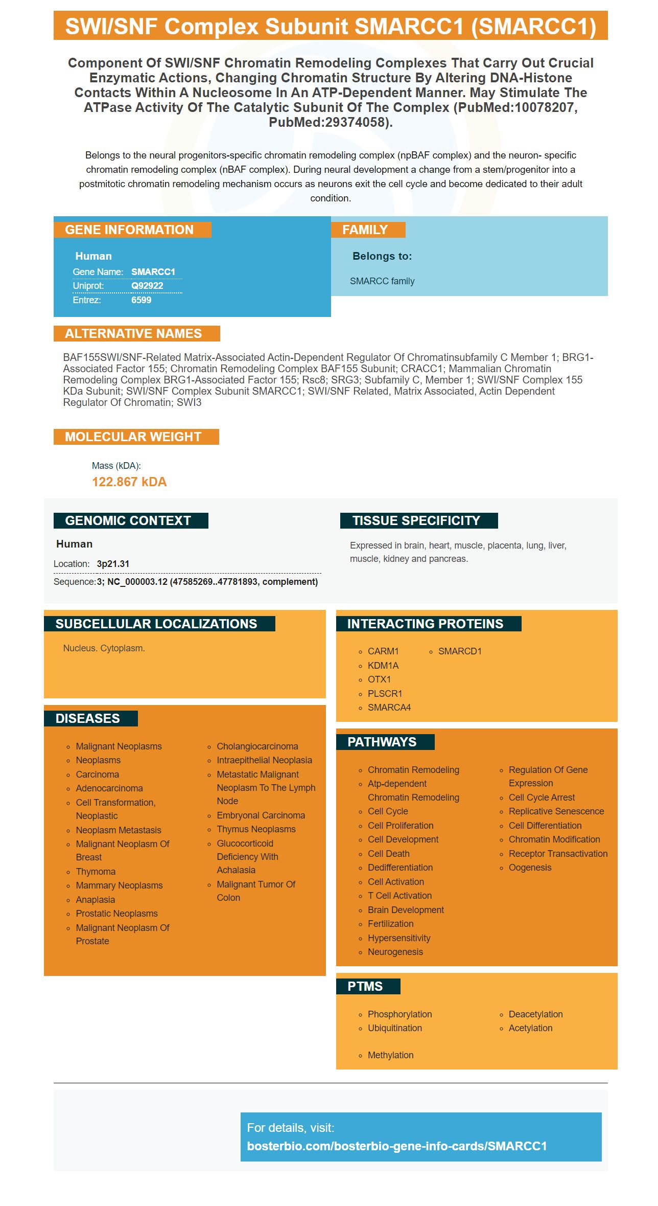
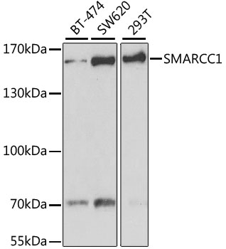
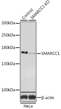
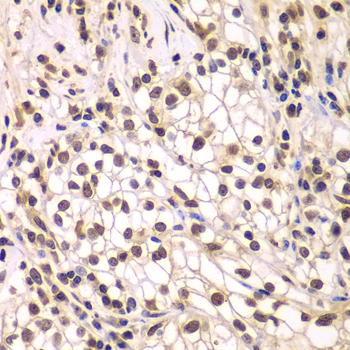
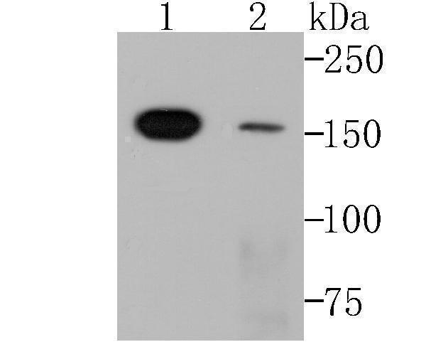
Facts about SWI/SNF complex subunit SMARCC1.
Belongs to the neural progenitors-specific chromatin remodeling complex (npBAF complex) and the neuron- specific chromatin remodeling complex (nBAF complex). During neural development a change from a stem/progenitor into a postmitotic chromatin remodeling mechanism occurs as neurons exit the cell cycle and become dedicated to their adult condition.
Human | |
---|---|
Gene Name: | SMARCC1 |
Uniprot: | Q92922 |
Entrez: | 6599 |
Belongs to: |
---|
SMARCC family |
BAF155SWI/SNF-related matrix-associated actin-dependent regulator of chromatinsubfamily C member 1; BRG1-associated factor 155; chromatin remodeling complex BAF155 subunit; CRACC1; mammalian chromatin remodeling complex BRG1-associated factor 155; Rsc8; SRG3; subfamily c, member 1; SWI/SNF complex 155 kDa subunit; SWI/SNF complex subunit SMARCC1; SWI/SNF related, matrix associated, actin dependent regulator of chromatin; SWI3
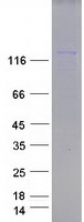
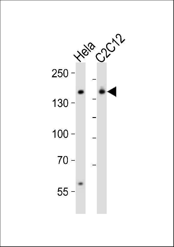
Mass (kDA):
122.867 kDA
Human | |
---|---|
Location: | 3p21.31 |
Sequence: | 3; NC_000003.12 (47585269..47781893, complement) |
Expressed in brain, heart, muscle, placenta, lung, liver, muscle, kidney and pancreas.
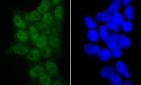
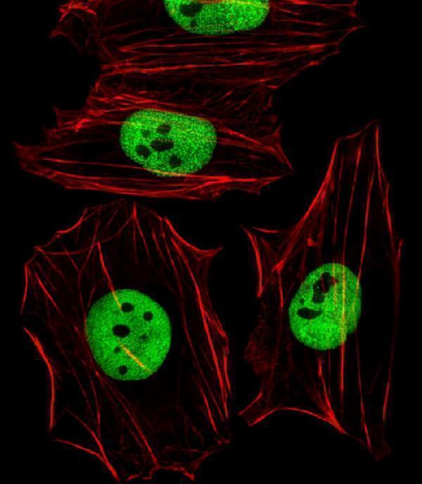
Nucleus. Cytoplasm.
Here are the Benefits of Using the SMARCC1 Marker in Western blots and ELISA kits. The SMARCC1 marker has many uses, including immunohistochemistry. We will discuss the various applications of ELISA kits and Western blots for SOX4, SMARCA4, TGFBR2, and SOX4.
Recent research revealed that the SMARCC1 indicator, which is a member the intranuclear SWI/SNF compound, could be used as a prognostic tool for PC progression. Tissue microarrays allowed for the immunostaining of prostate cancer cell lines for the SMARCC1 gene. A total of 327 PC specimens were analysed, while 30 benign prostate tissue samples were used as controls. The results showed that SMARCC1 staining in both PC and benign prostatic tissue specimens was minimal.
The SMARCC1 marker can also be used for prostate carcinoma diagnosis and follow-up. The time it changes and how quickly, as well as its effectiveness, will determine the effectiveness of this marker. It must be changed within three months of the initial treatment. The degree of change is linked to response. Different levels indicate different levels or response. A 20% decrease in CA19-9 is associated to a better PFS for pancreatic cancer.
Smarcc1 expression is not well-studied in PCa. However it may be of benefit to patients who are at higher risk for developing this disease. Recent studies show that Smarcc1 has a higher rate of survival than other cancers. It is also statistically significant in cancer-specific survival. The SMARCC1 gene is not found in all tumors so it is not a reliable test to diagnose PC.
Two cell lines, HCC1143 & HCC1954, were used to perform Western blots for TGFBR2, SOX4, SMARCA4 and TGFBR2. SMARCA4 was found to be 1.6-fold more abundant at the TGFBR2 enhancer in both cell lines than SOX4 mRNA. The overlapping band data were derived from different experimental settings and were analyzed within the contexts of human breast-cancer.
The method involves a series incubations with various immunochemicals reagents followed by several washes that remove unbound substances. Washing steps are crucial to reduce background noise and increase signal–to-noise. However, excessive washing can result in a high background, while too much washing can decrease the sensitivity and elute antibodies. Different buffers can be used in order to reduce background or increase specificity.
There are two primary methods of detection used for western blots. Although they are more flexible in documenting methods, enzyme-conjugated antibodies are less sensitive. The simplest method to see the signal development is to use Chromogenic Substrates. However, this method can cause the chromogenic band to fade over time as the blot dries. Permanent replicas can be made by scanning or photocopying chromogenic western-blot results.
The basic WB method requires the diluting of both primary and secondary antibodies. This is a common and reliable technique, and almost all commercial antibodies have been validated using it. This method also minimizes the risk of unwanted background interference by blocking some proteins. A good blocking buffer can help enhance the signal-to-noise ratio, and increase sensitivity of the assay.
Although WB results can be very reliable, they are not always accurate. Sometimes the immunoreactivity for a particular protein may not be correct. It is important to understand the protein's mass and the relationship between antibody binding and the blotting intensity. These are three fundamental aspects of the WB. A high-quality antibody may be highly specific and have minimal cross-reactivity. For successful results, it is important to have a low level of denaturation.
SOX4 not only influences PI3K/Akt signaling but also TGFBR2 expression. Both SOX4 mRNA levels and protein levels were decreased by siSOX4 treatment. However, V5 protein expression did not change. The results show that SOX4 plays an important role in regulating PI3K/Akt signals in TNBC. To find out more about these proteins you can order western blots of SOX4, SMARCA4, or TGFBR2 to aid your research.
Another effective way to determine the concentration of protein in a cell is to use different enzymes. This technique utilizes a different enzyme with a distinct wavelength, and allows the detection of more than one protein simultaneously. Chemifluorescence meanwhile uses an enzyme for the creation of an active fluorophor. Gold conjugation is another method. This produces a dark brown stain.
There are many types ELISA Kits. They can be direct, sandwich, or competitive. Direct ELISA uses one antibody per assay step, while sandwich ELISAs use 2 antibodies. The primary antibody binds to the antigen and provides a signal, whereas indirect ELISA uses a pre-coated antibody. The detection step is where the sandwich ELISA differs from direct. Sandwich ELISAs utilize two antibodies to detect the target protein in a sample, while direct ELISAs use one antibody.
ELISA is a common method of testing for enzymes. The most commonly used enzymes are horseradish peroxidase (alkaline phosphatase), nitrophenol, and horseradish phosphatase. Other enzymes such as b-galactosidase, acetylcholinesterase, and catalase have been used as labeling agents in ELISAs. The assay's sensitivity and the instrumentation that detects the signal will affect the choice of substrate.
The SMARCC1 mark is often used for the detection of reproductivehormones in the contexts of species conservation efforts. These levels can be measured using an ELISA kit. This is especially useful when assessing the levels of reproductive hormonal in a population. ELISA can also monitor the levels of these hormones for conservation purposes. Students can make an ELISA kit in a laboratory setting using a paper model containing reproductive hormones.
ELISA commercial kits come in 96 and 384-well plate formats. However, it is possible to adapt easily to larger sample size. They are easy to use and take very little time. They can detect a variety samples. These ELISA kits can detect a variety of samples because they use enzyme and substrate-based reaction.
SMARCC1 is an human gene that has an estimated amino acid length 1105 and a mass 122.9 kDa. It is located within the cytoplasm and in the nuclear subcellular region and is thought be involved in chromatin reconstruction. Its amino acid sequences match those surrounding Gly975 in human SMARCC1/BAF155.
ELISA is a highly sensitive and specific method for the detection of a specific protein in a complex mixture. Engvall-Perlmann developed it in 1971. This allows the ELISA assay to be fast, easy, and reproducible. It's also extremely reproducible and accurate, so the results will be reliable and consistent.
Sandwich ELISA is one the most popular types of ELISA. Sandwich ELISA uses two antibodies to detect the target protein. The capture antibody is coated on the microplate, while the detection antibody binds to an additional epitope on the target protein. The target antigen reacts then with the paired antibody, generating signal proportional in magnitude to the amount present in the sample.
PMID: 8804307 by Wang W., et al. Diversity and specialization of mammalian SWI/SNF complexes.
PMID: 9744861 by Sif S., et al. Mitotic inactivation of a human SWI/SNF chromatin remodeling complex.