This website uses cookies to ensure you get the best experience on our website.


- Table of Contents
Plan chromogenic SMOX IHC in paraffin sections using the catalog antibody (datasheet A05519-2). Assess cytoplasmic and nuclear staining, with Leydig cells as a high-staining reference (HPA tissue IHC).

Expected localisation, validated protocols, controls and antibodies — the at-a-glance facts below, then the full design guide.
| Expected localisation | Cytoplasmic and nuclear staining in most tissues (HPA tissue IHC) | |
| Staining pattern | Cytoplasmic and nuclear staining; Leydig cells high (HPA tissue IHC) | |
| Antigen retrieval | EDTA pH 8.0 HIER, heat-mediated (datasheet A05519-2) | |
| Positive control | Testis+4 more · see all | |
| Negative control | None in HPA (detected in all 45 tissues); use no-primary + isotype controls |
| Fixation | Matched tissue-IHC evidence does not establish the fixation claim. Validate the specimen-specific method before use. (selected-SKU IHC image A05519-2) | |
| Caveat | Staining and RNA show medium consistency (HPA tissue IHC) | |
| Regulation | Stimulus-specific regulation unreported (UniProt) | |
| Isoform / epitope | Six isoforms; epitope coverage across isoforms is unknown (UniProt) |
The catalog antibody uses EDTA pH 8.0 heat retrieval (datasheet A05519-2). The published IHC protocols below describe paraffin tissues, mouse oral mucosa, and lung cancer sections (PMC4345146; PMC9414028; PMC8339854).
| Sample | Paraffin-embedded human diffuse large B cell lymphoma tissue; fixative not specified (datasheet A05519-2) |
| Fixation | Image fixative and duration unreported (datasheet A05519-2); verify before use. |
| Sectioning | 4–5 µm sections on charged slides (standard) |
| Deparaffinisation | Xylene, graded ethanol series to water (standard) |
| Antigen retrieval | Heat retrieval: EDTA pH 8.0 (datasheet A05519-2); 20 min, 95–100 °C (standard) |
| Peroxidase block | 3% H2O2, 10 min, room temperature (standard) |
| Blocking | 10% goat serum (datasheet A05519-2) |
| Primary antibody | Rabbit anti-SMOX, 2-5 μg/ml (datasheet A05519-2) |
| Primary incubation | Overnight at 4 °C (datasheet A05519-2) |
| Detection | HRP-conjugated secondary, DAB chromogen (datasheet A05519-2) |
| Counterstain | Hematoxylin, blue, dehydrate and mount (standard) |
| Expected result | SMOX-positive staining in leydig cells of testis (HPA tissue IHC: High). HPA tissue profile: Cytoplasmic and nuclear expression in most tissues. No signal in the no-primary control. |
SMOX is reported in the cytoplasm and nucleus (UniProt Q9NWM0; HPA tissue IHC). In paraffin sections, expect staining across many tissues, with particularly strong staining in testicular Leydig cells and medium staining in several listed cell populations (HPA tissue IHC). HPA rates its tissue IHC pattern Approved, with medium consistency between staining and RNA data (HPA tissue IHC). SMOX has no transmembrane segment (UniProt Q9NWM0 topology).
| Leydig cells show strong nuclear and/or cytoplasmic staining, while nearby structures remain distinguishable. | This is a useful positive reference: HPA reports High staining in Leydig cells and a cytoplasmic and nuclear pattern across most tissues (HPA tissue IHC). Assess the expected cells and compartments together; strong color alone does not establish specificity (standard IHC practice). |
| Signal is confined to a sharp cell-surface rim or extracellular material, with no nuclear or cytoplasmic staining. | That distribution does not match the reported tissue pattern (HPA tissue IHC) or the absence of a transmembrane segment (UniProt Q9NWM0 topology). Review morphology and controls before interpreting it as SMOX; HPA ICC-IF also reports a nuclear-membrane location, which should not be confused with a cell-surface rim (HPA subcellular ICC-IF). |
| An unexpected cell population stains more strongly than the documented positive cells. | Compare against the cell-specific references: Leydig cells are High; adipocytes, selected glandular and respiratory epithelial cells, glia, and cerebellar granular-layer cells are Medium (HPA tissue IHC). Unexpected staining warrants a cross-reactivity check, but broad distribution alone is insufficient evidence of artefact because SMOX is widely expressed (UniProt Q9NWM0). |
| Color spreads across cells and tissue spaces, obscuring cell borders and nuclei. | Treat this as background until controls show otherwise (standard IHC practice). HPA describes cellular cytoplasmic and nuclear expression, not an uninterpretable tissue-wide haze (HPA tissue IHC). Compare a no-primary control and inspect whether the chromogen is restricted to identifiable cells (standard IHC practice). |
| The known-positive Leydig cells show no convincing signal. | HPA reports High staining in these cells, making an entirely blank result a reason to review assay performance before scoring other tissues negative (HPA tissue IHC). Check the positive control, antibody application, retrieval, and detection steps as general IHC troubleshooting; the supplied sources do not establish an SMOX-specific retrieval or fixation requirement. |
| Tissue and cell reference | Use the named cell population when judging intensity: Leydig cells are High; adipocytes, adrenal, appendix and breast glandular cells, bronchial respiratory epithelial cells, caudate glia, and cerebellar granular-layer cells are Medium (HPA tissue IHC). Cervical glandular, smooth-muscle, and bone-marrow hematopoietic cells are listed Low, not absent (HPA tissue IHC). |
| Compartment | Tissue IHC reports cytoplasmic and nuclear expression in most tissues (HPA tissue IHC), consistent with the UniProt cytoplasm and nucleus annotation (UniProt Q9NWM0). A nucleus-only or cytoplasm-only view of an individual cell should be assessed against tissue morphology and controls before being called incorrect; the sources do not prescribe a fixed nuclear-to-cytoplasmic ratio. |
| Antibody evidence | HPA047117 is Approved for IHC, and HPA describes medium consistency between antibody staining and RNA expression (HPA antibodies; HPA tissue IHC). This supports using the published pattern as a reference, while leaving room to investigate discordant staining; the supplied IHC record does not claim the Enhanced validation category (HPA antibodies). |
| Isoforms and protein features | UniProt lists 6 SMOX isoforms, a single chain spanning residues 1–555, no signal peptide or propeptide, no annotated glycosylation sites, and no transmembrane segment (UniProt Q9NWM0). The payload does not identify the antibody epitope, so these features cannot establish which isoforms it detects or predict an SMOX-specific retrieval response. |
| Does ICC-IF define the paraffin IHC pattern? | HPA ICC-IF places SMOX mainly in the nucleoplasm, with additional nuclear-membrane, vesicle, and cytosol locations; images are listed for A-549, SiHa, and U2OS (HPA subcellular ICC-IF). This is useful compartment context, but the ICC-Supported antibody HPA060198 differs from the IHC-Approved antibody HPA047117 (HPA antibodies). Interpret paraffin staining against the tissue IHC record. |
| Situation | Likely cause | Next action |
|---|---|---|
| Positive-control Leydig cells are blank or barely visible. | The run may have failed, or the chosen conditions may provide inadequate detection; High Leydig-cell staining is the HPA reference (HPA tissue IHC). | First verify the positive-control section, antibody and detection reagents, and that the retrieval and staining steps ran as intended (standard IHC practice). Do not infer an SMOX-specific retrieval or fixation sensitivity from this result. |
| All tissue structures, including spaces without identifiable cells, develop color. | Nonspecific background or endogenous detection activity can obscure cellular staining (standard IHC practice). | Review the no-primary control, blocking, washing, and chromogen development (standard IHC practice). If using an HRP detection system, check the endogenous-peroxidase control (standard IHC practice); retain only interpretable nuclear or cytoplasmic cellular signal (HPA tissue IHC). |
| Only a sharp cell-surface outline stains. | A surface-only pattern conflicts with the reported cytoplasmic and nuclear tissue staining (HPA tissue IHC) and SMOX lacking a transmembrane segment (UniProt Q9NWM0 topology). | Check morphology and no-primary staining, then compare with a documented positive section (standard IHC practice; HPA tissue IHC). Keep the ICC-IF nuclear-membrane observation distinct from a plasma-membrane outline (HPA subcellular ICC-IF). |
| Unexpected cells stain while the documented positive population also stains. | SMOX is widely expressed (UniProt Q9NWM0), so location alone cannot classify the extra signal; cross-reactivity remains possible (standard IHC practice). | Compare cell identity, compartment, and intensity with HPA's named tissue observations, then inspect controls (HPA tissue IHC; standard IHC practice). Report unexpected staining as uncertain if those checks cannot resolve it. |
| Low-staining tissue is scored as definitively SMOX-negative. | HPA lists cervical glandular cells, smooth-muscle cells, and bone-marrow hematopoietic cells as Low, while listing no negative tissue examples in this payload (HPA tissue IHC). | Record weak versus absent signal explicitly and judge the section alongside a positive control (standard IHC practice; HPA tissue IHC). Avoid designating an HPA Low population as a validated negative control. |
| An ICC-IF localization image appears inconsistent with the paraffin section. | The records use different modalities and antibodies: HPA060198 is ICC Supported, whereas HPA047117 is IHC Approved (HPA antibodies). | Use the tissue IHC pattern for the paraffin-section decision and the ICC-IF record as compartment context (HPA tissue IHC; HPA subcellular ICC-IF). Review the paraffin controls before calling a discrepancy biological (standard IHC practice). |
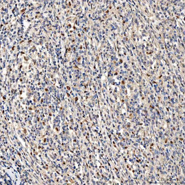
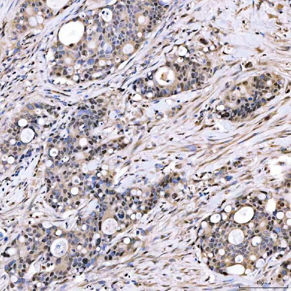
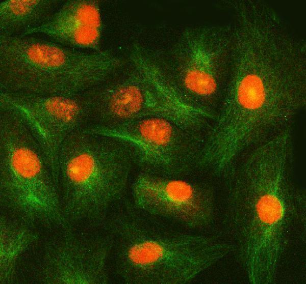


Comprehensive Human Protein Atlas IHC scoring per tissue (reliability: Approved — Medium consistency between antibody staining and RNA expression data.). Rows are taken directly from the HPA tissue chart — click any row's HPA link to view the source.
| Tissue | Cell type | Level | Evidence | Source |
|---|---|---|---|---|
| Testis | Leydig cells | High | Protein (IHC) | HPA → |
| Adipose tissue | Adipocytes | Medium | Protein (IHC) | HPA → |
| Adrenal gland | Glandular cells | Medium | Protein (IHC) | HPA → |
| Appendix | Glandular cells | Medium | Protein (IHC) | HPA → |
| Breast | Glandular cells | Medium | Protein (IHC) | HPA → |
| Tissue | Cell type | Level | Evidence | Source |
|---|---|---|---|---|
| None in HPA: SMOX is detected in all 45 scored tissues. Use a no-primary (secondary-only) and an isotype control instead. | ||||
Troubleshoot SMOX staining in paraffin sections by checking retrieval, cell compartment, controls, and scoring against the reported IHC pattern.
The IHC-validated anti-SMOX antibody has paraffin-section images from human tumors and an IF/ICC image from A549 cells; listed reactivity includes Human, Mouse, and Rat (catalog IHC/IF captions; catalog reactivity).
A05519-2 has IHC images from human paraffin sections of diffuse large B cell lymphoma, duodenal papilla adenocarcinoma, endometrioid adenocarcinoma, and glioblastoma (catalog IHC captions). The same SKU has an IF/ICC image in A549 cells and lists Human, Mouse, and Rat reactivity (catalog IF caption; catalog reactivity).
Which to pick: For tissue IHC, choose A05519-2: its own caption shows a human lymphoma paraffin section with EDTA retrieval at pH 8.0; the fixative is unreported (A05519-2 IHC caption). For IF/ICC, the same SKU has an A549-cell IF image and lists both applications (A05519-2 IF caption; catalog applications). For cross-species work, A05519-2 lists Human, Mouse, and Rat reactivity, while the supplied IHC images are human and the IF image is A549; clonality is unreported (catalog reactivity; A05519-2 IHC/IF captions; catalog clone field).