This website uses cookies to ensure you get the best experience on our website.


- Table of Contents
Plan chromogenic SMURF2 IHC-P with catalog antibody A02585 at 2 μg/mL (datasheet). Use testicular spermatogonia as a high-staining reference and assess cytoplasmic staining across tissues (HPA tissue IHC).

Expected localisation, validated protocols, controls and antibodies — the at-a-glance facts below, then the full design guide.
| Expected localisation | Cytoplasmic staining in most tissues (HPA tissue IHC) | |
| Staining pattern | Cytoplasmic in most tissues; high in spermatogonia (HPA tissue IHC) | |
| Antigen retrieval | Tris-EDTA pH 9.0 HIER, 95–98 °C, 20 min (rule: nuclear antigen) | |
| Positive control | Testis+4 more · see all | |
| Negative control | Adipose tissue+4 more · see all |
| Fixation | Keep fixation conditions consistent across sections. (standard IHC practice; not target-specific) | |
| Caveat | Some cell types, including adipocytes, lack detectable staining (HPA tissue IHC) | |
| Regulation | SMAD7 promotes SMURF2 autodegradation (UniProt) | |
| Isoform / epitope | No isoforms reported; one 1–748 chain (UniProt) |
The catalog antibody protocol is paired with published SMURF2 IHC methods for breast cancer tissues (PMC3918234), human osteoarthritic cartilage (PMC2636703), and pancreatic cancer tissue microarrays (PMC12133423).
| Sample | Tissue sections; selected-image fixative not specified (standard IHC workflow) |
| Fixation | Image fixative and duration unreported (datasheet A02585); verify before use. |
| Sectioning | 4–5 µm sections on charged slides (standard) |
| Deparaffinisation | Xylene, graded ethanol series to water (standard) |
| Antigen retrieval | Heat-induced epitope retrieval in Tris-EDTA buffer, pH 9.0, 20 min at 95–98 °C (standard rule: nuclear antigen) |
| Peroxidase block | 3% H2O2, 10 min, room temperature (standard) |
| Blocking | 10% normal serum of the secondary host, 30 min, room temperature (standard) |
| Primary antibody | Rabbit anti-SMURF2, 2 μg/mL (datasheet A02585) |
| Primary incubation | Overnight at 4 °C (standard) |
| Detection | HRP-polymer secondary, DAB chromogen 5–10 min (standard) |
| Counterstain | Hematoxylin, blue, dehydrate and mount (standard) |
| Expected result | SMURF2-positive staining in spermatogonia cells of testis (HPA tissue IHC: High). HPA tissue profile: Cytoplasmic expression in most tissues. No signal in the no-primary control. |
In paraffin-section IHC, expect predominantly cytoplasmic SMURF2 staining across many tissues, with high staining in testicular spermatogonia and medium staining in several other listed cell populations (HPA: tissue IHC). UniProt also places SMURF2 in the nucleus and at the cell membrane or membrane rafts, despite reporting no transmembrane segment (UniProt Q9HAU4: subcellular location and topology). The HPA tissue-IHC assessment is Enhanced, with medium consistency between antibody staining and RNA expression (HPA: tissue IHC).
| Cytoplasmic staining is strongest in spermatogonia; adrenal glandular cells, Purkinje cells, and kidney tubular cells show more moderate staining. | This fits the reported high spermatogonial and medium staining in those other cell populations (HPA: tissue IHC). Compare compartments and cell identities within each section; the listed levels describe observations, not a required intensity ratio between separately processed slides (HPA: tissue IHC; general IHC practice). |
| Staining is exclusively nuclear, or a sharp membrane rim dominates nearly every cell, with little of the expected cytoplasmic pattern. | Check whether the pattern is reproducible and antibody dependent before calling it SMURF2: HPA describes predominantly cytoplasmic tissue staining, while UniProt also lists nuclear and membrane locations (HPA: tissue IHC; UniProt Q9HAU4: subcellular location). Nuclear signal alone is therefore not proof of an artefact; a widespread membrane rim should not be treated as evidence of a transmembrane protein (UniProt Q9HAU4: topology). |
| Strong staining appears in adipocytes, cervical squamous epithelium, or lymph-node germinal-center cells. | These specific cell populations were reported as not detected, making a strong result discordant with the HPA reference pattern (HPA: tissue IHC). Investigate antibody cross-reactivity or endogenous chromogen-generating activity with appropriate controls before assigning the signal to SMURF2 (general IHC practice). |
| Color coats extracellular spaces, section edges, or many unrelated cell populations without a defined intracellular pattern. | Treat this as background until controls show otherwise: nonspecific binding, insufficient washing, and endogenous detection activity can obscure a cellular chromogenic signal (general IHC practice). The reference pattern is cytoplasmic expression in most tissues, not diffuse staining throughout the section (HPA: tissue IHC). |
| Spermatogonia are unstained in a section selected as a positive control. | A negative result conflicts with the reported high spermatogonial staining (HPA: tissue IHC). Check that the expected cells are present, then assess retrieval, antibody and detection performance with controls (general IHC practice). A failed run cannot establish that this specimen lacks SMURF2. |
| Tissue and cell selection | SMURF2 is widely expressed with low RNA tissue specificity, but staining differs by cell population: spermatogonia are high, several listed populations are medium, and selected populations are undetected (UniProt Q9HAU4: tissue specificity; HPA: tissue IHC). Name the cells being scored rather than treating an entire organ as uniformly positive or negative. |
| Compartment and protein topology | HPA reports mostly cytoplasmic tissue staining; UniProt also lists nucleus, cell membrane, and membrane rafts, with cytoplasmic localization in the presence of SMAD7 (HPA: tissue IHC; UniProt Q9HAU4: subcellular location). SMURF2 has no transmembrane segment, so a membrane-associated signal does not imply membrane-spanning topology (UniProt Q9HAU4: topology). |
| Antibody evidence and its limit | HPA lists antibody HPA071508 as Enhanced for IHC, while its tissue summary reports medium agreement between antibody staining and RNA expression (HPA: antibody validation; HPA: tissue IHC). Use that evidence to anchor expected patterns, while checking discordant cell populations with run controls (general IHC practice). |
| IF/ICC Q: What pattern should I expect? | A: HPA reports supported localization to nuclear speckles in ICC-IF images from BJ, U-251MG, and U2OS cells (HPA: subcellular ICC-IF). This observation comes from cultured-cell fluorescence imaging; interpret paraffin-section chromogenic staining against the tissue-IHC pattern instead (HPA: subcellular ICC-IF; HPA: tissue IHC). |
| Epitope and retrieval evidence | UniProt lists an intact 748-aa chain, no signal peptide or propeptide, and no listed glycosylation sites; the supplied evidence does not map the IHC antibody epitope or establish SMURF2-specific retrieval or fixation sensitivity (UniProt Q9HAU4: processing and glycosylation; HPA: antibody validation). Treat retrieval optimization as a general IHC workflow decision, not a predicted SMURF2 effect (general IHC practice). |
| Situation | Likely cause | Next action |
|---|---|---|
| No staining in the expected positive cells | The run may have inadequate antigen exposure, inactive detection reagents, or missing target cells; these are general IHC possibilities, not established SMURF2-specific fixation effects (general IHC practice). | Confirm spermatogonia are present in the control section and assess the antibody, retrieval, and detection steps with run controls (HPA: High in spermatogonia; general IHC practice). |
| Strong staining in a reference undetected population | A strong signal in adipocytes or cervical squamous cells conflicts with the HPA observations; antibody cross-reactivity or endogenous detection activity are possibilities (HPA: Not detected in those cells; general IHC practice). | Compare a no-primary control and a known-positive section, then check endogenous enzyme blocking for chromogenic detection before scoring the signal as SMURF2 (general IHC practice). |
| Diffuse color obscures cell boundaries | Nonspecific reagent binding, incomplete washing, or detection background can reduce contrast (general IHC practice). | Check the no-primary control; review blocking, washes, antibody working concentration, and chromogen development while retaining a positive control (general IHC practice). |
| A nuclear pattern conflicts with the expected tissue-IHC image | Nuclear localization is biologically plausible, but HPA's paraffin-tissue summary is predominantly cytoplasmic; a nuclear-only result needs verification (UniProt Q9HAU4: subcellular location; HPA: tissue IHC). | Compare matched positive and no-primary controls, confirm cell identity, and evaluate reproducibility before interpreting nuclear signal as SMURF2 (general IHC practice). |
| Membrane-edge color dominates the section | UniProt permits membrane-raft association, but SMURF2 has no transmembrane segment; uniform edge staining may also reflect technical background (UniProt Q9HAU4: subcellular location and topology; general IHC practice). | Check whether staining follows specific cells rather than section edges; compare controls and the expected cytoplasmic tissue pattern (HPA: tissue IHC; general IHC practice). |
| IF/ICC nuclear speckles and tissue-IHC cytoplasmic staining seem inconsistent | The observations come from different assay contexts: HPA supports nuclear speckles in cultured-cell ICC-IF and describes cytoplasmic expression in most tissue-IHC samples (HPA: subcellular ICC-IF; HPA: tissue IHC). | Record assay, specimen, and compartment separately; score the paraffin section against its tissue-IHC reference and assess the IF/ICC image against its own subcellular reference (HPA: tissue IHC; HPA: subcellular ICC-IF). |


![HPA ICC-IF BJ [Human fibroblast] — SMURF2 (HPA071508)](https://images.proteinatlas.org/71508/1533_A8_1_blue_red_green.jpg)
![HPA ICC-IF BJ [Human fibroblast] — SMURF2 (HPA071508)](https://images.proteinatlas.org/71508/1533_A8_2_blue_red_green.jpg)
Comprehensive Human Protein Atlas IHC scoring per tissue (reliability: Enhanced — Medium consistency between antibody staining and RNA expression data.). Rows are taken directly from the HPA tissue chart — click any row's HPA link to view the source.
| Tissue | Cell type | Level | Evidence | Source |
|---|---|---|---|---|
| Testis | Spermatogonia cells | High | Protein (IHC) | HPA → |
| Adrenal gland | Glandular cells | Medium | Protein (IHC) | HPA → |
| Caudate | Neuronal cells | Medium | Protein (IHC) | HPA → |
| Cerebellum | Purkinje cells | Medium | Protein (IHC) | HPA → |
| Duodenum | Glandular cells | Medium | Protein (IHC) | HPA → |
| Tissue | Cell type | Level | Evidence | Source |
|---|---|---|---|---|
| Adipose tissue | Adipocytes | Not detected | Protein (IHC) | HPA → |
| Cervix | Squamous epithelial cells | Not detected | Protein (IHC) | HPA → |
| Epididymis | Glandular cells | Not detected | Protein (IHC) | HPA → |
| Esophagus | Squamous epithelial cells | Not detected | Protein (IHC) | HPA → |
| Liver | Cholangiocytes | Not detected | Protein (IHC) | HPA → |
Troubleshoot SMURF2 staining in paraffin sections by checking retrieval, cellular compartment, controls and scoring before interpreting chromogenic signal.
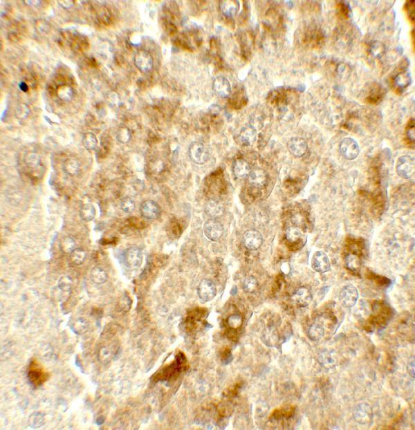
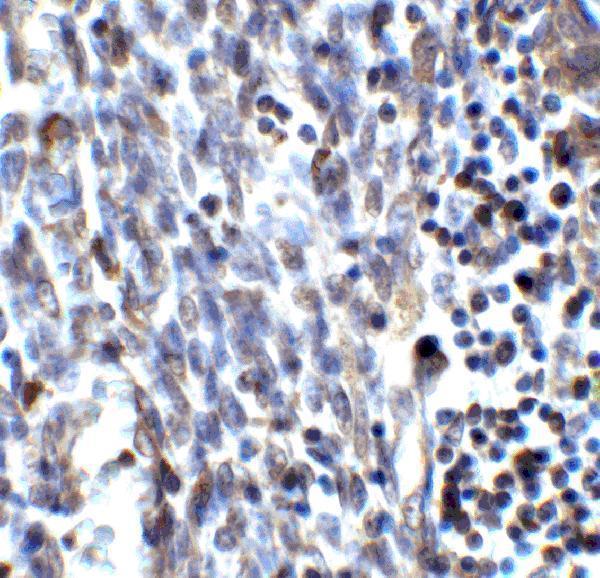
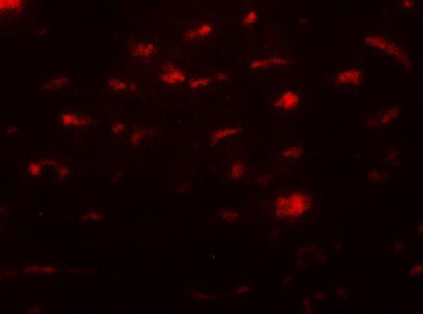
A02585 has IHC and IF images from mouse liver and human uterus (catalog: image alts); listed reactivity includes human, mouse and rat (catalog: reactivity).
A02585 will render with its mouse liver IHC figure at 5 μg/mL (card: image alt). Additional IHC and IF images show mouse liver and human uterus (catalog: IHC and IF image alts).
Which to pick: For paraffin tissue IHC, choose A02585 because IHC-P is listed (catalog: applications); its own IHC captions show mouse liver and human uterus, but do not report the fixative (catalog: IHC image alts). For IF, A02585 has tissue images at 20 μg/mL (catalog: IF image alts); ICC validation is unreported (catalog: applications and image alts). For cross-species work, A02585 lists human, mouse and rat reactivity (catalog: reactivity), though its IHC images cover human and mouse only (catalog: IHC image alts); it is rabbit hosted, with clonality unreported (catalog: host and clone).