This website uses cookies to ensure you get the best experience on our website.


- Table of Contents
Plan paraffin section IHC for SNAPIN using its reported cytoplasmic tissue pattern and high staining in lung macrophages (HPA tissue IHC). This guide covers fixation consistency, antibody dilution, chromogenic detection, and interpretation, with medium IHC–RNA consistency in mind (HPA tissue IHC).

Expected localisation, validated protocols, controls and antibodies — the at-a-glance facts below, then the full design guide.
| Expected localisation | General cytoplasmic tissue staining (HPA tissue IHC) | |
| Staining pattern | Cytoplasmic staining, high in lung macrophages (HPA tissue IHC) | |
| Antigen retrieval | Citrate pH 6.0 HIER, heat-mediated (datasheet A03491) | |
| Positive control | Adipose tissue+4 more · see all | |
| Negative control | Smooth muscle |
| Fixation | Keep fixation consistent across paraffin sections (standard IHC practice; not target-specific) | |
| Caveat | Medium IHC–RNA consistency; verify specificity (HPA tissue IHC) | |
| Regulation | No expression regulator established (UniProt) | |
| Isoform / epitope | 0 annotated isoforms; epitope dependence unknown (UniProt) |
The catalog antibody’s IHC-P protocol is accompanied by 3 published SNAPIN IHC protocols covering mouse pancreas, human liver cancer specimens, and rat brain (PMC8237857; PMC12775592; PMC12338511).
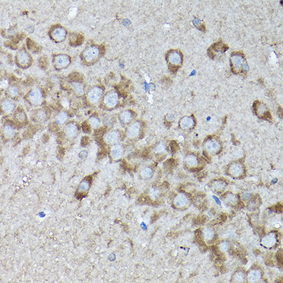
| Sample | Paraffin-embedded rat brain tissue; fixative not specified (datasheet A03491) |
| Fixation | Image fixative and duration unreported (datasheet A03491); verify before use. |
| Sectioning | 4–5 µm sections on charged slides (standard) |
| Deparaffinisation | Xylene, graded ethanol series to water (standard) |
| Antigen retrieval | Heat retrieval: Citrate pH 6.0 (datasheet A03491); 20 min, 95–100 °C (standard) |
| Peroxidase block | 3% H2O2, 10 min, room temperature (standard) |
| Blocking | 10% normal serum of the secondary host, 30 min, room temperature (standard) |
| Primary antibody | Rabbit anti-SNAPIN, 1:50-1:200 (datasheet A03491) |
| Primary incubation | Overnight at 4 °C (standard) |
| Detection | HRP-polymer secondary, DAB chromogen 5–10 min (standard) |
| Counterstain | Hematoxylin, blue, dehydrate and mount (standard) |
| Expected result | SNAPIN-positive staining in adipocytes of adipose tissue (HPA tissue IHC: High). HPA tissue profile: General cytoplasmic expression. No signal in the no-primary control. |
SNAPIN should appear mainly cytoplasmic in tissue IHC, with signal in several cell types, including lung macrophages, cerebellar Purkinje cells, and kidney glomerular cells (HPA: general cytoplasmic expression; High in each listed cell type). Its reported membrane associations include Golgi, lysosomal, and vesicle membranes, despite no transmembrane segment (UniProt O95295: subcellular location; topology). HPA rates the tissue IHC pattern Approved, with medium consistency against RNA and external verification pending (HPA: reliability).
| Cytoplasmic staining in lung macrophages, with a clear cell-associated pattern. | This fits the reported high staining in macrophages and general cytoplasmic IHC profile (HPA: lung macrophages High; tissue IHC profile). Judge the labeled cells and surrounding background separately; a dark field alone does not establish a convincing cellular pattern (general IHC practice). |
| Cytoplasmic signal in Purkinje cells or cells in kidney glomeruli. | Both are reported high-staining populations and can serve as tissue-pattern references (HPA: cerebellum Purkinje cells High; kidney cells in glomeruli High). The reference is supportive, not definitive, because HPA reports only medium consistency with RNA and pending external verification (HPA: reliability description). |
| Predominantly strong nuclear staining without a convincing cytoplasmic component. | Treat this as discordant with the general tissue IHC pattern and review specificity and detection background (HPA: general cytoplasmic expression). Do not automatically reject discrete nucleolar signal in IF/ICC: HPA reports that additional location with uncertain confidence (HPA: subcellular location). |
| Prominent staining of smooth muscle cells while expected cytoplasmic staining is weak. | Smooth muscle cells are reported as not detected, so this distribution raises concern for cross-reactivity or endogenous chromogenic activity (HPA: smooth muscle cells Not detected; general IHC practice). Check morphology and detection controls before calling the smooth muscle signal SNAPIN-specific (general IHC practice). |
| Widespread diffuse color obscures cell boundaries, or no signal appears in a reported high-staining population. | Diffuse color limits localization scoring; absent signal in a reported high-staining population makes the run hard to interpret (general IHC practice; HPA: lung macrophages High). Review controls and the detection workflow before treating either appearance as a true expression result (general IHC practice). |
| Compartment and topology | SNAPIN is annotated in cytosol, the perinuclear region, and on Golgi, lysosomal, and vesicle membranes; it has no transmembrane segment (UniProt O95295: subcellular location; topology). Interpret tissue IHC primarily against HPA's general cytoplasmic pattern, without requiring one sharply defined organelle pattern in chromogenic sections (HPA: tissue IHC profile; general IHC practice). |
| Choice of tissue reference | Adipocytes, adrenal glandular cells, bone marrow hematopoietic cells, ovary follicle cells, lung macrophages, Purkinje cells, and kidney glomerular cells are reported High (HPA: tissue IHC). Smooth muscle cells are reported Not detected (HPA: tissue IHC). These are observed patterns, not guarantees for every specimen or run (HPA: Approved; medium consistency; external verification pending). |
| Antibody evidence | The listed antibody HPA046974 is Approved for IHC; HPA's tissue IHC reliability description says external verification is pending (HPA: antibody validation; tissue IHC reliability). Compare compartment and cell distribution with the HPA observations, and keep uncertain cases provisional rather than treating an Approved label as independent proof of specificity (general IHC practice). |
| Processing and modified residues | UniProt lists no signal peptide, propeptide, annotated isoforms, or glycosylation sites, and lists several modified residues (UniProt O95295: processing; isoforms; glycosylation; modified residues). Those annotations do not identify the antibody epitope or predict an antigen-retrieval condition or fixation sensitivity; no target-specific effect is established by the supplied sources. |
| IF/ICC Q: What localization is reported? | A: Golgi apparatus is the approved main location in HPA ICC-IF; nucleoli are an additional uncertain location (HPA: subcellular). That IF/ICC observation can inform compartment review, while tissue IHC interpretation follows HPA's general cytoplasmic profile (HPA: tissue IHC profile). |
| Situation | Likely cause | Next action |
|---|---|---|
| No stain in lung macrophages or another reported High population. | The run may have insufficient detectable signal, or the chosen cells may be absent from the examined area (general IHC practice; HPA: lung macrophages High). | Confirm the expected cells on the counterstained section, then review the run's positive control, antigen-retrieval and detection records (general IHC practice). Do not infer that SNAPIN is absent from this single negative slide. |
| Smooth muscle cells stain prominently. | This conflicts with HPA's Not detected observation and may reflect cross-reactivity or endogenous detection activity (HPA: smooth muscle cells Not detected; general IHC practice). | Check a no-primary control and the chromogenic detection controls; compare the same section's expected cytoplasmic populations before assigning the smooth muscle color to SNAPIN (general IHC practice; HPA: tissue IHC profile). |
| Color is diffuse across tissue or obscures cell borders. | Background from blocking, washing, or detection can prevent compartment assignment (general IHC practice). | Review no-primary control, blocking, washes, and detection exposure; score SNAPIN only where cell-associated cytoplasmic signal remains distinguishable (general IHC practice; HPA: general cytoplasmic expression). |
| Signal is mainly nuclear in the chromogenic section. | The distribution is discordant with HPA's general cytoplasmic tissue IHC profile; IF/ICC nucleolar localization is reported with uncertain confidence (HPA: tissue IHC profile; subcellular location). | Review morphology, counterstain, and no-primary control before interpreting the nuclear color; seek a convincing cytoplasmic component in a reported positive population (general IHC practice; HPA: tissue IHC). |
| Reported High and Not detected populations look equally stained. | Poor contrast between the populations weakens the tissue-pattern evidence and may indicate background or a specificity problem (HPA: High populations; smooth muscle cells Not detected; general IHC practice). | Score cell types separately, inspect no-primary control, and compare intensity with HPA's population-specific observations; flag an unresolved distribution as inconclusive (general IHC practice; HPA: tissue IHC). |
| Punctate or perinuclear cytoplasmic staining is hard to classify. | UniProt reports perinuclear, Golgi, lysosomal, and vesicle-associated locations, while HPA summarizes tissue IHC as generally cytoplasmic (UniProt O95295: subcellular location; HPA: tissue IHC profile). | Record the observed compartment and cell type without assigning a specific organelle from chromogenic morphology alone; compare with HPA's reported positive populations (general IHC practice; HPA: tissue IHC). |


Comprehensive Human Protein Atlas IHC scoring per tissue (reliability: Approved — Medium consistency between antibody staining and RNA expression data. Pending external verification.). Rows are taken directly from the HPA tissue chart — click any row's HPA link to view the source.
| Tissue | Cell type | Level | Evidence | Source |
|---|---|---|---|---|
| Adipose tissue | Adipocytes | High | Protein (IHC) | HPA → |
| Adrenal gland | Glandular cells | High | Protein (IHC) | HPA → |
| Bone marrow | Hematopoietic cells | High | Protein (IHC) | HPA → |
| Cerebellum | Purkinje cells | High | Protein (IHC) | HPA → |
| Endometrium | Glandular cells | Medium | Protein (IHC) | HPA → |
| Tissue | Cell type | Level | Evidence | Source |
|---|---|---|---|---|
| Smooth muscle | Smooth muscle cells | Not detected | Protein (IHC) | HPA → |
Troubleshoot SNAPIN staining in paraffin sections using the catalog antibody’s retrieval condition, known subcellular locations, and tissue expression evidence (datasheet A03491; UniProt O95295; HPA tissue IHC).
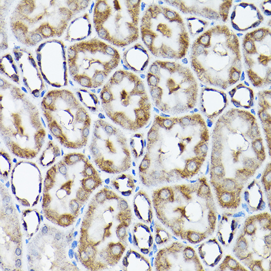
A03491 has real IHC images from paraffin-embedded rat brain and kidney (catalog IHC image captions). Its listed reactivity is Human, Mouse, and Rat (catalog: reactivity); no IF images are provided (catalog: IF image alts).
A03491 will render with its rat brain IHC figure (catalog: IHC image caption). A second caption documents paraffin-embedded rat kidney staining; the listed applications are IHC and WB (catalog: IHC image captions; applications).
Which to pick: Choose A03491 for paraffin-section tissue IHC: it is a rabbit polyclonal antibody with rat brain and kidney staining shown at 1:200 after citrate pH 6.0 retrieval (catalog: host; IHC image captions). Its listed Human, Mouse, and Rat reactivity supports cross-species selection, although the supplied IHC images show rat tissue only (catalog: reactivity; IHC image captions). No IF/ICC-validated SKU is supplied here (catalog: applications; IF image alts); the IHC captions report paraffin embedding but do not report the fixative (catalog: IHC image captions).