This website uses cookies to ensure you get the best experience on our website.


- Table of Contents
Plan chromogenic SNCA IHC in paraffin sections using CNS neuropil and marrow cell staining as reference patterns (HPA tissue IHC). This guide covers controls, localisation and a 1:50 starting dilution for the IHC-validated antibody (datasheet M00215-1).

Expected localisation, validated protocols, controls and antibodies — the at-a-glance facts below, then the full design guide.
| Expected localisation | Presynaptic terminals; cytoplasm, membrane and nucleus (UniProt) | |
| Staining pattern | High in CNS neuropil, peripheral nerves and marrow cell subsets (HPA tissue IHC) | |
| Antigen retrieval | EDTA pH 8.0 HIER, heat-mediated (datasheet M00215-1) | |
| Positive control | Bone marrow+3 more · see all | |
| Negative control | Adipose tissue+4 more · see all |
| Fixation | Keep fixation consistent across sections (standard IHC practice; not target-specific) | |
| Caveat | Glia may be negative despite strong CNS neuropil staining (HPA tissue IHC) | |
| Regulation | Enriched in brain and presynaptic terminals (UniProt) | |
| Isoform / epitope | Three isoforms; epitope coverage depends on antibody specificity (UniProt) |
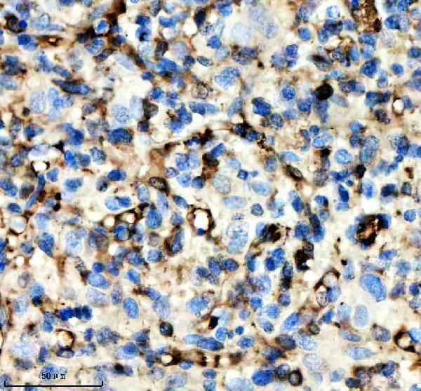
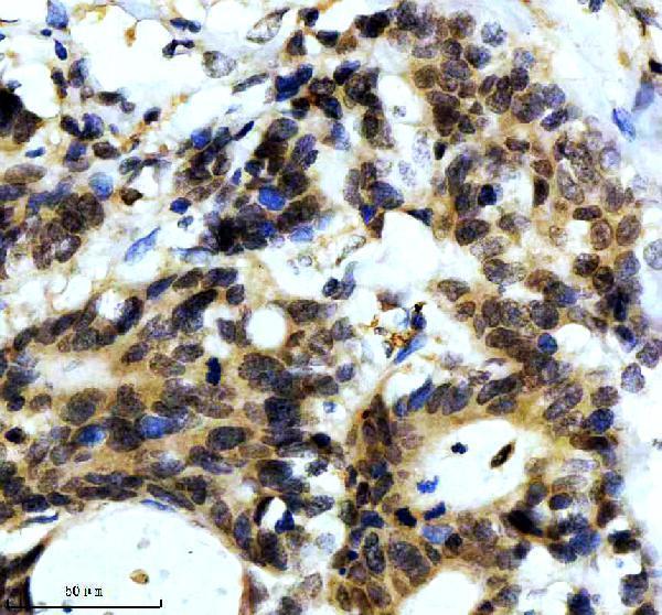
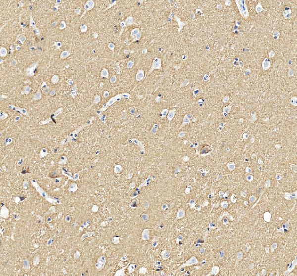
The catalog antibody has a paraffin-section protocol (datasheet: M00215-1). Published IHC examples cover mouse brain and spinal cord (PMC4986551), human brain (PMC4549856), and lung adenocarcinoma slides (PMC9009002).
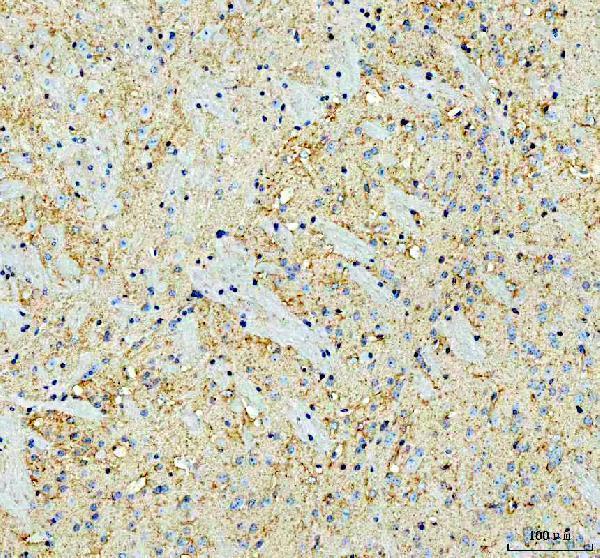
| Sample | Paraffin-embedded human glioma tissue; fixative not specified (datasheet M00215-1) |
| Fixation | Image fixative and duration unreported (datasheet M00215-1); verify before use. |
| Sectioning | 4–5 µm sections on charged slides (standard) |
| Deparaffinisation | Xylene, graded ethanol series to water (standard) |
| Antigen retrieval | Heat retrieval: EDTA pH 8.0 (datasheet M00215-1); 20 min, 95–100 °C (standard) |
| Peroxidase block | 3% H2O2, 10 min, room temperature (standard) |
| Blocking | 10% goat serum (datasheet M00215-1) |
| Primary antibody | Rabbit monoclonal (clone AGA-19) anti-SNCA, 1:50 (datasheet M00215-1) |
| Primary incubation | Overnight at 4 °C (datasheet M00215-1) |
| Detection | HRP-conjugated secondary, DAB chromogen (datasheet M00215-1) |
| Counterstain | Hematoxylin, blue, dehydrate and mount (standard) |
| Expected result | SNCA-positive staining in hematopoietic cells of bone marrow (HPA tissue IHC: High). HPA tissue profile: High expression in CNS and peripheral nerves, as well as subsets of cells in bone marrow. No signal in the no-primary control. |
SNCA is enriched at presynaptic terminals in the central nervous system and is also annotated in cytoplasm, membrane, nucleus, synapses and axons; it has no transmembrane segment (UniProt P37840). In tissue IHC, expect strong neuropil staining in cerebral cortex, staining of cerebellar granular-layer processes, and staining in subsets of bone-marrow hematopoietic cells (HPA: Enhanced tissue reliability; High cortical neuropil and bone-marrow staining; Medium cerebellar process staining).
| Cortical neuropil stains strongly while neuronal processes are more apparent than cell bodies. | This fits the reported High cortical neuropil signal and presynaptic enrichment (HPA: High in cerebral-cortex neuropil; UniProt P37840: presynaptic terminals). Interpret staining by structure as well as intensity; a field of positive processes need not show uniformly positive nuclei or somata. |
| Staining is confined to a compartment inconsistent with the sampled structure, with no expected neuropil or process signal. | Reassess localisation before calling the section positive: cytoplasm, membrane, nucleus, synapse and axon are annotated, so nuclear signal alone is not proof of artefact (UniProt P37840). A compartment-only pattern that misses the expected tissue structures raises concern for an IHC artefact (HPA: cortical neuropil and cerebellar processes). |
| Strong signal appears in cells reported as unstained, such as adipocytes or duodenal glandular cells. | Those specific cell populations are Not detected in HPA tissue IHC (HPA: adipose adipocytes; duodenal glandular cells). Check cross-reactivity and endogenous detection activity as possible explanations (general IHC practice). Do not label an entire tissue negative when its documented negative call applies to a named cell type. |
| A diffuse chromogenic haze covers positive and expected-negative structures alike. | A uniform haze obscures the contrast needed to recognize High cortical neuropil and Not detected adipocytes (HPA: cerebral-cortex neuropil; adipose adipocytes). Treat the pattern as background until the expected anatomical contrast is visible; review blocking, detection background and primary-antibody concentration (general IHC practice). |
| A cerebral-cortex positive control shows no neuropil staining. | That conflicts with the reported High cortical neuropil staining (HPA: cerebral cortex). First check that the expected structure is present on the section, then review the staining run and the catalog antibody's IHC-P directions (general IHC practice). An absent control signal makes negative calls in test sections unreliable. |
| Anatomical compartment | SNCA is enriched in presynaptic terminals and annotated in synapses and axons (UniProt P37840). Score signal in neuropil and processes rather than requiring a uniformly filled neuronal soma (HPA: cortical neuropil; cerebellar granular-layer processes). |
| Tissue and cell-type contrast | HPA reports High signal in cortical neuropil, bone-marrow hematopoietic cells and kidney glomerular cells, Medium signal in cerebellar granular-layer processes, and Not detected signal in listed adipocytes (HPA: tissue IHC). These calls apply to the specified structures or cells. |
| Antibody validation | Two listed antibodies have Enhanced IHC status (HPA: HPA005459 and CAB010877). The tissue profile also has Enhanced reliability, reflecting consistency with RNA data; HPA notes staining in structures outside its annotations, so review the image when a structure is uncertain (HPA: tissue IHC). |
| Protein forms and epitope information | Three isoforms and several modified residues are annotated, including phosphoserine at position 129 (UniProt P37840). The supplied records give no antibody epitope or isoform specificity, so they cannot predict which form an IHC signal represents (UniProt P37840; HPA: antibody summary). |
| Retrieval and fixation | The supplied sources report no target-specific fixation sensitivity or retrieval condition (UniProt P37840; HPA: tissue IHC). Use the catalog antibody's IHC-P instructions for a starting workflow; assess retrieval changes with positive and expected-negative controls (general IHC practice). |
| Situation | Likely cause | Next action |
|---|---|---|
| No signal in cerebral-cortex neuropil | The positive control failed, the expected structure is absent, or a staining step failed (HPA: High cortical neuropil; general IHC practice). | Confirm neuropil is present; check reagent sequence, detection and the catalog antibody's IHC-P instructions before interpreting test sections (general IHC practice). |
| Signal fills the slide without anatomical contrast | Nonspecific binding or detection background can obscure structure-specific staining (general IHC practice). | Review blocking, washes and primary-antibody concentration; compare a control omitting primary antibody with the expected cortical neuropil pattern (general IHC practice; HPA: High cortical neuropil). |
| Adipocytes or duodenal glandular cells stain strongly | These named cells are Not detected in the HPA tissue profile; cross-reactivity or endogenous detection activity is possible (HPA: named negative cells; general IHC practice). | Inspect cell identity and the control omitting primary antibody, then review detection blocking and antibody conditions (general IHC practice). |
| Only nuclei stain in a cortical section | Nucleus is an annotated SNCA location, but nuclear-only staining does not reproduce the reported cortical neuropil pattern (UniProt P37840; HPA: High cortical neuropil). | Check the cortical positive control and inspect staining in neuropil; do not classify nuclear localisation alone as artefact (UniProt P37840; general IHC practice). |
| Cerebellar signal is weaker than cortical signal | HPA rates granular-layer processes Medium and cortical neuropil High; the comparison may reflect the reported tissue pattern (HPA: cerebellum; cerebral cortex). | Score the named structures separately. Investigate a failed run if the cortical positive control is also absent (HPA: tissue IHC; general IHC practice). |
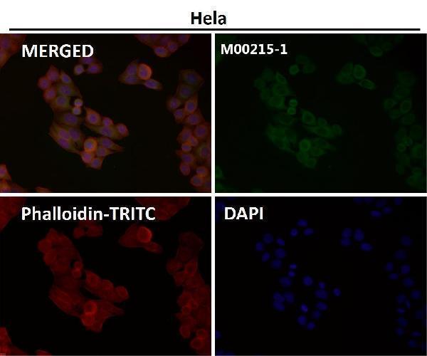
| What should an IF/ICC image look like? | The supplied HPA subcellular record gives no main location or cell lines with ICC-IF images (HPA: subcellular record). | Use the separate IF/ICC guide for assay design. This tissue IHC pattern cannot establish an IF/ICC image pattern or protocol (HPA: tissue IHC and subcellular records). |


Comprehensive Human Protein Atlas IHC scoring per tissue (reliability: Enhanced — High consistency between antibody staining and RNA expression data. Antibody staining in cells/structures not annotated, view images.). Rows are taken directly from the HPA tissue chart — click any row's HPA link to view the source.
| Tissue | Cell type | Level | Evidence | Source |
|---|---|---|---|---|
| Bone marrow | Hematopoietic cells | High | Protein (IHC) | HPA → |
| Cerebral cortex | Neuropil | High | Protein (IHC) | HPA → |
| Kidney | Cells in glomeruli | High | Protein (IHC) | HPA → |
| Cerebellum | Processes in granular layer | Medium | Protein (IHC) | HPA → |
| Tissue | Cell type | Level | Evidence | Source |
|---|---|---|---|---|
| Adipose tissue | Adipocytes | Not detected | Protein (IHC) | HPA → |
| Adrenal gland | Glandular cells | Not detected | Protein (IHC) | HPA → |
| Appendix | Glandular cells | Not detected | Protein (IHC) | HPA → |
| Breast | Adipocytes | Not detected | Protein (IHC) | HPA → |
| Bronchus | Respiratory epithelial cells | Not detected | Protein (IHC) | HPA → |
Troubleshoot SNCA staining by checking retrieval, compartment, cell identity and detection controls before comparing signal across paraffin sections.
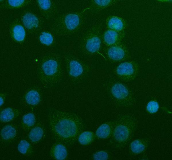
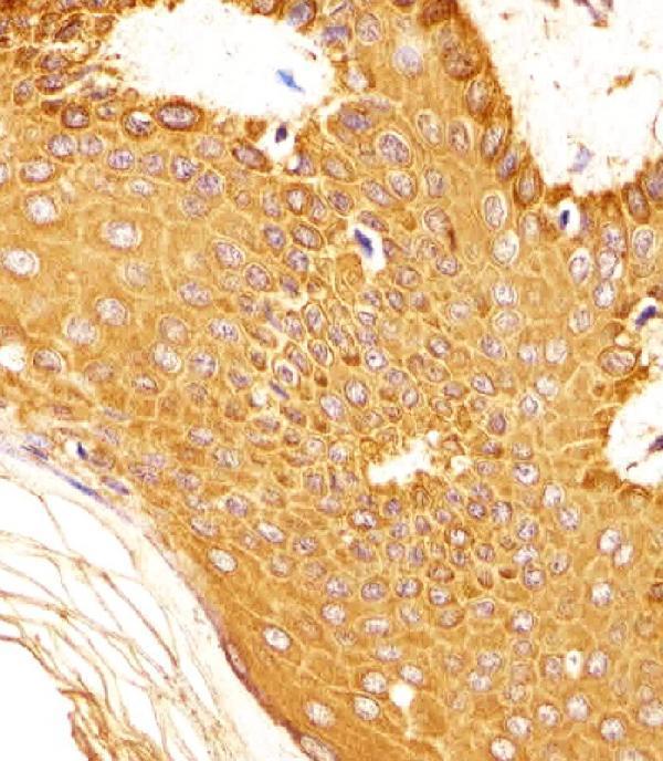
Anti-SNCA IHC examples cover human glioma, colon cancer, brain and skin, plus mouse and rat brain (catalog IHC captions); IF/ICC is illustrated in SiHa cells (A00215-3 IF caption).
M00215-1 shows paraffin-section IHC in human glioma and colon cancer and mouse brain (M00215-1 IHC captions); A00215-3 shows paraffin-section IHC in human, mouse and rat brain (A00215-3 IHC captions). M00215-2 shows IHC-P in formaldehyde-fixed human skin (M00215-2 IHC caption).
Which to pick: For tissue IHC, M00215-1 is a rabbit monoclonal option with human and mouse paraffin-section examples; the captions do not report the fixative (catalog: M00215-1 clone and IHC captions). For IF/ICC, choose A00215-3, which lists both applications and has a SiHa-cell IF example; it also has the broadest illustrated IHC species coverage, with human, mouse and rat brain examples whose fixative is unreported (catalog: A00215-3 applications, IF caption and IHC captions). For paraffin-section human skin IHC-P, choose M00215-2 (M00215-2 IHC caption). The selected M00215-1 tissue-IHC caption documents paraffin sections, but does not specify the fixative (selected-SKU IHC image M00215-1).