SND1 / Staphylococcal nuclease domain-containing protein 1 · IHC design guide

Design Immunohistochemistry for SND1

Plan chromogenic SND1 IHC in paraffin sections using the IHC-validated antibody (datasheet M02602-2). Compare staining with HPA’s variable cytoplasmic tissue profile, including high staining in placental cytotrophoblasts and absent staining in adipocytes (HPA tissue IHC).

Evidence assembled Oct 2026 · For research use; verify linked source records and product datasheet before use
Immunohistochemistry protocol sheet for SND1 (IHC for SND1): expected localisation Variable cytoplasmic tissue staining (HPA tissue IHC), antibody M02602-2, validated IHC image, and IHC protocol steps
Printable SND1 IHC protocol sheet — expected localisation Variable cytoplasmic tissue staining (HPA tissue IHC), antibody M02602-2, controls and protocol steps. Open the full SND1 IHC guide →

SND1 Immunohistochemistry Experimental Design Guide

Expected localisation, validated protocols, controls and antibodies — the at-a-glance facts below, then the full design guide.

Must know before staining
Expected localisation Variable cytoplasmic tissue staining (HPA tissue IHC)
Staining pattern Variable cytoplasmic staining across cell types (HPA tissue IHC)
Antigen retrieval EDTA pH 8.0 HIER, heat-mediated (datasheet M02602-2)
Positive control ⓘ Bone marrow+4 more · see all
Negative control ⓘ Adipose tissue+3 more · see all
Important caveats
Reasons your staining may differ from the expected pattern.
Fixation Keep fixation consistent across sections. (standard IHC practice; not target-specific)
Caveat Adipocytes may show no staining (HPA tissue IHC)
Regulation Variable tissue expression (HPA tissue IHC)
Isoform / epitope 0 isoforms annotated; epitope differences unreported (UniProt)
Section 1

Recommended SND1 IHC & IF Protocols

The catalog antibody’s IHC-P protocol is accompanied by four published SND1 IHC protocols (datasheet: M02602-2; PMC10895293; PMC11390458; PMC4050181; PMC10518936).

Recommended immunohistochemistry (IHC-P) protocol parameters
SampleParaffin-embedded human liver cancer tissue; fixative not specified (datasheet M02602-2)
FixationImage fixative and duration unreported (datasheet M02602-2); verify before use.
Sectioning4–5 µm sections on charged slides (standard)
DeparaffinisationXylene, graded ethanol series to water (standard)
Antigen retrievalHeat retrieval: EDTA pH 8.0 (datasheet M02602-2); 20 min, 95–100 °C (standard)
Peroxidase block3% H2O2, 10 min, room temperature (standard)
Blocking10% goat serum (datasheet M02602-2)
Primary antibodyMouse monoclonal (clone 6G3B4) anti-SND1, 2 μg/ml (datasheet M02602-2)
Primary incubationOvernight at 4 °C (datasheet M02602-2)
DetectionStreptavidin-biotin complex (SABC), DAB chromogen (datasheet M02602-2)
CounterstainHematoxylin, blue, dehydrate and mount (standard)
Expected resultSND1-positive staining in hematopoietic cells of bone marrow (HPA tissue IHC: High). HPA tissue profile: Ubiquitous cytoplasmic expression at variable levels. No signal in the no-primary control.
💡Decision noteStart with heat-mediated EDTA retrieval at pH 8.0 (datasheet: M02602-2); citrate buffer is a published alternative (PMC10895293).
Section 2

What Is the Expected SND1 Staining Pattern?

SND1 should appear predominantly cytoplasmic in paraffin-section IHC, with variable intensity across tissues (HPA tissue IHC: ubiquitous cytoplasmic expression; Enhanced reliability, with medium staining–RNA consistency). Strong examples include bone-marrow hematopoietic cells, endometrial glandular cells and lung alveolar type II cells (HPA tissue IHC: High). SND1 has no transmembrane segment; UniProt also lists nuclear and melanosome locations (UniProt Q7KZF4: topology and subcellular location).

What am I looking at on my slide?
Clear cytoplasmic staining in hematopoietic, glandular or alveolar type II cells.This fits the reported IHC pattern; compare intensity within the identified cell population, since SND1 staining varies by tissue (HPA tissue IHC: ubiquitous cytoplasmic expression; High in bone marrow, endometrium and lung).
Predominantly nuclear staining with little cytoplasmic signal.Recheck staining specificity and slide interpretation against a known-positive cytoplasmic control (HPA tissue IHC: cytoplasmic pattern; standard IHC practice). Nuclear SND1 is biologically possible: UniProt reports nuclear colocalization with STAT6 in IL-4-stimulated cells (UniProt Q7KZF4: subcellular location).
Strong staining in adipocytes, cardiomyocytes or skeletal myocytes.Treat an isolated strong result cautiously: these cells were reported as not detected in the HPA tissue IHC survey (HPA tissue IHC: adipocytes, cardiomyocytes and myocytes). Check antibody specificity and endogenous chromogen activity before assigning it to SND1 (standard IHC practice).
Uniform haze extends through cells and surrounding section.Diffuse background obscures the reported cell-associated cytoplasmic pattern (HPA tissue IHC: cytoplasmic expression). Review blocking, antibody concentration, washing and the detection-only control as general IHC checks; haze alone cannot establish target localization (standard IHC practice).
No staining in a selected known-positive population.A blank hematopoietic, endometrial glandular or pancreatic exocrine population conflicts with the reported High examples (HPA tissue IHC: bone marrow, endometrium and pancreas). Confirm the cell population, then check retrieval, antibody and detection performance before calling the sample negative (standard IHC practice).
💡Expected SND1 appearanceCall a convincing positive when identifiable cells show predominantly cytoplasmic chromogen, with strong signal possible in HPA High populations; diffuse haze or dominant staining in HPA not-detected cells needs investigation (HPA tissue IHC: cytoplasmic expression; High and Not detected examples).
How each factor affects the staining
Choice of tissue and cell populationUse a reported High population to judge whether the assay can detect SND1; use adipocytes or myocytes as contrasting populations within an appropriate section (HPA tissue IHC: High in bone marrow and endometrium; Not detected in adipose and skeletal muscle). HPA observations are reference patterns, not absolute exclusion rules.
Antibody validationHPA rates IHC staining Enhanced for HPA002529 and HPA002632, and Supported for CAB019323 (HPA antibodies: IHC status). These ratings support comparison with the reported pattern; they do not prove that every unexpected signal in a new specimen is specific.
Retrieval and epitope accessOptimizing antigen retrieval is a general paraffin-section IHC step (standard IHC practice). Neither source supplies an SND1-specific retrieval response or fixation-sensitivity result; do not infer one from staining levels, topology or modified residues (HPA tissue IHC; UniProt Q7KZF4).
Intracellular location and antibody epitopeSND1 has no transmembrane segment or signal peptide, and UniProt lists four TNase-like domains plus a Tudor domain (UniProt Q7KZF4: topology and domains). The payload gives no antibody epitope coordinates, so these features cannot predict which antibody will stain a given preparation.
IF/ICC: What pattern should appear?Expect mainly cytosolic fluorescence in the HPA ICC-IF reference, where cytosol is the enhanced main location (HPA subcellular: Cytosol). Nuclear SND1 is also reported under IL-4 stimulation, so assess experimental context before treating nuclear fluorescence as specific (UniProt Q7KZF4: subcellular location).
Why is my staining missing, weak or wrong?
SituationLikely causeNext action
Known-positive tissue is blank.The High cell population may be absent from the viewed area, or the IHC workflow may have failed (HPA tissue IHC: High examples; standard IHC practice).Identify the expected cells on the counterstain, then verify retrieval, antibody exposure and detection with a working control section (standard IHC practice).
Signal is mostly nuclear.The result differs from HPA's usual cytoplasmic IHC pattern; nuclear localization can occur in IL-4-stimulated cells (HPA tissue IHC; UniProt Q7KZF4: subcellular location).Compare a known-positive cytoplasmic control and review the sample's stimulation context; assess antibody specificity before interpreting a nuclear-only result (standard IHC practice).
HPA not-detected cells stain strongly.Non-specific binding or endogenous detection activity is possible; HPA reports adipocytes, cardiomyocytes and myocytes as Not detected (HPA tissue IHC; standard IHC practice).Inspect cell identity, run a detection-only control and compare the result with a reported High population (standard IHC practice; HPA tissue IHC: High examples).
Whole section has diffuse brown background.Excess antibody, inadequate blocking or insufficient washing can raise background in chromogenic IHC (standard IHC practice).Review the antibody concentration and blocking and washing steps; use a detection-only control to locate background from the detection system (standard IHC practice).
Strong positive cells vary across tissues.Variable intensity is compatible with HPA's ubiquitous cytoplasmic profile; a low or not-detected HPA population is not a universal assay failure (HPA tissue IHC: profile and cell-level observations).Score comparable cell populations and include a reported High population when judging assay performance (HPA tissue IHC: High examples; standard IHC practice).
IF/ICC fluorescence seems exclusively nuclear.HPA's enhanced ICC-IF localization is cytosolic, while UniProt reports nuclear colocalization with STAT6 after IL-4 stimulation (HPA subcellular; UniProt Q7KZF4).Check stimulation context and compare cytosolic signal with an appropriate control before assigning the nuclear fluorescence to SND1 (standard IF practice).

Sample controls for SND1 IHC & IF

🧪Run endometrium first: glandular cells should stain for SND1 (HPA: High in endometrial glandular cells). Use skeletal muscle myocytes as the negative tissue (HPA: Not detected in myocytes); on the endometrial slide, internal comparator cells should show no signal above control background, but HPA does not identify a verified SND1-negative cell type there.
Positive control tissue: Bone marrow (Hematopoietic cells, HPA High)
Negative control tissue: Adipose tissue (HPA Not detected)
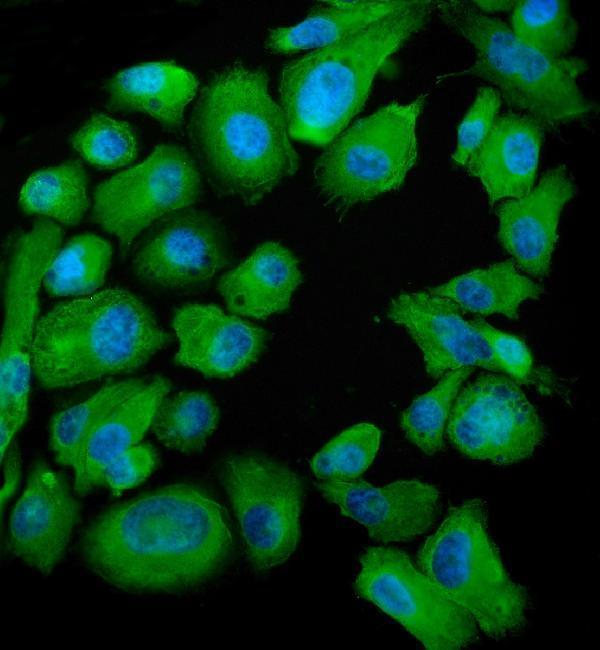
ICC-IF cell lines (HPA subcellular resource): HPA ICC-IF images show SND1 in A-431, U-251MG, U2OS, U2OS, siRNA 1 (10x), U2OS, siRNA 1 (40x), U2OS, scrambled (10x), U2OS, scrambled (40x), NIH 3T3, with annotated localisation: Cytosol (enhanced) (HPA subcellular).
Technical controls: Include a no-primary, secondary-only control, a matched mouse IgG isotype control, and SND1-knockout material as a biological negative (caption: mouse anti-SND1 primary). Block endogenous peroxidase and check for endogenous biotin background when using the caption’s biotinylated secondary, streptavidin complex, and DAB detection (caption: biotin-based chromogenic detection).
⚠️Feasibility: A target-specific fixation window or fixation effect is unreported, and the selected M02602-2 paraffin-section caption does not state a fixative (caption: fixative unreported). The caption uses heat retrieval in EDTA at pH 8.0, but it does not establish that retrieval is required or that frozen sections or IF/ICC are easier (caption: EDTA heat retrieval). No endometrium-specific artefact is reported in the supplied evidence; assess background with the controls above (HPA: High in endometrial glandular cells).

HPA tissue IHC evidence for SND1

Comprehensive Human Protein Atlas IHC scoring per tissue (reliability: Enhanced — Medium consistency between antibody staining and RNA expression data.). Rows are taken directly from the HPA tissue chart — click any row's HPA link to view the source.

Positive expression · recommended positive controls

TissueCell typeLevelEvidenceSource
Bone marrow Hematopoietic cells High Protein (IHC) HPA →
Cerebellum Cells in molecular layer High Protein (IHC) HPA →
Cerebral cortex Neuronal cells High Protein (IHC) HPA →
Endometrium Glandular cells High Protein (IHC) HPA →
Gallbladder Glandular cells High Protein (IHC) HPA →

Undetected expression · recommended negative controls

TissueCell typeLevelEvidenceSource
Adipose tissue Adipocytes Not detected Protein (IHC) HPA →
Heart muscle Cardiomyocytes Not detected Protein (IHC) HPA →
Skeletal muscle Myocytes Not detected Protein (IHC) HPA →
Soft tissue Chondrocytes Not detected Protein (IHC) HPA →
Section 3

Advanced SND1 IHC Tips

Troubleshooting SND1 staining in paraffin sections centers on retrieval, intracellular localization, background control, and cell-resolved interpretation of chromogenic signal.

Which retrieval conditions should I try first for weak SND1 staining?
Start with heat-mediated antigen retrieval in EDTA at pH 8.0 for the catalog antibody (datasheet M02602-2). The documented paraffin-section image used this retrieval before staining with 2 μg/ml primary antibody overnight at 4°C (datasheet M02602-2). If signal remains weak, compare controlled changes in heating duration or an alternative retrieval buffer on adjacent sections while keeping detection conditions constant (standard IHC practice). Include a positive tissue with a defined expected cell population, such as pancreatic exocrine glandular cells (HPA: High in pancreatic exocrine glandular cells). Assess staining within intact cells, since excessive retrieval can compromise section morphology and make apparent signal difficult to interpret (standard IHC practice).
Could fixation explain inconsistent SND1 staining between paraffin blocks?
Target-specific sensitivity of SND1 to fixative type or fixation duration is unknown from the supplied evidence (selected M02602-2 caption: fixative not stated). Record each block's fixative and processing history, then compare sections using the same retrieval, antibody concentration, and detection run (standard IHC practice). The catalog image documents a paraffin-embedded human liver cancer section, but does not establish that the tissue was formalin fixed (datasheet M02602-2). If staining differs, inspect morphology and include a consistently processed reference section before attributing the difference to SND1 abundance (standard IHC practice). Do not infer fixation sensitivity from ubiquitous expression or protein modifications (UniProt Q7KZF4; HPA: ubiquitous cytoplasmic expression).
Should I score nuclear SND1 staining alongside cytoplasmic staining?
Score cytoplasmic staining separately because tissue IHC shows ubiquitous cytoplasmic expression at variable levels, and cell imaging places SND1 in the cytosol (HPA: tissue IHC; HPA: subcellular). Nuclear localization is biologically plausible: SND1 is annotated in the nucleus and colocalizes with STAT6 there in IL-4-stimulated cells (UniProt Q7KZF4). Document the nuclear fraction and intensity rather than combining it with cytoplasmic signal in one score (standard IHC practice). A nuclear-only pattern in an otherwise weak section needs corroboration through controls and a matched condition before interpretation (standard IHC practice; HPA: cytosolic main location). Melanosome-fraction identification does not establish a routine chromogenic staining pattern (UniProt Q7KZF4).
How should I investigate discordant staining caused by epitope accessibility?
Check the antibody's stated immunogen or epitope before assigning a molecular explanation to a staining difference; the supplied caption does not identify it (datasheet M02602-2). UniProt lists 0 isoforms and describes four TNase-like domains plus a Tudor domain, so an isoform-specific explanation lacks support here (UniProt Q7KZF4). SND1 has no annotated transmembrane segment, while listed modified residues include phosphorylation and acetylation sites (UniProt Q7KZF4). Those annotations do not show that any modification blocks this antibody's epitope (UniProt Q7KZF4; datasheet M02602-2). Compare retrieval conditions and, where available, an independently validated antibody targeting a distinct region before concluding that SND1 expression changed (standard IHC practice).
How can I use IF to check a disputed SND1 IHC pattern?
Use IF as an orthogonal localization check, with its antibody and fixation conditions validated separately from the chromogenic tissue workflow (standard IF practice; datasheet M02602-2: paraffin IHC caption). Pair SND1 with a validated marker for the expected cell population, such as alveolar type II cells, to test cell identity (HPA: High in lung alveolar type II cells). Choose spectrally separated fluorophores and favor a far-red channel when tissue autofluorescence obscures shorter wavelengths (standard IF practice). Because SND1 is intracellular and has no annotated transmembrane segment, validate permeabilization for access to its cytosolic or nuclear epitope (UniProt Q7KZF4). Include single-label and secondary-only controls when comparing channels (standard IF practice).
How do I separate SND1 staining from chromogenic background?
Begin with a no-primary control and inspect whether staining follows tissue edges, damaged areas, or the expected cell interiors (standard IHC practice). The documented workflow used 10% goat serum, biotinylated goat anti-mouse secondary antibody, an avidin-biotin complex, and DAB (datasheet M02602-2). Apply an endogenous peroxidase block and check secondary-only signal as general controls for chromogenic detection (standard IHC practice). If diffuse staining persists, titrate the primary around the documented 2 μg/ml condition while holding retrieval and development time constant (datasheet M02602-2; standard IHC practice). Compare intact cells with the reported predominantly cytoplasmic tissue pattern before assigning weak brown haze to SND1 (HPA: tissue IHC).
What is a defensible way to quantify variable SND1 IHC signal? ⚠ ANSWER MARKED FOR VERIFICATION
Define the cell population and compartment before scoring, since SND1 tissue staining is broadly cytoplasmic but varies in level (HPA: tissue IHC). For intensity-based analysis, record the percentage of viable cells at each intensity and calculate an H-score consistently across sections (standard IHC practice). Alternatively, report % positive cells or positive-cell density per mm², with the threshold set from controls (standard IHC practice). Normalize counts to the relevant viable cell population or tissue area, and exclude necrosis, folds, and edges using prespecified rules (standard IHC practice). Report nuclear staining separately if assessed, given the cytosolic main location and conditional nuclear localization annotation (HPA: subcellular; UniProt Q7KZF4).
When is an unexpected SND1-positive area likely to be artefact?
Look first for staining inside intact cytoplasm, the predominant tissue pattern for SND1, rather than pigment confined to edges or damaged regions (HPA: tissue IHC; standard IHC practice). Check cell identity against the reference pattern: pancreatic exocrine glandular cells are reported High, while adipocytes are Not detected (HPA: tissue IHC). A nuclear signal warrants separate assessment because nuclear localization is annotated under IL-4 stimulation, whereas cytosol is the main observed cell location (UniProt Q7KZF4; HPA: subcellular). Exclude necrotic regions and section edges from interpretation, and investigate signal in no-primary controls for endogenous enzyme or detection background (standard IHC practice). Corroborate a discordant pattern with an independent control before calling it SND1 (standard IHC practice).
Boster reagents

Best SND1 / Staphylococcal nuclease domain-containing protein 1 IHC Antibodies

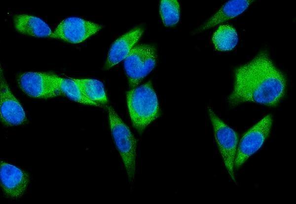
Three anti-SND1 antibodies have IHC images from human paraffin sections and IF/ICC images from PC-3 cells (catalog image captions). All list human, mouse and rat reactivity (catalog reactivity).

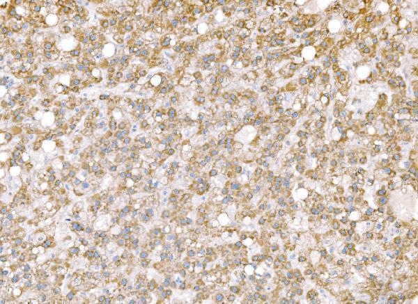
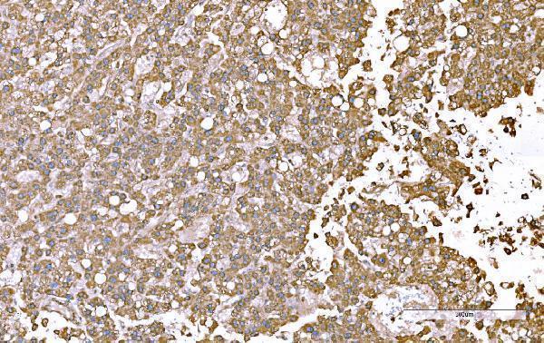
Real IHC data IHC analysis of SND1 using anti-SND1 antibody (M02602-2). SND1 was detected in a paraffin-embedded section of human liver cancer tissue. Heat mediated antigen retrieval was performed in EDTA buffer (pH 8.0, epitope retrieval solution). The tissue section was blocked with 10% goat serum. The tissue section was then incubated with 2 μg/ml mouse anti-SND1 Antibody (M02602-2) overnight at 4°C. Biotinylated goat anti-mouse IgG was used as secondary antibody and incubated for 30 minutes at 37°C. The tissue section was developed using Strepavidin-Biotin-Complex (SABC) (Catalog # SA1021) with DAB as the chromogen.
Anti-SND1 Antibody ® (monoclonal, 6G3B4)
Cat # M02602-2
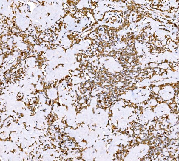
Real IHC data IHC analysis of SND1 using anti-SND1 antibody (M02602-1). SND1 was detected in a paraffin-embedded section of human Hodgkin's lymphoma tissue. Heat mediated antigen retrieval was performed in EDTA buffer (pH 8.0, epitope retrieval solution). The tissue section was blocked with 10% goat serum. The tissue section was then incubated with 2 μg/ml mouse anti-SND1 Antibody (M02602-1) overnight at 4°C. Biotinylated goat anti-mouse IgG was used as secondary antibody and incubated for 30 minutes at 37°C. The tissue section was developed using Strepavidin-Biotin-Complex (SABC) (Catalog # SA1021) with DAB as the chromogen.
Anti-SND1 Antibody ® (monoclonal, 5F5E9)
Cat # M02602-1
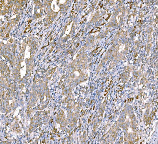
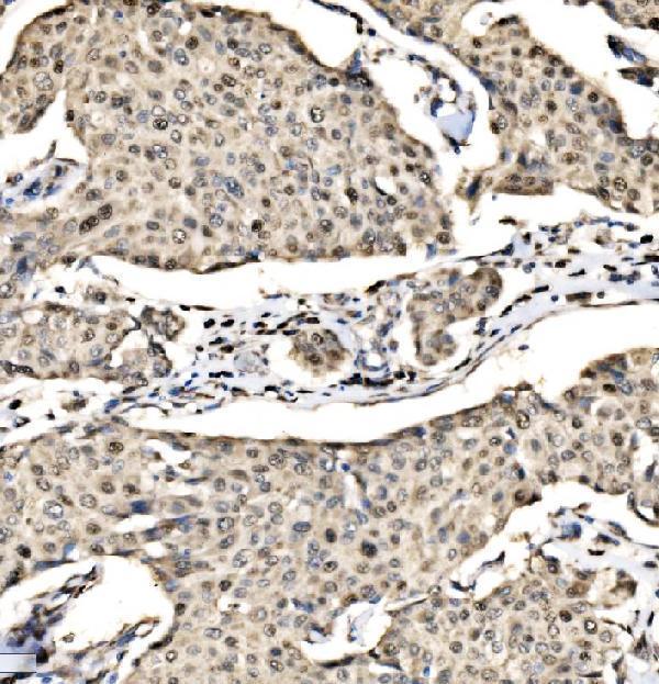
Real IHC data IHC analysis of SND1 using anti-SND1 antibody (A02602-3). SND1 was detected in paraffin-embedded section of human breast cancer tissue. Heat mediated antigen retrieval was performed in EDTA buffer (pH8.0, epitope retrieval solution). The tissue section was blocked with 10% goat serum. The tissue section was then incubated with 2μg/ml rabbit anti-SND1 Antibody (A02602-3) overnight at 4°C. Biotinylated goat anti-rabbit IgG was used as secondary antibody and incubated for 30 minutes at 37°C. The tissue section was developed using Strepavidin-Biotin-Complex (SABC) (Catalog # SA1022) with DAB as the chromogen.
Anti-SND1 Antibody ®
Cat # A02602-3

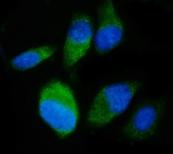
M02602-2 has an IHC image from human liver cancer, while M02602-1 has one from human Hodgkin’s lymphoma; both have IF/ICC images from PC-3 cells (respective catalog image captions). A02602-3 has an IHC image from human breast cancer and an IF/ICC image from PC-3 cells (catalog image captions).

Which to pick: For tissue IHC, choose the SKU whose own paraffin-section image matches your sample: M02602-2 for liver cancer, M02602-1 for Hodgkin’s lymphoma, or A02602-3 for breast cancer (respective catalog IHC captions); fixation is unreported in those captions. For IF/ICC, A02602-3 is a rabbit antibody with a documented 5 μg/ml PC-3 cell example; both mouse monoclonals also have PC-3 cell examples (respective catalog IF captions; catalog host/clone). For cross-species work, all three list human, mouse and rat reactivity, but the supplied IHC examples use human tissue (catalog reactivity; respective catalog IHC captions).

Each figure is that product's own IHC / IF validation image from its datasheet.

References

  1. UniProt Consortium. UniProt entry Q7KZF4 (SND1_HUMAN, Staphylococcal nuclease domain-containing protein 1).
  2. Human Protein Atlas. SND1 tissue IHC expression (reliability: Enhanced).
  3. Human Protein Atlas. SND1 subcellular location (ICC-IF): Localized to the cytosol..
  4. Human Protein Atlas. SND1 antibody validation summary (3 antibodies).
  5. [Identification and Analysis of SND1 as an Oncogene and Prognostic Biomarker for Lung Adenocarcinoma]. Zhongguo fei ai za zhi = Chinese journal of lung cancer 2024 — PMC10895293.
  6. Androgen receptor modulatory miR-1271-5p can promote hormone sensitive prostate cancer cell growth. Frontiers in oncology 2024 — PMC11390458.
  7. Staphylococcal nuclease domain containing-1 (SND1) promotes migration and invasion via angiotensin II type 1 receptor (AT1R) and TGFβ signaling. FEBS open bio 2014 — PMC4050181.
  8. SND1 aggravates mitochondrial damage, apoptosis and extracellular matrix degradation in IL-1β-stimulated chondrocytes via PINK1/BECN1 pathway. European journal of medical research 2023 — PMC10518936.
  9. PubMed PMID:7651391 — UniProt-cited evidence.
  10. PubMed PMID:15489334 — UniProt-cited evidence.
  11. PubMed PMID:12234934 — UniProt-cited evidence.