This website uses cookies to ensure you get the best experience on our website.


- Table of Contents
Plan SNRNP200 paraffin IHC around the observed nuclear tissue pattern (HPA tissue IHC). Start the catalog antibody at 2–5 μg/ml (datasheet: 2–5 μg/ml), and assess staining with a nuclear counterstain.

Expected localisation, validated protocols, controls and antibodies — the at-a-glance facts below, then the full design guide.
| Expected localisation | Nuclear staining in several cell types (HPA tissue IHC) | |
| Staining pattern | Nuclear staining across several cell types (HPA tissue IHC) | |
| Antigen retrieval | EDTA pH 8.0 HIER, heat-mediated (datasheet A04514-2) | |
| Positive control | Bronchus+4 more · see all | |
| Negative control | Adrenal gland+4 more · see all |
| Fixation | Keep fixation consistent across sections. (standard IHC practice; not target-specific) | |
| Caveat | Fallopian tube cilia show high signal; score nuclei separately (HPA tissue IHC) | |
| Regulation | No specific regulator reported (UniProt) | |
| Isoform / epitope | 2 isoforms; check epitope coverage (UniProt) |
The catalog antibody protocol is paired with published chromogenic SNRNP200 IHC methods for human spinal cord, motor cortex, and prostate sections (PMC9083243; PMC7000340).
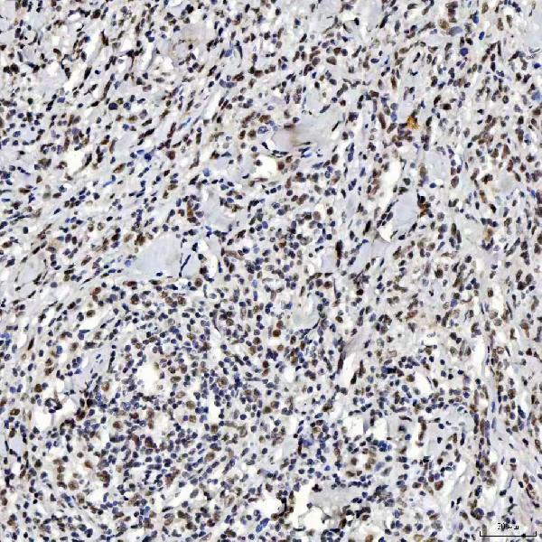
| Sample | Paraffin-embedded human diffuse large B cell lymphoma tissue; fixative not specified (datasheet A04514-2) |
| Fixation | Image fixative and duration unreported (datasheet A04514-2); verify before use. |
| Sectioning | 4–5 µm sections on charged slides (standard) |
| Deparaffinisation | Xylene, graded ethanol series to water (standard) |
| Antigen retrieval | Heat retrieval: EDTA pH 8.0 (datasheet A04514-2); 20 min, 95–100 °C (standard) |
| Peroxidase block | 3% H2O2, 10 min, room temperature (standard) |
| Blocking | 10% goat serum (datasheet A04514-2) |
| Primary antibody | Rabbit anti-SNRNP200, 2-5 μg/ml (datasheet A04514-2) |
| Primary incubation | Overnight at 4 °C (datasheet A04514-2) |
| Detection | HRP-conjugated secondary, DAB chromogen (datasheet A04514-2) |
| Counterstain | Hematoxylin, blue, dehydrate and mount (standard) |
| Expected result | SNRNP200-positive staining in ciliated cells (cell body) of bronchus (HPA tissue IHC: High). HPA tissue profile: Nuclear expression in several different cell types. No signal in the no-primary control. |
SNRNP200 is a nuclear spliceosome protein with no transmembrane segment (UniProt O75643). In paraffin-section IHC, expect nuclear staining in several cell types, including bronchial ciliated cells, placental trophoblastic cells and testicular seminiferous-duct cells reported as High (HPA tissue IHC). The tissue IHC assessment is Approved, with external verification pending (HPA antibody HPA029321).
| Distinct nuclear chromogen in bronchial ciliated cell bodies, with recognizable tissue structure. | This fits the reported nuclear profile and High staining in these cells (HPA tissue IHC). Judge the result by its nuclear location and the stained cell population; a dark deposit alone does not establish specific staining (general IHC practice). |
| Strong, widespread cytoplasmic or surface staining with little nuclear signal. | That is inconsistent with the dominant nuclear IHC profile (HPA tissue IHC; UniProt O75643). Review it as possible background or detection artefact (general IHC practice). HPA ICC-IF also reports uncertain cytosolic localization, so an isolated cytoplasmic signal is not definitive evidence either way (HPA subcellular). |
| Prominent staining in a cell population reported as Not detected, such as heart cardiomyocytes. | The result conflicts with that specific HPA tissue observation; it does not prove that every cardiomyocyte lacks SNRNP200 (HPA tissue IHC). Consider cross-reactivity or endogenous detection activity, then compare a negative reagent control and a known-positive cell population on the same run (general IHC practice). |
| Diffuse chromogen across nuclei, cytoplasm and tissue spaces, without clear cell boundaries. | This distribution cannot reliably be scored as nuclear SNRNP200 staining (HPA tissue IHC; general IHC practice). Examine background controls and tissue morphology before assigning intensity; diffuse deposition can obscure a genuine nuclear signal (general IHC practice). |
| No nuclear staining in an adequately represented, known-positive population, such as placental trophoblastic cells. | HPA reports High staining in those cells (HPA tissue IHC). Check that the expected cells are present, then review retrieval and detection controls as general IHC troubleshooting steps. A failed run cannot establish biological absence (general IHC practice). |
| Tissue and cell selection | Bronchial ciliated cell bodies, placental trophoblastic cells, seminiferous-duct cells and bladder urothelial cells are reported High; stomach glandular cells are Low (HPA tissue IHC). Compare like cell populations when interpreting intensity. These observations are examples, not a rule that every cell in each tissue stains equally (HPA tissue IHC). |
| Validation strength | The listed antibody, HPA029321, is Approved for IHC, with external verification pending (HPA antibody; HPA tissue IHC). Treat agreement with its reported pattern as supportive rather than conclusive. The supplied record does not assign it an Enhanced IHC validation status (HPA antibody). |
| Protein organization | SNRNP200 is nuclear, has no transmembrane segment, and has no signal peptide or propeptide reported (UniProt O75643). A membrane-dominant pattern would need independent confirmation. These features do not establish how formalin fixation or antigen retrieval affects its epitope (UniProt O75643). |
| Isoforms and antibody epitope | UniProt lists two isoforms (UniProt O75643). The supplied evidence gives no epitope position or isoform coverage for HPA029321, so do not attribute differing staining to an isoform or prescribe a target-specific retrieval condition from this record (HPA antibody; UniProt O75643). |
| IF/ICC Q: What localization should I expect? | A: Mainly nucleoplasmic staining is supported; cytosol and primary-cilium localization are marked uncertain (HPA subcellular). HPA also reports High staining in fallopian-tube cilia axonemes by tissue IHC (HPA tissue IHC). Assess any ciliary signal in its assay context; this IHC section supplies no IF/ICC protocol. |
| Situation | Likely cause | Next action |
|---|---|---|
| Known-positive cells have no visible nuclear chromogen. | The cause is unresolved; absent cells, an ineffective retrieval step or a failed detection step are possibilities (general IHC practice). | Confirm the expected cell population is present and inspect run controls before changing one IHC condition at a time (general IHC practice). Use an HPA High population as a reference, such as placental trophoblastic cells (HPA tissue IHC). |
| Most tissue areas look uniformly brown. | Nonspecific reagent binding or endogenous detection activity may produce background in chromogenic IHC (general IHC practice). | Compare a negative reagent control, check blocking and washing, and inspect whether chromogen follows cells or tissue spaces (general IHC practice). Score nuclear staining only where cell boundaries remain interpretable (HPA tissue IHC; general IHC practice). |
| Signal is mainly cytoplasmic, with weak or absent nuclear staining. | The distribution conflicts with the dominant nuclear pattern, although cytosolic localization is listed as uncertain in ICC-IF (HPA tissue IHC; HPA subcellular). | Check a nuclear positive population and a negative reagent control; review counterstain and morphology before calling cytoplasmic signal specific (general IHC practice). Do not use the uncertain ICC-IF location alone to validate IHC staining (HPA subcellular). |
| A reported Not detected population stains strongly. | Cross-reactivity or endogenous detection activity is possible; HPA's Not detected designation is an observation for that cell population (HPA tissue IHC; general IHC practice). | Verify cell identity, compare the negative reagent control, and assess an HPA High population in the same run (HPA tissue IHC; general IHC practice). Treat the discrepancy as unresolved until controls support an interpretation. |
| A low-staining tissue appears negative. | Low expression can be difficult to distinguish from background; stomach glandular cells are reported Low (HPA tissue IHC; general IHC practice). | First verify the run with an HPA High population, then score the low-staining cells against the negative reagent control (HPA tissue IHC; general IHC practice). Do not infer assay failure from a Low reference alone. |
| Prominent ciliary staining seems at odds with a nuclear result elsewhere. | HPA reports High fallopian-tube cilia-axoneme staining in tissue IHC, while its ICC-IF primary-cilium assignment is uncertain (HPA tissue IHC; HPA subcellular). | Record the tissue, cell compartment and assay separately; check controls and the accompanying nuclear pattern before assigning specificity (general IHC practice). The two HPA observations do not establish that every ciliary deposit is SNRNP200. |
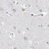
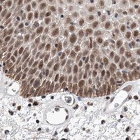
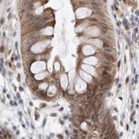
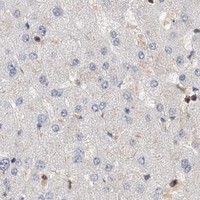

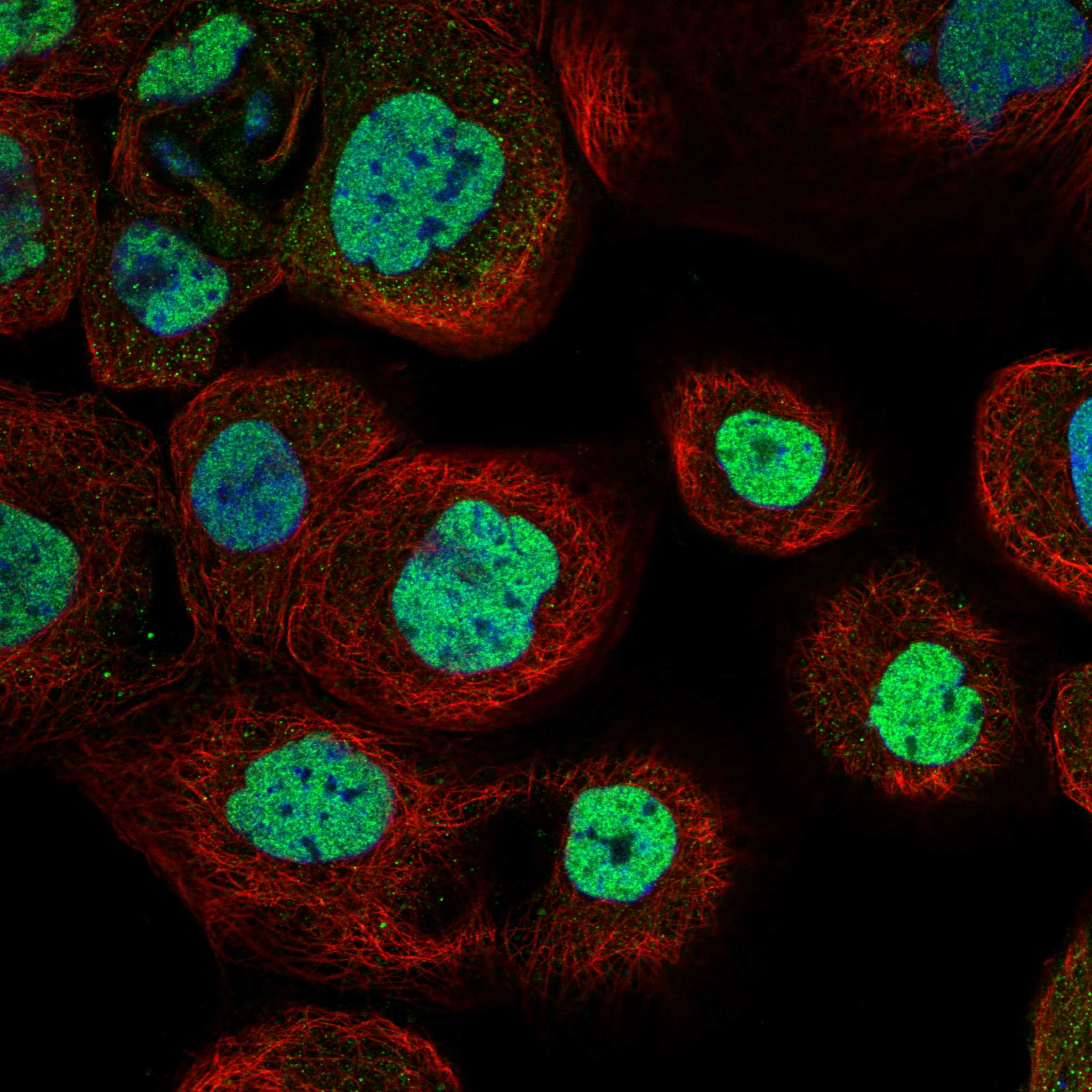
Comprehensive Human Protein Atlas IHC scoring per tissue (reliability: Approved — Pending external verification.). Rows are taken directly from the HPA tissue chart — click any row's HPA link to view the source.
| Tissue | Cell type | Level | Evidence | Source |
|---|---|---|---|---|
| Bronchus | Ciliated cells (cell body) | High | Protein (IHC) | HPA → |
| Fallopian tube | Ciliated cells (cilia axoneme) | High | Protein (IHC) | HPA → |
| Nasopharynx | Ciliated cells (cell body) | High | Protein (IHC) | HPA → |
| Placenta | Trophoblastic cells | High | Protein (IHC) | HPA → |
| Stomach | Glandular cells | Low | Protein (IHC) | HPA → |
| Tissue | Cell type | Level | Evidence | Source |
|---|---|---|---|---|
| Adrenal gland | Glandular cells | Not detected | Protein (IHC) | HPA → |
| Heart muscle | Cardiomyocytes | Not detected | Protein (IHC) | HPA → |
| Liver | Cholangiocytes | Not detected | Protein (IHC) | HPA → |
| Ovary | Ovarian stroma cells | Not detected | Protein (IHC) | HPA → |
| Parathyroid gland | Glandular cells | Not detected | Protein (IHC) | HPA → |
Troubleshoot SNRNP200 staining by checking nuclear localisation, the documented retrieval conditions, and cell-level controls before interpreting chromogenic signal.
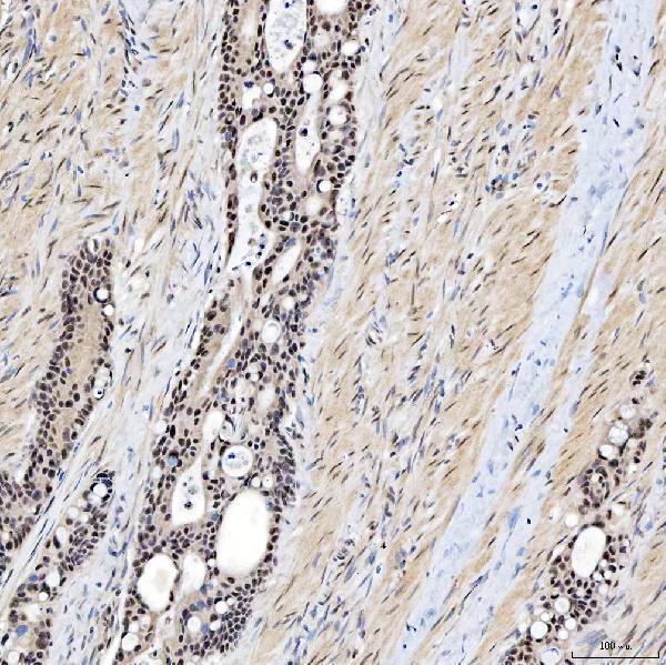
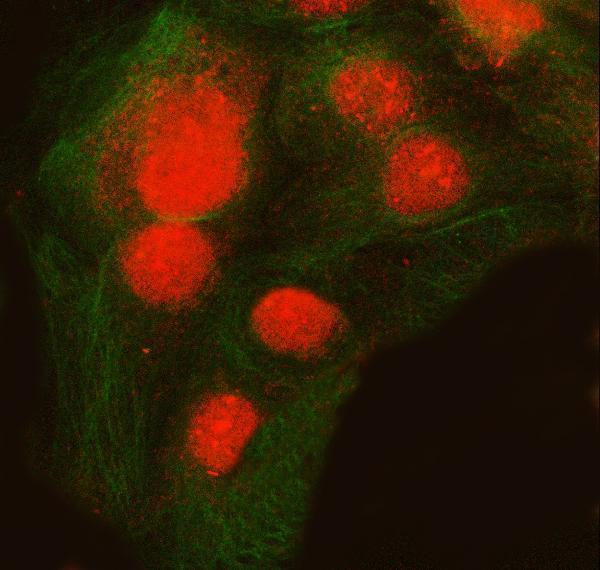
A04514-2 has IHC images from human paraffin sections and IF images from Caco-2 cells and a human paraffin section (catalog figure captions). Listed reactivity covers human, mouse, and rat (catalog).
A04514-2 has IHC images from human diffuse large B cell lymphoma, duodenal papilla adenocarcinoma, endometrioid adenocarcinoma, and glioblastoma paraffin sections (A04514-2 IHC captions). It is also listed for IF, with images from Caco-2 cells and a human intestinal cancer paraffin section (A04514-2 applications; IF captions).
Which to pick: For tissue IHC, choose A04514-2: its own caption documents a human paraffin section, EDTA pH 8.0 retrieval, and 2 μg/ml primary antibody; the fixative is unreported (A04514-2 IHC caption). For IF/ICC, the same rabbit antibody is listed for IF and shown in cell and tissue images; clonality is unreported (A04514-2 catalog; IF captions). For mouse or rat samples, A04514-2 lists reactivity, while its supplied IHC images document human sections (A04514-2 catalog reactivity; IHC captions).