This website uses cookies to ensure you get the best experience on our website.


- Table of Contents
Plan SNRPA paraffin-section IHC around the broad nuclear staining reported in tissues (HPA tissue IHC). Use the IHC-validated antibody’s documented conditions and compare staining across samples with consistent fixation.

Expected localisation, validated protocols, controls and antibodies — the at-a-glance facts below, then the full design guide.
| Expected localisation | Nuclear staining across tissues (HPA tissue IHC) | |
| Staining pattern | Broad nuclear staining across tissue cell types (HPA tissue IHC) | |
| Antigen retrieval | EDTA pH 8.0 HIER, heat-mediated (datasheet A08780-2) | |
| Positive control | Caudate+4 more · see all | |
| Negative control | None in HPA (detected in all 45 tissues); use no-primary + isotype controls |
| Fixation | Keep paraffin-section fixation consistent across samples. (standard IHC practice; not target-specific) | |
| Caveat | Vaginal squamous cells show low staining (HPA tissue IHC) | |
| Regulation | Low tissue specificity (HPA tissue RNA) | |
| Isoform / epitope | 0 isoforms annotated; chain spans residues 2–282 (UniProt) |
The catalog antibody protocol uses EDTA pH 8.0 heat retrieval (datasheet A08780-2). The published SNRPA IHC protocols below provide tissue preparation, staining, and detection details where reported (cited PMC methods).
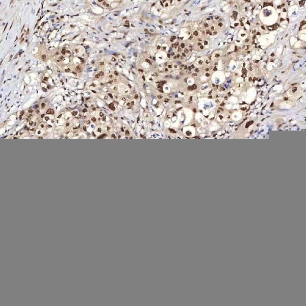
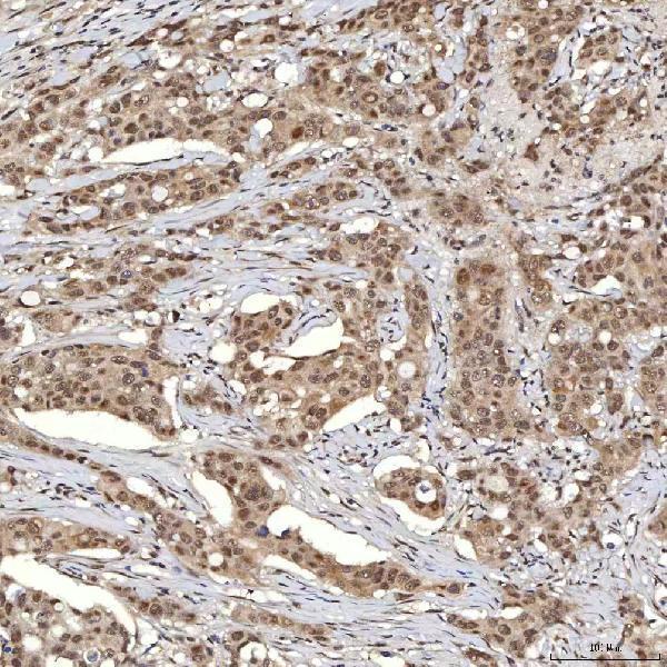
| Sample | Paraffin-embedded human appendix adenocarcinoma tissue; fixative not specified (datasheet A08780-2) |
| Fixation | Image fixative and duration unreported (datasheet A08780-2); verify before use. |
| Sectioning | 4–5 µm sections on charged slides (standard) |
| Deparaffinisation | Xylene, graded ethanol series to water (standard) |
| Antigen retrieval | Heat retrieval: EDTA pH 8.0 (datasheet A08780-2); 20 min, 95–100 °C (standard) |
| Peroxidase block | 3% H2O2, 10 min, room temperature (standard) |
| Blocking | 10% goat serum (datasheet A08780-2) |
| Primary antibody | Rabbit anti-SNRPA, 2-5 μg/ml (datasheet A08780-2) |
| Primary incubation | Overnight at 4 °C (datasheet A08780-2) |
| Detection | HRP-conjugated secondary, DAB chromogen (datasheet A08780-2) |
| Counterstain | Hematoxylin, blue, dehydrate and mount (standard) |
| Expected result | SNRPA-positive staining in neuronal cells of caudate (HPA tissue IHC: High). HPA tissue profile: Ubiquitous nuclear expression. No signal in the no-primary control. |
SNRPA should stain nuclei, consistent with its nuclear localisation and lack of a transmembrane segment (UniProt P09012: subcellular location and topology). HPA describes ubiquitous nuclear expression with Supported tissue IHC reliability, meaning high consistency between antibody staining and RNA expression (HPA tissue IHC). Expect nuclear staining across many cell types, with particularly high staining in the specific cells listed by HPA; intensity need not be uniform across tissues (HPA tissue IHC).
| Distinct nuclear chromogen in kidney tubular cells, with nuclei distinguishable from surrounding cytoplasm. | This fits the expected compartment and a documented high staining cell population (UniProt P09012: nucleus; HPA tissue IHC: High in kidney tubular cells). Judge the pattern in identifiable cells, rather than treating all brown tissue as positive. |
| Predominantly cytoplasmic, membranous, or extracellular colour with little nuclear staining. | That distribution conflicts with the reported nuclear location and absence of a transmembrane segment (UniProt P09012: location and topology). Treat it as a suspect IHC pattern and assess background, detection, and tissue morphology before assigning it to SNRPA. |
| Strong colour in an unexpected cell population while the documented cells lack clear nuclear staining. | The mismatch warrants checking antibody cross-reactivity or endogenous detection activity (general IHC practice). HPA reports ubiquitous nuclear expression, so an unlisted cell type alone does not prove a false positive; compare compartment and local cell identity together (HPA tissue IHC). |
| Diffuse colour over nuclei, cytoplasm, and tissue spaces, without crisp cell boundaries. | The distribution is difficult to score as specific nuclear staining (UniProt P09012: nucleus). Diffuse signal can reflect nonspecific binding or detection background; inspect controls and chromogen development before interpreting intensity (general IHC practice). |
| No nuclear signal in an adequately preserved kidney section containing identifiable tubular cells. | This is discordant with HPA's high tubular cell staining (HPA tissue IHC: Kidney, Cells in tubules, High). First check section quality and assay controls; the supplied sources do not establish a SNRPA specific fixation or retrieval failure. |
| Compartment used for scoring | Score nuclei: UniProt places SNRPA in the nucleus, and HPA reports supported nucleoplasmic localisation by ICC-IF (UniProt P09012: location; HPA subcellular). Cytoplasmic colour alone does not match either observation. |
| Cell and tissue choice | HPA reports high staining in kidney tubular cells, lung alveolar type I cells, and several listed brain cell populations, but low staining in vaginal squamous epithelial cells (HPA tissue IHC). Use the documented cell population when comparing sections. |
| Strength of tissue evidence | The tissue IHC profile has Supported reliability and low RNA tissue specificity (HPA tissue IHC). These support a broadly nuclear expectation, while the listed High and Low observations remain cell specific rather than universal intensity standards. |
| Antibody evidence by application | HPA lists IHC as Supported for HPA046440 and CAB004652; HPA054834 has ICC Supported without an IHC status (HPA antibodies). Apply an antibody's stated evidence to its own application; the supplied record gives no catalog dilution or IHC protocol. |
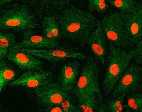
| IF/ICC: what localisation should appear? | Expect nucleoplasmic fluorescence: HPA reports supported nucleoplasmic localisation and images in A-431, U-251MG, and U2OS cells (HPA subcellular). This is an interpretation reference for IF/ICC, not an IHC protocol or a claim of equal antibody performance. |
| Target specific preparation sensitivity | No target specific fixation or antigen retrieval effect is supplied (UniProt P09012; HPA tissue IHC). Select and assess retrieval through the IHC assay's controls (general IHC practice); do not infer fixation sensitivity from topology, modifications, or tissue staining level. |
| Situation | Likely cause | Next action |
|---|---|---|
| Kidney tubular nuclei show no chromogen. | The result conflicts with a documented high staining population (HPA tissue IHC: Kidney, Cells in tubules, High); the cause cannot be assigned from this record. | Confirm tubules and preserved nuclei, then inspect positive control performance, antibody application, detection reagents, and retrieval conditions (general IHC practice). Do not assume a SNRPA specific fixation effect. |
| Colour is mainly cytoplasmic or membranous. | The compartment conflicts with nuclear SNRPA and its lack of a transmembrane segment (UniProt P09012: location and topology); background or cross-reactivity is possible (general IHC practice). | Compare a reagent control and an HPA documented positive cell population; score only convincing nuclear signal, then reassess blocking and detection background (HPA tissue IHC; general IHC practice). |
| The whole section is diffusely brown. | Diffuse colour obscures the nuclear pattern expected for SNRPA (UniProt P09012: nucleus). Nonspecific binding, endogenous detection activity, or excessive chromogen development can cause broad colour (general IHC practice). | Inspect the reagent control, check endogenous activity blocking appropriate to the detection chemistry, and review antibody concentration and chromogen development (general IHC practice). |
| An unexpected cell population stains strongly. | Cross-reactivity or endogenous detection activity is possible (general IHC practice), but HPA's ubiquitous nuclear profile means cell identity alone cannot establish an artefact (HPA tissue IHC). | Verify cell identity and nuclear localisation; compare documented high staining cells and reagent controls before rejecting the result (HPA tissue IHC; general IHC practice). |
| A vaginal squamous epithelial sample looks weak beside kidney tubules. | HPA reports low staining in vaginal squamous epithelial cells and high staining in kidney tubular cells (HPA tissue IHC). That difference can be consistent with the reported profile. | Assess each cell population against its own expected level and confirm that the kidney control stains nuclei; avoid calling the lower level a technical failure solely from this comparison (HPA tissue IHC). |
| Two antibody results disagree, or ICC images appear clearer than the IHC section. | HPA records application specific statuses: two antibodies have IHC Supported, while HPA054834 has ICC Supported without an IHC status (HPA antibodies). Different application evidence does not resolve an IHC discrepancy. | Check the exact antibody identity and IHC validation status, then compare nuclear patterns using the same tissue and appropriate assay controls (HPA antibodies; general IHC practice). |


Comprehensive Human Protein Atlas IHC scoring per tissue (reliability: Supported — High consistency between antibody staining and RNA expression data.). Rows are taken directly from the HPA tissue chart — click any row's HPA link to view the source.
| Tissue | Cell type | Level | Evidence | Source |
|---|---|---|---|---|
| Caudate | Neuronal cells | High | Protein (IHC) | HPA → |
| Cerebellum | Cells in molecular layer | High | Protein (IHC) | HPA → |
| Cerebral cortex | Glial cells | High | Protein (IHC) | HPA → |
| Hippocampus | Glial cells | High | Protein (IHC) | HPA → |
| Kidney | Cells in tubules | High | Protein (IHC) | HPA → |
| Tissue | Cell type | Level | Evidence | Source |
|---|---|---|---|---|
| None in HPA: SNRPA is detected in all 45 scored tissues. Use a no-primary (secondary-only) and an isotype control instead. | ||||
Use nuclear localisation and matched tissue controls to troubleshoot SNRPA staining in paraffin sections (UniProt P09012; HPA: ubiquitous nuclear expression).
The catalog antibody has paraffin-section IHC images from human tumors and an IF image from human cells (catalog: A08780-2 image captions); its listed reactivity includes human, mouse, and rat (catalog: A08780-2 reactivity).
A08780-2 has IHC images from human paraffin sections and IF images from human A549 cells and an intestinal cancer section (catalog: A08780-2 image captions). M08780-1 lists IF/ICC among its applications and human, mouse, and rat reactivity, but provides no IHC or IF image (catalog: M08780-1 applications, reactivity, image captions).
Which to pick: Choose A08780-2 for tissue IHC: it lists IHC for human, mouse, and rat at 2–5 μg/ml (catalog: A08780-2 dilution data), and its own IHC captions document EDTA retrieval at pH 8.0 in human paraffin sections; the fixative is unreported (catalog: A08780-2 IHC image captions). For IF/ICC, A08780-2 has its own IF images and a listed 5 μg/ml concentration (catalog: A08780-2 IF image captions and dilution data); M08780-1 is a rabbit monoclonal, clone 18S46, with listed IF/ICC use at 1:50–1:200 but no supplied IF image (catalog: M08780-1 clone, applications, dilution data, image captions). For mouse or rat tissue IHC, A08780-2 is the listed option, although its supplied IHC images show human sections (catalog: A08780-2 dilution data and IHC image captions).