This website uses cookies to ensure you get the best experience on our website.


- Table of Contents
Plan SNRPA1 staining in paraffin sections using its ubiquitous nuclear tissue pattern as the expected result (HPA tissue IHC). This guide covers fixation consistency, the catalog antibody’s 2–5 μg/ml IHC range (datasheet A10655-1), chromogenic detection, and interpretation of low signal in muscle cells (HPA tissue IHC).

Expected localisation, validated protocols, controls and antibodies — the at-a-glance facts below, then the full design guide.
| Expected localisation | Ubiquitous nuclear staining (HPA tissue IHC) | |
| Staining pattern | Widespread nuclear staining across tissue cell types (HPA tissue IHC) | |
| Antigen retrieval | EDTA pH 8.0 HIER, heat-mediated (datasheet A10655-1) | |
| Positive control | Adrenal gland+4 more · see all | |
| Negative control | None in HPA (detected in all 45 tissues); use no-primary + isotype controls |
| Fixation | Keep paraffin-section fixation consistent between samples. (standard IHC practice; not target-specific) | |
| Caveat | Low signal in cardiomyocytes and skeletal myocytes (HPA tissue IHC) | |
| Regulation | Tissue-specific regulation unannotated (UniProt) | |
| Isoform / epitope | 0 isoforms annotated; chain spans residues 2–255 (UniProt) |
The catalog antibody’s IHC-P protocol is paired here with one published lung tissue IHC protocol (PMC10435942: methods).
| Sample | Paraffin-embedded human glioblastoma tissue; fixative not specified (datasheet A10655-1) |
| Fixation | Image fixative and duration unreported (datasheet A10655-1); verify before use. |
| Sectioning | 4–5 µm sections on charged slides (standard) |
| Deparaffinisation | Xylene, graded ethanol series to water (standard) |
| Antigen retrieval | Heat retrieval: EDTA pH 8.0 (datasheet A10655-1); 20 min, 95–100 °C (standard) |
| Peroxidase block | 3% H2O2, 10 min, room temperature (standard) |
| Blocking | 10% goat serum (datasheet A10655-1) |
| Primary antibody | Rabbit anti-SNRPA1, 2-5 μg/ml (datasheet A10655-1) |
| Primary incubation | Overnight at 4 °C (datasheet A10655-1) |
| Detection | HRP-conjugated secondary, DAB chromogen (datasheet A10655-1) |
| Counterstain | Hematoxylin, blue, dehydrate and mount (standard) |
| Expected result | SNRPA1-positive staining in glandular cells of adrenal gland (HPA tissue IHC: High). HPA tissue profile: Ubiquitous nuclear expression. No signal in the no-primary control. |
SNRPA1 is a nuclear spliceosome protein with no transmembrane segment (UniProt P09661). In paraffin-section IHC, expect nuclear staining across many cell types, including glandular, respiratory epithelial and neuronal cells (HPA tissue IHC: ubiquitous nuclear expression). HPA rates the tissue pattern Supported because paired antibodies show similar staining and have external characterization data (HPA tissue IHC: reliability Supported).
| Distinct nuclear staining in glandular, respiratory epithelial or neuronal cells. | This fits the expected compartment and the High staining reported for those cell types in selected tissues (UniProt P09661: nucleus; HPA tissue IHC: High). Compare nuclei within the tissue rather than requiring every cell to have identical intensity (general IHC interpretation). |
| Predominantly cytoplasmic or membranous staining, with little nuclear signal. | Treat a compartment shift as suspect: UniProt places SNRPA1 in the nucleus, and HPA describes ubiquitous nuclear tissue staining (UniProt P09661; HPA tissue IHC). Review the counterstain and detection controls before calling it target signal (general IHC practice). |
| Strong staining in a cell population expected to show little signal. | Consider cross-reactivity or endogenous detection activity, especially if the stain lies outside nuclei (general IHC practice; UniProt P09661: nucleus). HPA reports Low staining in cardiomyocytes and skeletal myocytes, but lists no negative tissue; staining there alone does not prove an artefact (HPA tissue IHC). |
| Diffuse colour over nuclei, cytoplasm and surrounding tissue. | Broad background obscures the nuclear pattern and is insufficient to score as specific SNRPA1 staining (HPA tissue IHC: ubiquitous nuclear expression; general IHC interpretation). Check the reagent-omission control, blocking, washes and chromogen development as general detection variables (general IHC practice). |
| No nuclear signal in a selected known-positive section. | First verify tissue identity and an intact counterstain, then check the IHC-validated antibody and detection controls (general IHC practice). HPA reports High staining in adrenal gland glandular cells and bronchus respiratory epithelial cells; an absent signal there warrants troubleshooting, not an immediate biological-negative call (HPA tissue IHC). |
| Where should the signal be scored? | Score nuclei in paraffin-section IHC: SNRPA1 is nuclear and participates in U2 spliceosome function (UniProt P09661). Nuclear speckles and bodies are additional ICC-IF locations; their visibility is not a requirement for chromogenic tissue IHC (HPA subcellular ICC-IF; general IHC interpretation). |
| Which tissue contrasts are supported? | Selected glandular, respiratory epithelial and neuronal populations are High, while cardiomyocytes and skeletal myocytes are Low (HPA tissue IHC). The record supplies no negative tissue, so Low muscle staining is a relative contrast rather than an absence standard (HPA tissue IHC). |
| How strong is the antibody evidence? | HPA rates the tissue profile Supported and lists HPA045622 and HPA048499 as Supported for IHC (HPA tissue IHC; HPA antibodies). Agreement between antibodies supports the pattern, but a new experiment still needs its own controls (general IHC practice). |
| Does processing predict a staining difference? | UniProt records no signal peptide or transmembrane segment, one annotated chain spanning residues 2–255, and no annotated isoforms (UniProt P09661). These annotations do not identify the catalog antibody's epitope or establish a fixation effect; interpret any altered staining with experimental controls (general IHC practice). |
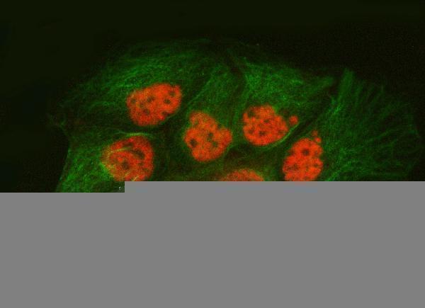
| What should IF/ICC show? | HPA reports a supported main location in the nucleoplasm, with additional supported nuclear speckles and nuclear bodies in ICC-IF (HPA subcellular ICC-IF). This provides a localisation comparison for the separate IF/ICC guide; it does not specify an IHC protocol or require those structures to resolve in tissue sections (general IHC interpretation). |
| Situation | Likely cause | Next action |
|---|---|---|
| No stain in a High HPA cell population. | The assay may have failed, or the sampled section may lack the expected cells (general IHC practice). | Confirm tissue morphology and cell identity; run a known-positive section, reagent controls and the IHC-validated antibody's stated IHC-P conditions (HPA tissue IHC: High; general IHC practice). |
| Colour appears mainly outside nuclei. | The compartment conflicts with the documented nuclear pattern; nonspecific binding or detection background is possible (UniProt P09661; HPA tissue IHC; general IHC practice). | Compare with the nuclear counterstain and reagent-omission control; score SNRPA1 only where signal tracks nuclei (general IHC practice). |
| The whole section shows diffuse brown colour. | Excess background or endogenous detection activity can obscure cellular localisation (general chromogenic IHC practice). | Inspect omission controls, blocking and wash steps, and chromogen development; reassess whether nuclear contrast remains (general IHC practice). |
| Strong stain appears in cardiomyocytes or myocytes. | HPA describes these cells as Low, although neither is established as negative (HPA tissue IHC). | Compare nuclear localisation and intensity with a High reference population; investigate cross-reactivity or endogenous detection activity if the pattern is discordant (HPA tissue IHC; general IHC practice). |
| Nuclear staining varies across the section. | Cell composition or assay variation may affect the comparison; HPA gives a tissue profile rather than a uniform-intensity rule (HPA tissue IHC; general IHC interpretation). | Compare like cell types in well-preserved areas and inspect controls before assigning a biological difference (general IHC practice). |
| Speckles or nuclear bodies are not resolved. | Those additional locations come from ICC-IF images, whereas the tissue IHC profile is reported as nuclear (HPA subcellular ICC-IF; HPA tissue IHC). | Judge the chromogenic section by convincing nuclear staining in expected cells; use the separate IF/ICC guide when subnuclear localisation is the experimental question (HPA tissue IHC; general IHC interpretation). |




Comprehensive Human Protein Atlas IHC scoring per tissue (reliability: Supported — Paired antibodies with high similarity and external characterization data supports antibody staining.). Rows are taken directly from the HPA tissue chart — click any row's HPA link to view the source.
| Tissue | Cell type | Level | Evidence | Source |
|---|---|---|---|---|
| Adrenal gland | Glandular cells | High | Protein (IHC) | HPA → |
| Appendix | Glandular cells | High | Protein (IHC) | HPA → |
| Breast | Glandular cells | High | Protein (IHC) | HPA → |
| Bronchus | Respiratory epithelial cells | High | Protein (IHC) | HPA → |
| Caudate | Neuronal cells | High | Protein (IHC) | HPA → |
| Tissue | Cell type | Level | Evidence | Source |
|---|---|---|---|---|
| None in HPA: SNRPA1 is detected in all 45 scored tissues. Use a no-primary (secondary-only) and an isotype control instead. | ||||
Use nuclear staining, matched controls and the documented paraffin-section conditions to troubleshoot SNRPA1 chromogenic IHC.
A10655-1 has IHC images from human paraffin sections and IF images from human sections, mouse brain, and Caco-2 cells; M10655 lists IHC for human, mouse, and rat (catalog image captions; applications/reactivity).
A10655-1 has illustrated IHC in human glioblastoma, larynx squamous cell carcinoma, liver cancer, and lung adenocarcinoma paraffin sections, plus IF in Caco-2 cells and human and mouse sections (A10655-1 image captions). M10655 lists IHC and human, mouse, and rat reactivity, but provides no IHC image caption in this payload (M10655 catalog applications/reactivity; image captions).
Which to pick: Choose A10655-1 for tissue IHC: its human paraffin-section captions document EDTA pH 8.0 retrieval and 2 μg/ml primary antibody; the fixative is unreported (A10655-1 IHC image captions). Choose A10655-1 for IF/ICC because those applications and corresponding images are listed; M10655 lists IHC only and is a rabbit monoclonal alternative for tissue IHC (catalog applications; M10655 clone field). For cross-species work, both list human, mouse, and rat reactivity, while A10655-1 additionally has illustrated mouse IF; the supplied IHC images are human only (catalog reactivity; A10655-1 image captions).