This website uses cookies to ensure you get the best experience on our website.


- Table of Contents
Plan SNRPD3 paraffin-section IHC around widespread nuclear staining (HPA tissue IHC), using breast glandular cells or bronchial respiratory epithelial cells as high-staining controls (HPA tissue IHC). The catalog antibody's IHC dilution is 2–5 μg/ml (datasheet: A09533-1); score nuclear staining with consistent fixation and appropriate controls (standard IHC practice).

Expected localisation, validated protocols, controls and antibodies — the at-a-glance facts below, then the full design guide.
| Expected localisation | Nuclear staining (HPA tissue IHC) | |
| Staining pattern | Widespread nuclear staining across tissue cell types (HPA tissue IHC) | |
| Antigen retrieval | EDTA pH 8.0 HIER, heat-mediated (datasheet A09533-1) | |
| Positive control | Breast+4 more · see all | |
| Negative control | None in HPA (detected in all 44 tissues); use no-primary + isotype controls |
| Fixation | Keep fixation consistent across sections (standard IHC practice; not target-specific) | |
| Caveat | Broad expression limits negative-tissue controls (HPA tissue IHC) | |
| Regulation | Low tissue specificity (HPA tissue RNA) | |
| Isoform / epitope | 2 isoforms; epitope coverage is unknown (UniProt) |
The catalog antibody’s IHC-P protocol is accompanied by a published SNRPD3 chromogenic IHC protocol for esophageal squamous cell cancer tissue microarrays (PMC9949073).
| Sample | Paraffin-embedded mouse brain tissue; fixative not specified (datasheet A09533-1) |
| Fixation | Image fixative and duration unreported (datasheet A09533-1); verify before use. |
| Sectioning | 4–5 µm sections on charged slides (standard) |
| Deparaffinisation | Xylene, graded ethanol series to water (standard) |
| Antigen retrieval | Heat retrieval: EDTA pH 8.0 (datasheet A09533-1); 20 min, 95–100 °C (standard) |
| Peroxidase block | 3% H2O2, 10 min, room temperature (standard) |
| Blocking | 10% goat serum (datasheet A09533-1) |
| Primary antibody | Rabbit anti-SNRPD3, 2-5 μg/ml (datasheet A09533-1) |
| Primary incubation | Overnight at 4 °C (datasheet A09533-1) |
| Detection | HRP-conjugated secondary, DAB chromogen (datasheet A09533-1) |
| Counterstain | Hematoxylin, blue, dehydrate and mount (standard) |
| Expected result | SNRPD3-positive staining in glandular cells of breast (HPA tissue IHC: High). HPA tissue profile: Ubiquitous nuclear expression. No signal in the no-primary control. |
SNRPD3 should show widespread nuclear staining in IHC, including strong staining in several glandular and respiratory epithelial cell populations (HPA tissue IHC: ubiquitous nuclear expression; High in listed populations). Cytosolic staining is biologically plausible because snRNP assembly begins in the cytosol before nuclear transport (UniProt P62318: subcellular location). SNRPD3 has no transmembrane segment (UniProt P62318: topology). HPA rates the tissue IHC pattern Supported, with medium consistency between staining and RNA expression (HPA tissue IHC: reliability).
| Clear nuclear signal in breast glandular cells, bronchial respiratory epithelial cells, or pancreatic exocrine glandular cells (HPA tissue IHC: High in each). | This fits the reported widespread nuclear pattern (HPA tissue IHC: ubiquitous nuclear expression). Compare nuclei with adjacent cells and background on the same section; intensity alone does not establish antibody specificity (general IHC practice). |
| Predominantly cytoplasmic staining with little or no nuclear signal in cells expected to stain (HPA tissue IHC: ubiquitous nuclear expression). | Review specificity and detection conditions before calling the pattern positive (general IHC practice). Some cytosolic signal is plausible, especially in IF, but nucleoplasm is the main supported IF location (HPA subcellular ICC-IF: nucleoplasm main; cytosol additional). |
| Strong color in structures outside the expected cellular pattern, such as pigment, debris, or tissue edges (general IHC practice). | Cross-reactivity, endogenous detection activity, or a preparation artefact may explain it (general IHC practice). SNRPD3 expression is widespread, so staining in an unlisted cell type alone does not prove an off-target result (HPA tissue IHC: ubiquitous nuclear expression). |
| Diffuse color across cells and extracellular spaces obscures nuclear boundaries (general IHC practice). | Interpret the section as high background until controls clarify the signal (general IHC practice). The reported tissue pattern is nuclear; diffuse extracellular color is not evidence of SNRPD3 localization (HPA tissue IHC: ubiquitous nuclear expression). |
| No nuclear signal in a positive reference section containing breast glandular cells or bronchial respiratory epithelial cells (HPA tissue IHC: High in both). | Check that tissue, primary antibody, detection reagents, and counterstain were processed as intended (general IHC practice). HPA's High designation provides a reference expectation, not a guarantee for every specimen or staining run (HPA tissue IHC: High; reliability Supported). |
| Tissue and cell population (HPA tissue IHC). | HPA reports High staining in breast and fallopian tube glandular cells, bronchial respiratory epithelial cells, and pancreatic exocrine glandular cells; it reports Low staining in oral squamous epithelium and salivary glandular cells (HPA tissue IHC: listed levels). Use the named cell population when comparing sections. |
| Strength of the tissue evidence (HPA tissue IHC: Supported). | The nuclear profile has medium consistency between antibody staining and RNA expression (HPA tissue IHC: reliability description). HPA lists no negative tissue in this payload, so the Low populations are weaker comparators, not established negative controls (HPA tissue IHC: Low; negative list empty). |
| Nuclear and cytosolic distribution (UniProt P62318; HPA subcellular ICC-IF). | UniProt places SNRPD3 in nucleus and cytosol and describes cytosolic snRNP assembly before nuclear transport (UniProt P62318: subcellular location). IF shows nucleoplasm as the main supported location, with nuclear bodies and cytosol additionally supported (HPA subcellular ICC-IF: locations). |
| Available antibody validation (HPA antibodies: HPA001170). | HPA labels HPA001170 Supported for both IHC and ICC (HPA antibodies: HPA001170 validation). This supports using its reported pattern as a comparison; it does not by itself establish that another antibody has the same specificity (general IHC practice). |
| IF/ICC interpretation (HPA subcellular ICC-IF). | Q: Should IF show only nuclei? A: Expect mainly nucleoplasm, while nuclear bodies and cytosol can also be seen (HPA subcellular ICC-IF: supported locations). The IF observations guide compartment interpretation here; this IHC section provides no IF protocol option. |
| Situation | Likely cause | Next action |
|---|---|---|
| The positive reference section is blank (HPA tissue IHC: High in breast glandular cells and bronchial respiratory epithelial cells). | A staining run or detection failure is possible (general IHC practice); HPA does not establish a target-specific fixation effect (HPA tissue IHC: source scope). | Verify the reference cell population and inspect run controls, primary antibody handling, and detection reagents (general IHC practice). Do not infer fixation sensitivity from the blank result alone. |
| Staining is strongest in cytoplasm and nuclei are faint (HPA tissue IHC: ubiquitous nuclear expression). | The distribution may reflect background or a specificity problem, although cytosolic SNRPD3 is plausible (general IHC practice; UniProt P62318: subcellular location). | Compare with a reported High tissue population and check the no-primary control; assess whether discrete nuclear signal remains (HPA tissue IHC: High in listed populations; general IHC practice). |
| Color appears broadly in extracellular spaces or at section edges (general IHC practice). | Nonspecific staining or detection background may obscure cellular localization (general IHC practice). | Inspect the no-primary control, blocking, washes, and detection steps, then score only interpretable cells (general IHC practice). HPA's reported pattern is widespread nuclear expression (HPA tissue IHC: profile). |
| Unexpected cells stain strongly while expected nuclei are weak (HPA tissue IHC: ubiquitous nuclear expression). | Cross-reactivity or endogenous detection activity is possible, but HPA provides no negative tissue here (general IHC practice; HPA tissue IHC: negative list empty). | Identify the stained cell type, check no-primary and detection controls, and compare a named High population before assigning the signal to SNRPD3 (general IHC practice; HPA tissue IHC: listed High populations). |
| Oral squamous epithelium looks weaker than breast glandular cells (HPA tissue IHC: Low and High, respectively). | This difference agrees with the reported staining levels and need not indicate a failed run (HPA tissue IHC: listed levels). | Score each named cell population against its reported level and the run controls (HPA tissue IHC: listed levels; general IHC practice). Do not use the Low population as a proven negative control (HPA tissue IHC: negative list empty). |
| IF shows nuclear bodies or cytosol alongside nucleoplasm (HPA subcellular ICC-IF: supported locations). | These are reported additional locations; nucleoplasm remains the main supported IF location (HPA subcellular ICC-IF: locations). | Interpret IF compartment staining using its own guide and controls (general IF practice). For this IHC section, compare chromogenic tissue staining with HPA's widespread nuclear profile (HPA tissue IHC: profile). |
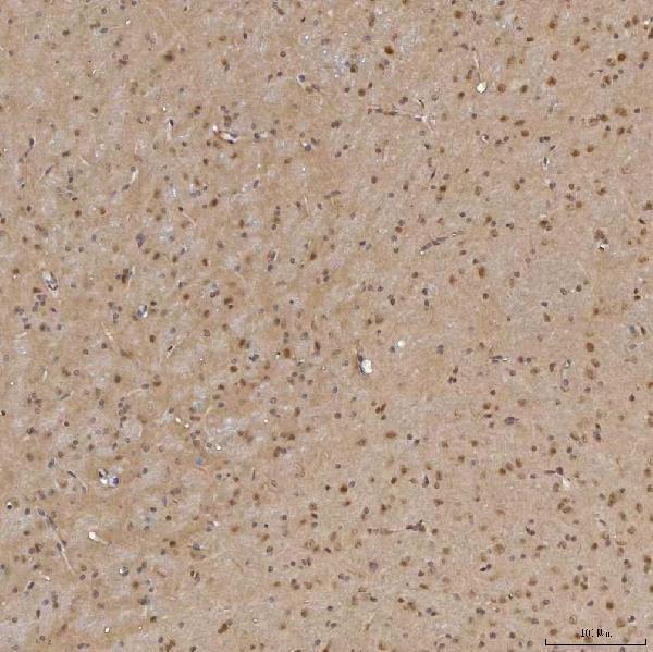
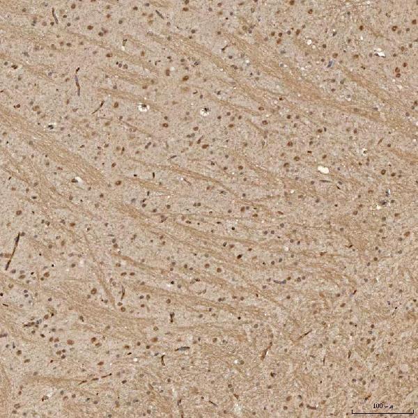
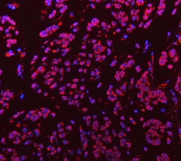


Comprehensive Human Protein Atlas IHC scoring per tissue (reliability: Supported — Medium consistency between antibody staining and RNA expression data.). Rows are taken directly from the HPA tissue chart — click any row's HPA link to view the source.
| Tissue | Cell type | Level | Evidence | Source |
|---|---|---|---|---|
| Breast | Glandular cells | High | Protein (IHC) | HPA → |
| Bronchus | Respiratory epithelial cells | High | Protein (IHC) | HPA → |
| Caudate | Glial cells | High | Protein (IHC) | HPA → |
| Cerebral cortex | Neuropil | High | Protein (IHC) | HPA → |
| Epididymis | Glandular cells | High | Protein (IHC) | HPA → |
| Tissue | Cell type | Level | Evidence | Source |
|---|---|---|---|---|
| None in HPA: SNRPD3 is detected in all 44 scored tissues. Use a no-primary (secondary-only) and an isotype control instead. | ||||
Troubleshoot chromogenic SNRPD3 IHC in paraffin sections using the catalog antibody’s tissue staining conditions and expected cellular localisation.
A09533-1 has IHC images from paraffin sections of mouse and rat brain, human breast cancer and glioblastoma, plus an IF image from human breast cancer (catalog image captions).
A09533-1 has IHC images from paraffin sections of mouse and rat brain, human breast cancer and glioblastoma (catalog IHC image captions). A09533-1 also has an IF image from a paraffin section of human breast cancer; its listed reactivity is human, mouse and rat (catalog IF image caption; catalog reactivity).
Which to pick: For tissue IHC, choose A09533-1: it is a rabbit antibody listed for IHC, with paraffin-section images using EDTA retrieval at pH 8.0 and 2 μg/ml primary antibody (catalog applications; catalog IHC image captions). For IF, A09533-1 has an image from a human breast cancer paraffin section at 5 μg/ml; ICC validation is not reported (catalog IF image caption; catalog applications). For cross-species IHC, A09533-1 lists human, mouse and rat reactivity and has paraffin-section images from all three species; the captions do not report the fixative (catalog reactivity; catalog IHC image captions).