SNX16 / Sorting nexin-16 · IHC design guide

Design Immunohistochemistry for SNX16

Plan SNX16 paraffin IHC with the catalog antibody at 2–5 μg/ml (datasheet A11003-3). Use the reported glandular and peritubular staining as tissue references, and assess cytoplasmic and nuclear signal (HPA tissue IHC).

Evidence assembled Oct 2026 · For research use; verify linked source records and product datasheet before use
Immunohistochemistry protocol sheet for SNX16 (IHC for SNX16): expected localisation Cytoplasmic and nuclear tissue staining (HPA tissue IHC), antibody A11003-3, validated IHC image, and IHC protocol steps
Printable SNX16 IHC protocol sheet — expected localisation Cytoplasmic and nuclear tissue staining (HPA tissue IHC), antibody A11003-3, controls and protocol steps. Open the full SNX16 IHC guide →

SNX16 Immunohistochemistry Experimental Design Guide

Expected localisation, validated protocols, controls and antibodies — the at-a-glance facts below, then the full design guide.

Must know before staining
Expected localisation Cytoplasmic and nuclear tissue staining (HPA tissue IHC)
Staining pattern Glandular and peritubular cells show cytoplasmic and nuclear staining (HPA tissue IHC)
Antigen retrieval EDTA pH 8.0 HIER, heat-mediated (datasheet A11003-3)
Positive control ⓘ Duodenum+4 more · see all
Negative control ⓘ None in HPA (detected in all 45 tissues); use no-primary + isotype controls
Important caveats
Reasons your staining may differ from the expected pattern.
Fixation Keep fixation conditions consistent across paraffin sections. (standard IHC practice; not target-specific)
Caveat Medium agreement between staining and RNA warrants validation (HPA tissue IHC)
Regulation Stimulus-dependent regulation is unreported (UniProt)
Isoform / epitope 2 isoforms; no transmembrane segment; epitope coverage unknown (UniProt)
Section 1

Recommended SNX16 IHC & IF Protocols

The catalog antibody’s IHC-P protocol (datasheet A11003-3) is paired with one published Snx16 IHC method (PMC3082179).

Recommended immunohistochemistry (IHC-P) protocol parameters
SampleParaffin-embedded human liver tissue; fixative not specified (datasheet A11003-3)
FixationImage fixative and duration unreported (datasheet A11003-3); verify before use.
Sectioning4–5 µm sections on charged slides (standard)
DeparaffinisationXylene, graded ethanol series to water (standard)
Antigen retrievalHeat retrieval: EDTA pH 8.0 (datasheet A11003-3); 20 min, 95–100 °C (standard)
Peroxidase block3% H2O2, 10 min, room temperature (standard)
Blocking10% goat serum (datasheet A11003-3)
Primary antibodyRabbit anti-SNX16, 2-5 μg/ml (datasheet A11003-3)
Primary incubationOvernight at 4 °C (datasheet A11003-3)
DetectionHRP-conjugated secondary, DAB chromogen (datasheet A11003-3)
CounterstainHematoxylin, blue, dehydrate and mount (standard)
Expected resultSNX16-positive staining in glandular cells of duodenum (HPA tissue IHC: High). HPA tissue profile: General cytoplasmic and nuclear expression. No signal in the no-primary control.
💡Decision noteStart with heat-mediated EDTA retrieval at pH 8.0 (datasheet A11003-3); the published method uses fluorescent secondaries (PMC3082179).
Section 2

What Is the Expected SNX16 Staining Pattern?

SNX16 is associated with early and late endosome membranes, cytoplasm, and lysosomes, with no transmembrane segment (UniProt P57768). Expect cytoplasmic staining in glandular cells of duodenum, rectum, and small intestine, and in testis peritubular cells (HPA: High). HPA also reports general cytoplasmic and nuclear tissue staining; its IHC assessment is Approved, with medium agreement between staining and RNA data and external verification pending (HPA tissue IHC).

What am I looking at on my slide?
Cytoplasmic staining in intestinal glandular cells or testis peritubular cells, with locally punctate detail.These are high-staining cell populations (HPA: High). Puncta are consistent with endosomal or lysosomal association (UniProt P57768), but tissue IHC alone does not identify individual organelles.
Strong staining restricted to nuclei, with little cytoplasmic signal.Assess cautiously: HPA reports a general cytoplasmic and nuclear IHC profile (HPA tissue IHC), while UniProt assigns SNX16 to cytoplasm and endosomal or lysosomal membranes (UniProt P57768). Nuclear staining alone cannot verify the expected trafficking-associated pattern.
Strong signal mainly in a cell population outside the documented high-staining populations.Compare its location with adjacent glandular or peritubular cells (HPA: High in those cells). Unexpected signal may reflect cross-reactivity or endogenous detection activity; HPA reports low tissue specificity, so location alone cannot establish an off-target result (HPA tissue IHC).
Diffuse chromogen across cells and surrounding tissue, without discernible cellular boundaries.This does not resolve the reported cytoplasmic and nuclear tissue pattern (HPA tissue IHC). General IHC background can arise from detection reagents, inadequate blocking, or an overly concentrated primary antibody; a no-primary control helps localise its source.
No signal in the documented high-staining cells.Treat the run as inconclusive before calling the specimen SNX16-negative: duodenal, rectal, and small-intestinal glandular cells and testis peritubular cells stain High in HPA tissue IHC. Check controls and reagent performance, then review retrieval and detection conditions as general IHC variables.
💡Expected SNX16 appearanceA convincing positive shows cytoplasmic staining, potentially with vesicular detail, in High-staining intestinal glandular or testis peritubular cells (HPA tissue IHC; UniProt P57768); diffuse, structure-free deposit is a background warning, while nuclear staining requires cautious interpretation because HPA also reports it (HPA tissue IHC).
How each factor affects the staining
Tissue and cell selectionDuodenum, rectum, and small intestine glandular cells and testis peritubular cells are documented High examples (HPA tissue IHC). HPA lists low-staining populations but no negative tissue control; weak signal there does not establish assay failure.
Compartment evidenceEndosomal and lysosomal association supports a cytoplasmic, potentially vesicular pattern (UniProt P57768). HPA tissue IHC also describes nuclear staining; its presence should be recorded without assigning it to a defined organelle (HPA tissue IHC).
Antibody evidenceHPA024730, HPA024731, and HPA024817 have Approved IHC status (HPA antibodies). HPA024730 has Supported ICC status, but the tissue profile has medium agreement with RNA data and awaits external verification (HPA tissue IHC).
Isoforms and processingUniProt lists 2 isoforms, a single 1–344 chain, and no signal peptide or propeptide (UniProt P57768). Without an antibody epitope map, these entries cannot predict isoform coverage or justify a tissue-specific staining difference.
IF/ICC cross-checkHPA ICC-IF supports vesicles and labels plasma membrane localisation uncertain (HPA subcellular). This can inform compartment interpretation; it does not supply an IHC-P protocol or establish that plasma membrane staining is an expected tissue IHC result.
Why is my staining missing, weak or wrong?
SituationLikely causeNext action
No staining in a positive reference section.The run may have failed; HPA documents High staining in intestinal glandular and testis peritubular cells (HPA tissue IHC).Check the reference section, primary antibody, detection reagents, and chromogen. Then review retrieval as a general IHC variable; SNX16-specific fixation or retrieval sensitivity is unreported in the supplied sources.
Signal is weak but confined to glia, adipocytes, or another listed low-staining population.HPA lists low staining for caudate glia and adipose adipocytes (HPA tissue IHC); a weak result there is not an informative positive-control failure.Assess a documented high-staining cell population on a reference section (HPA tissue IHC), using the same IHC run for comparison.
Nuclei dominate the staining.The result is ambiguous because HPA reports nuclear as well as cytoplasmic tissue staining (HPA tissue IHC), while the documented trafficking compartments are cytoplasmic (UniProt P57768).Record nuclear and cytoplasmic scores separately. Check a no-primary control and compare cellular distribution in a documented high-staining tissue before interpreting the nuclear signal.
Broad tissue haze or precipitate obscures cells.General IHC causes include nonspecific reagent binding, endogenous detection activity, or chromogen deposit; this appearance cannot be assigned to SNX16 from the supplied evidence.Inspect the no-primary control and detection-only background. Review blocking, washing, chromogen development, and primary dilution as general workflow checks.
Unexpected cell types stain more strongly than documented high-staining cells.Cross-reactivity or endogenous activity is possible, but HPA reports low tissue specificity and its IHC profile awaits external verification (HPA tissue IHC).Compare cell morphology and a no-primary control; confirm the spatial pattern with an independently validated antibody if available. Do not treat an unfamiliar cell pattern alone as proof of specificity.
A continuous cell-edge signal is interpreted as the main positive pattern.HPA ICC-IF calls plasma membrane localisation uncertain and vesicles supported (HPA subcellular); UniProt places SNX16 at endosomal and lysosomal membranes (UniProt P57768).Recheck focus and cellular boundaries, then score cytoplasmic signal separately. Treat a dominant cell-edge pattern as unresolved unless corroborated by an independent specificity check.

Sample controls for SNX16 IHC & IF

🧪Run duodenum first and assess its glandular cells for SNX16 staining (HPA: High in duodenal glandular cells). HPA lists no negative tissue and detects SNX16 in all 45 scored tissues, so use no-primary and isotype controls for background; treat adjacent nonglandular cells as an internal comparison only if they stain less, not as presumed SNX16-negative cells (HPA: no negative rows; detected in all 45 tissues).
Positive control tissue: Duodenum (Glandular cells, HPA High)
Negative control tissue: None in HPA: SNX16 is detected in all 45 scored tissues. Use a no-primary (secondary-only) and an isotype control instead.
ICC-IF cell lines (HPA subcellular resource): HPA ICC-IF images show SNX16 in A-431, BJ [Human fibroblast], U2OS, KOLF2.1J, with annotated localisation: Vesicles (supported), Plasma membrane (uncertain) (HPA subcellular).
Technical controls: Include a no-primary, secondary-only control, a concentration-matched rabbit IgG isotype control, and SNX16-knockout material as a biological specificity control where available (caption: rabbit primary antibody). For duodenal chromogenic IHC, block endogenous peroxidase and check the controls for background staining (HPA: duodenal glandular cells; caption: HRP/DAB detection).
⚠️Feasibility: No target-specific fixation window or fixation effect is reported, and the selected A11003-3 paraffin-section caption does not state a fixative (caption: fixative not stated). Heat retrieval in EDTA at pH 8.0 is a documented starting condition, but retrieval dependency is unreported (caption: EDTA heat retrieval). The supplied evidence does not establish whether frozen sections or IF are easier; if using IF in duodenum, assess tissue autofluorescence with a no-primary control (HPA: duodenal glandular cells).

HPA tissue IHC evidence for SNX16

Comprehensive Human Protein Atlas IHC scoring per tissue (reliability: Approved — Medium consistency between antibody staining and RNA expression data. Pending external verification.). Rows are taken directly from the HPA tissue chart — click any row's HPA link to view the source.

Positive expression · recommended positive controls

TissueCell typeLevelEvidenceSource
Duodenum Glandular cells High Protein (IHC) HPA →
Rectum Glandular cells High Protein (IHC) HPA →
Small intestine Glandular cells High Protein (IHC) HPA →
Testis Peritubular cells High Protein (IHC) HPA →
Adrenal gland Glandular cells Medium Protein (IHC) HPA →

Undetected expression · recommended negative controls

TissueCell typeLevelEvidenceSource
None in HPA: SNX16 is detected in all 45 scored tissues. Use a no-primary (secondary-only) and an isotype control instead.
Section 3

Advanced SNX16 IHC Tips

Use the catalog antibody’s liver IHC conditions as a starting point, then check staining against SNX16’s reported cell types and compartments.

Which retrieval conditions should I try first for SNX16 paraffin sections?
Start with heat-mediated retrieval in EDTA, pH 8.0 (datasheet A11003-3). The catalog antibody’s liver section was processed this way, then incubated at 2 μg/ml overnight at 4°C (datasheet A11003-3). If staining is weak, compare a shorter or longer heating interval on adjacent sections while holding antibody concentration and detection constant; the caption does not report a retrieval duration (datasheet A11003-3). Include a section processed without primary antibody and inspect tissue edges, since uneven heating and edge staining can make a retrieval change appear successful (standard IHC practice).
How should I assess whether fixation is suppressing SNX16 staining?
Target-specific fixation sensitivity is unknown: the selected liver image describes a paraffin-embedded section but does not state its fixative or fixation duration (datasheet A11003-3). Record those details for each block, and compare sections from similarly processed tissue before changing the EDTA, pH 8.0 retrieval condition (datasheet A11003-3). If fixation differs across samples, run them in the same staining batch and include a reference section to separate processing variation from staining variation (standard IHC practice). Do not treat a stronger signal after retrieval as proof of a particular fixation effect; compare preserved morphology and background alongside the signal (standard IHC practice).
What staining pattern is plausible for SNX16 in chromogenic IHC?
Expect cytoplasmic staining with a possible punctate component: SNX16 is reported at early and late endosome membranes and lysosomes, as well as in cytoplasm (UniProt P57768 subcellular location). The protein has no transmembrane segment, so a crisp, continuous cell-surface rim alone needs careful review (UniProt P57768 topology). HPA tissue IHC describes general cytoplasmic and nuclear expression, which broadens the patterns to examine but does not establish that every nuclear signal is specific (HPA tissue IHC profile; HPA reliability: Approved, pending external verification). Compare stained cells with their neighbors and a no-primary control at the same DAB development time before assigning compartments (standard IHC practice).
Can this antibody distinguish SNX16 isoforms or a phosphorylation-dependent epitope?
SNX16 has 2 reported isoforms, and its PX domain spans residues 105–218 (UniProt P57768 isoforms and domains). Phosphoserine at residue 222 is recorded, but the supplied antibody evidence does not map its epitope or establish phosphorylation-dependent recognition (UniProt P57768 modified residues; datasheet A11003-3). Therefore, score the stain as SNX16 immunoreactivity without assigning it to an isoform or phosphorylation state; the liver caption demonstrates tissue staining under its stated conditions, not epitope identity (datasheet A11003-3). If that distinction matters, seek documented epitope information and compare validated independent reagents on adjacent sections before making an isoform-specific claim (standard IHC validation practice).
How should I plan IF follow-up to a positive SNX16 IHC result?
Use IF as a separate validation experiment: the selected antibody evidence describes paraffin-section chromogenic IHC, while HPA reports vesicular localisation in its cellular imaging (datasheet A11003-3; HPA subcellular summary). For multiplexing, pair SNX16 with a validated marker of the expected cell type, such as glandular cells when studying duodenum, and inspect whether puncta fall within those cells (HPA tissue IHC: High in duodenal glandular cells; UniProt P57768 subcellular location). Choose a fluorophore channel with low autofluorescence in the specimen and include single-stain controls (standard IF practice). Because the antibody epitope’s membrane-facing side is unreported, establish accessibility before optimizing mild permeabilisation for a cytosolic-facing epitope (datasheet A11003-3; standard IF practice).
How can I reduce diffuse or misleading DAB background?
First compare the stain with a no-primary section and review whether background concentrates at edges, damaged tissue, or pigment deposits (standard IHC practice). The selected liver protocol uses 10% goat serum, a peroxidase-conjugated secondary, and DAB; its caption does not describe an endogenous peroxidase block (datasheet A11003-3). Add an appropriate peroxidase block as a general chromogenic IHC step, then optimize blocking and washes while keeping the 2 μg/ml primary condition as the initial reference (standard IHC practice; datasheet A11003-3). If diffuse signal persists, titrate primary antibody downward on adjacent sections and judge improvement by retained cellular pattern, not by lower background alone (standard IHC practice).
How should I quantify SNX16 staining across paraffin sections? ⚠ ANSWER MARKED FOR VERIFICATION
Define the cell population and compartment before scoring, because HPA reports general cytoplasmic and nuclear tissue staining while UniProt places SNX16 in cytoplasm and endosomal or lysosomal membranes (HPA tissue IHC profile; UniProt P57768 subcellular location). Within matched regions, record an H-score from intensity and percent positive cells, or report positive-cell density per mm² when cell abundance varies (standard IHC quantification practice). Normalize counts to viable tissue area or the number of the relevant cells, and keep section thickness, imaging, and DAB development consistent (standard IHC practice). Analyze compartments separately and exclude folds, necrosis, and edge staining under a prespecified rule (standard IHC practice).
How do I distinguish credible SNX16 staining from artefact?
Give greatest weight to a reproducible cellular pattern in an expected population: HPA reports high staining in duodenal, rectal, and small-intestinal glandular cells (HPA tissue IHC). Cytoplasmic or punctate staining fits SNX16’s reported endosomal, lysosomal, and cytoplasmic locations, while an isolated sharp membrane rim warrants additional scrutiny (UniProt P57768 subcellular location and topology). Check suspicious signal against neighboring cells and a no-primary section, especially at section edges, necrotic regions, and sites of endogenous enzyme activity (standard IHC practice). HPA rates its tissue IHC data Approved with medium RNA–staining consistency and pending external verification, so confirm consequential interpretations independently (HPA tissue IHC reliability).
Boster reagents

Best SNX16 / Sorting nexin-16 IHC Antibodies

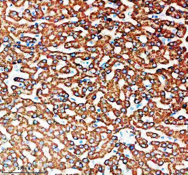
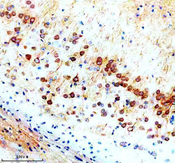
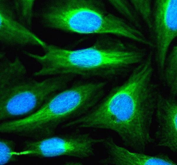
The IHC-validated anti-SNX16 antibody has paraffin-section images from human liver and mouse and rat brain (catalog: IHC image captions), plus an IF/ICC image from U2OS cells (catalog: IF image caption).

Real IHC data IHC analysis of SNX16 using anti-SNX16 antibody (A11003-3). SNX16 was detected in a paraffin-embedded section of human liver tissue. Heat mediated antigen retrieval was performed in EDTA buffer (pH 8.0, epitope retrieval solution). The tissue section was blocked with 10% goat serum. The tissue section was then incubated with 2 μg/ml rabbit anti-SNX16 Antibody (A11003-3) overnight at 4°C. Peroxidase Conjugated Goat Anti-rabbit IgG was used as secondary antibody and incubated for 30 minutes at 37°C. The tissue section was developed using HRP Conjugated Rabbit IgG Super Vision Assay Kit (Catalog # SV0002) with DAB as the chromogen.
Anti-SNX16 Antibody ®
Cat # A11003-3

A11003-3 has IHC images from paraffin-embedded human liver and mouse and rat brain sections (catalog: A11003-3 IHC image captions). It also has an IF/ICC image from U2OS cells (catalog: A11003-3 IF image caption).

Which to pick: Choose A11003-3 for paraffin-section IHC: its human liver image used EDTA retrieval at pH 8.0 and 2 μg/ml primary antibody (catalog: A11003-3 human liver IHC image caption). Choose the same SKU for IF/ICC in U2OS cells, as shown in its fluorescence image (catalog: A11003-3 IF image caption). For cross-species tissue IHC, it lists human, mouse and rat reactivity and shows IHC images for each; its clonality and the tissue fixative are unreported (catalog: A11003-3 reactivity, clone field and IHC image captions).

Each figure is that product's own IHC / IF validation image from its datasheet.

References

  1. UniProt Consortium. UniProt entry P57768 (SNX16_HUMAN, Sorting nexin-16).
  2. Human Protein Atlas. SNX16 tissue IHC expression (reliability: Approved).
  3. Human Protein Atlas. SNX16 subcellular location (ICC-IF): Localized to the plasma membrane and vesicles..
  4. Human Protein Atlas. SNX16 antibody validation summary (3 antibodies).
  5. SNX16 is required for hepatocellular carcinoma survival via modulating the EGFR-AKT signaling pathway. Scientific reports 2024 — PMC11161632.
  6. SNX16 activates c-Myc signaling by inhibiting ubiquitin-mediated proteasomal degradation of eEF1A2 in colorectal cancer development. Molecular oncology 2020 — PMC6998659.
  7. SNX16 aggravates AngII-induced cardiac hypertrophy in mice via EGFR transactivation. Communications biology 2025 — PMC12770542.
  8. A presynaptic endosomal trafficking pathway controls synaptic growth signaling. The Journal of cell biology 2011 — PMC3082179.
  9. PubMed PMID:14702039 — UniProt-cited evidence.
  10. PubMed PMID:17974005 — UniProt-cited evidence.
  11. PubMed PMID:16421571 — UniProt-cited evidence.