This website uses cookies to ensure you get the best experience on our website.


- Table of Contents
Plan SNX4 staining in paraffin sections using the IHC-validated antibody, with tissue controls and chromogenic detection. Compare cytoplasmic and nuclear staining with the tissue profile while accounting for its uncertain reliability (HPA tissue IHC).

Expected localisation, validated protocols, controls and antibodies — the at-a-glance facts below, then the full design guide.
| Expected localisation | Tissue: cytoplasmic/nuclear (HPA tissue IHC); molecular: early endosome/ERC (UniProt) | |
| Staining pattern | Cytoplasmic and nuclear staining in several tissues (HPA tissue IHC) | |
| Antigen retrieval | EDTA pH 8.0 HIER, heat-mediated (datasheet A08783-2) | |
| Positive control | Placenta+4 more · see all | |
| Negative control | Esophagus+4 more · see all |
| Fixation | Keep fixation consistent across sections. (standard IHC practice; not target-specific) | |
| Caveat | Tissue staining has low concordance with RNA expression (HPA tissue IHC) | |
| Regulation | Expression regulation is unannotated (UniProt) | |
| Isoform / epitope | 2 isoforms; epitope coverage is unspecified (UniProt) |
The catalog antibody uses heat-mediated EDTA pH 8.0 retrieval (datasheet A08783-2). The published paraffin-section brain protocol below uses fluorescent detection (PMC5251330).
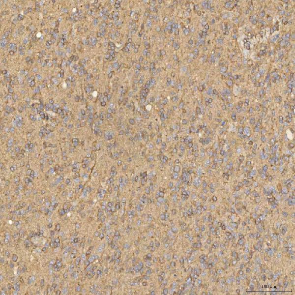
| Sample | Paraffin-embedded human glioblastoma tissue; fixative not specified (datasheet A08783-2) |
| Fixation | Image fixative and duration unreported (datasheet A08783-2); verify before use. |
| Sectioning | 4–5 µm sections on charged slides (standard) |
| Deparaffinisation | Xylene, graded ethanol series to water (standard) |
| Antigen retrieval | Heat retrieval: EDTA pH 8.0 (datasheet A08783-2); 20 min, 95–100 °C (standard) |
| Peroxidase block | 3% H2O2, 10 min, room temperature (standard) |
| Blocking | 10% goat serum (datasheet A08783-2) |
| Primary antibody | Rabbit anti-SNX4, 2-5 μg/ml (datasheet A08783-2) |
| Primary incubation | Overnight at 4 °C (datasheet A08783-2) |
| Detection | HRP-conjugated secondary, DAB chromogen (datasheet A08783-2) |
| Counterstain | Hematoxylin, blue, dehydrate and mount (standard) |
| Expected result | SNX4-positive staining in trophoblastic cells of placenta (HPA tissue IHC: High). HPA tissue profile: Cytoplasmic and nuclear expression in several tissues. No signal in the no-primary control. |
SNX4 is associated with the early endosome membrane and a juxtanuclear endocytic recycling compartment; it has no transmembrane segment (UniProt O95219 topology and subcellular location). In paraffin-section IHC, prioritize cytoplasmic staining in placental trophoblastic cells or splenic red-pulp cells, both reported as High (HPA tissue IHC). HPA also reports nuclear staining in several tissues, but rates its tissue IHC evidence Uncertain because staining and RNA expression have low consistency (HPA tissue IHC).
| Cytoplasmic staining is strongest in placental trophoblastic cells or splenic red-pulp cells (HPA tissue IHC: High in both). | This is the most useful reported positive-tissue pattern, although its specificity remains unverified (HPA tissue IHC: Uncertain). A granular or juxtanuclear emphasis would fit SNX4's annotated endosomal locations; routine chromogenic IHC may not resolve individual endosomes (UniProt O95219 subcellular location; general IHC practice). |
| Signal is predominantly nuclear, with little cytoplasmic staining (HPA tissue IHC: cytoplasmic and nuclear expression reported in several tissues). | Do not classify every nuclear signal as an artefact: HPA reports it, while UniProt places SNX4 at endosomal membranes and a juxtanuclear recycling compartment (HPA tissue IHC; UniProt O95219 subcellular location). Predominantly nuclear staining therefore needs independent specificity checks, especially given HPA's Uncertain reliability rating (HPA tissue IHC). |
| Strong staining appears in esophageal squamous epithelium or heart cardiomyocytes (HPA tissue IHC: Not detected in these cells). | That distribution conflicts with the reported tissue IHC pattern; investigate antibody cross-reactivity, endogenous chromogenic activity, or nonspecific detection before scoring it as SNX4 (HPA tissue IHC; general IHC practice). A reported Not detected result is a comparison control, not proof that every specimen or assay must be negative (HPA tissue IHC: Uncertain). |
| Broad, fairly uniform color covers tissue and the surrounding section rather than defined cells. | Interpret this as background until controls establish otherwise (general IHC practice). Examine a no-primary control for detection-system signal, then review blocking, washes, antibody concentration, and chromogen development (general IHC practice). The distribution alone cannot establish an SNX4-specific signal, particularly with an Uncertain HPA tissue IHC profile (HPA tissue IHC). |
| Placental trophoblastic cells or splenic red-pulp cells show no detectable staining (HPA tissue IHC: High in both). | First check that the section contains the expected cells and that a same-run positive control developed (general IHC practice). Review the IHC-validated antibody's documented dilution and retrieval conditions if available; none are supplied here. An absent signal may reflect assay performance, while HPA's Uncertain rating limits how firmly these tissues can define a required positive result (HPA tissue IHC). |
| Compartment and topology | SNX4 is annotated at early endosome membranes and a juxtanuclear recycling compartment, with no transmembrane segment (UniProt O95219 topology and subcellular location). Judge cytoplasmic localization in cells, not membrane-outline staining alone; chromogenic resolution may blur small puncta (general IHC practice). |
| Tissue distribution and confidence | Placental trophoblastic cells and splenic red-pulp cells are High, while several other cell populations are Medium, Low, or Not detected (HPA tissue IHC). HPA labels the tissue IHC profile Uncertain because antibody staining and RNA data have low consistency, so use these contrasts as provisional guides (HPA tissue IHC). |
| Antibody validation | The listed antibody, HPA005709, has an Uncertain IHC validation status (HPA antibodies). Interpret unexpected compartments or cell types with controls and, when feasible, independent confirmation (general IHC practice). The supplied record gives no epitope, dilution, retrieval condition, or catalog-antibody protocol to specify here. |
| Isoform coverage | UniProt lists two SNX4 isoforms (UniProt O95219 isoforms). With no epitope information in the supplied record, coverage of both isoforms cannot be inferred; a negative stain should therefore be interpreted alongside antibody documentation and assay controls (general IHC practice). |
| Situation | Likely cause | Next action |
|---|---|---|
| Expected positive tissue is blank (HPA tissue IHC: High in placental trophoblastic cells and splenic red-pulp cells). | The relevant cells may be absent from the section, or the staining run may have failed (general IHC practice). | Verify morphology and a same-run positive control; then check the antibody's documented IHC dilution, retrieval, and detection steps if those instructions are available (general IHC practice). |
| Signal is mainly nuclear (HPA tissue IHC: nuclear expression reported in several tissues). | The distribution is less concordant with annotated endosomal localization, though nuclear IHC staining has been reported (UniProt O95219 subcellular location; HPA tissue IHC). | Compare nuclear and cytoplasmic staining in reported positive cells, review no-primary controls, and seek independent confirmation before assigning nuclear signal to SNX4 (HPA tissue IHC: Uncertain; general IHC practice). |
| Reported Not detected cells stain strongly, such as esophageal squamous epithelium (HPA tissue IHC). | Possible antibody cross-reactivity, nonspecific binding, or endogenous detection activity (general IHC practice). | Inspect a no-primary control and assess blocking and detection chemistry; compare cell-level distribution with a reported positive tissue in the same run (HPA tissue IHC; general IHC practice). |
| Color is diffuse or persists outside cells. | Excess background can follow nonspecific detection or overly strong development (general IHC practice). | Compare no-primary and tissue controls, then adjust blocking, washes, antibody concentration, or chromogen development according to the assay instructions (general IHC practice). Do not score diffuse color as a localized SNX4 result. |
| Staining varies across tissues or between runs. | HPA reports low consistency between antibody staining and RNA expression and rates tissue IHC reliability Uncertain; technical variation may also contribute (HPA tissue IHC; general IHC practice). | Score the same cell types with matched controls and consistent processing, recording compartment, intensity, and background separately; avoid treating a single discordant section as definitive (general IHC practice). |
| IF/ICC: should SNX4 form visible endosomal puncta? | Endosomal and juxtanuclear localization is annotated, but HPA supplies no ICC-IF images or confirmed main IF location for this record (UniProt O95219 subcellular location; HPA subcellular). | Treat puncta as a localization hypothesis, not a validated IF pattern; assess antibody specificity and compare with an endosomal marker if planning the separate IF/ICC experiment (UniProt O95219 subcellular location; HPA subcellular; general IF practice). |
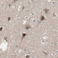

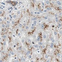
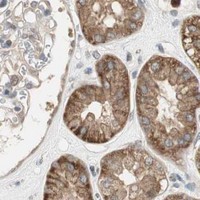
Comprehensive Human Protein Atlas IHC scoring per tissue (reliability: Uncertain — Low consistency between antibody staining and RNA expression data. Pending external verification.). Rows are taken directly from the HPA tissue chart — click any row's HPA link to view the source.
| Tissue | Cell type | Level | Evidence | Source |
|---|---|---|---|---|
| Placenta | Trophoblastic cells | High | Protein (IHC) | HPA → |
| Spleen | Cells in red pulp | High | Protein (IHC) | HPA → |
| Adrenal gland | Glandular cells | Medium | Protein (IHC) | HPA → |
| Bone marrow | Hematopoietic cells | Medium | Protein (IHC) | HPA → |
| Bronchus | Respiratory epithelial cells | Medium | Protein (IHC) | HPA → |
| Tissue | Cell type | Level | Evidence | Source |
|---|---|---|---|---|
| Esophagus | Squamous epithelial cells | Not detected | Protein (IHC) | HPA → |
| Heart muscle | Cardiomyocytes | Not detected | Protein (IHC) | HPA → |
| Lymph node | Germinal center cells | Not detected | Protein (IHC) | HPA → |
| Oral mucosa | Squamous epithelial cells | Not detected | Protein (IHC) | HPA → |
| Ovary | Ovarian stroma cells | Not detected | Protein (IHC) | HPA → |
Troubleshoot SNX4 staining in paraffin sections by checking retrieval, compartment, controls and scoring before interpreting chromogenic signal.
A08783-2 has real IHC data from human paraffin sections; no IF/ICC data are supplied (catalog IHC captions; catalog applications and images).
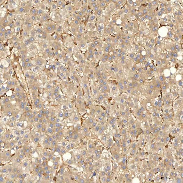
A08783-2 has IHC images from human paraffin sections of glioblastoma, liver cancer, thyroid papillary carcinoma, and colon adenocarcinoma (catalog IHC captions). Human, mouse, and rat reactivity is listed, while the IHC dilution applies to human samples (catalog reactivity and dilution).
Which to pick: Choose A08783-2 for human paraffin-section chromogenic IHC: its own glioblastoma image shows EDTA pH 8 retrieval, 2 μg/ml rabbit primary antibody, and HRP/DAB detection; the fixative is unreported (A08783-2 IHC caption). No IF/ICC-validated SKU is provided, and clonality is unreported (catalog applications, images, and clone field). A08783-2 lists mouse and rat reactivity, but the supplied paraffin-section IHC images and 2–5 μg/ml IHC recommendation cover human samples (catalog reactivity and dilution; A08783-2 IHC captions).