This website uses cookies to ensure you get the best experience on our website.


- Table of Contents
Plan SNX6 IHC on paraffin sections using the catalog antibody at 2–5 μg/ml (datasheet A06545). Assess cytoplasmic staining in glandular cells and kidney tubules, bearing in mind the low consistency between antibody staining and RNA expression (HPA tissue IHC).

Expected localisation, validated protocols, controls and antibodies — the at-a-glance facts below, then the full design guide.
| Expected localisation | Cytoplasmic staining in several tissues (HPA tissue IHC) | |
| Staining pattern | Cytoplasmic staining in glandular and kidney tubule cells (HPA tissue IHC) | |
| Antigen retrieval | EDTA pH 8.0 HIER, heat-mediated (datasheet A06545) | |
| Positive control | Breast+4 more · see all | |
| Negative control | Adipose tissue+4 more · see all |
| Fixation | Matched tissue-IHC evidence does not establish the fixation claim. Validate the specimen-specific method before use. (selected-SKU IHC image A06545); target-specific effects are unknown. | |
| Caveat | IHC staining has low consistency with tissue RNA expression (HPA tissue IHC) | |
| Regulation | Tissue expression regulation is not annotated (UniProt) | |
| Isoform / epitope | 2 isoforms; residue 1 is absent in a processed chain; epitope unknown (UniProt) |
The catalog antibody provides the IHC-P starting protocol (datasheet A06545); the published option below describes SNX6 staining in mouse brain sections (PMC5323044).
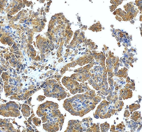
| Sample | Paraffin-embedded human ovarian cancer tissue; fixative not specified (datasheet A06545) |
| Fixation | Image fixative and duration unreported (datasheet A06545); verify before use. |
| Sectioning | 4–5 µm sections on charged slides (standard) |
| Deparaffinisation | Xylene, graded ethanol series to water (standard) |
| Antigen retrieval | Heat retrieval: EDTA pH 8.0 (datasheet A06545); 20 min, 95–100 °C (standard) |
| Peroxidase block | 3% H2O2, 10 min, room temperature (standard) |
| Blocking | 10% goat serum (datasheet A06545) |
| Primary antibody | Rabbit anti-SNX6, 2-5 μg/ml (datasheet A06545) |
| Primary incubation | Overnight at 4 °C (datasheet A06545) |
| Detection | HRP-conjugated secondary, DAB chromogen (datasheet A06545) |
| Counterstain | Hematoxylin, blue, dehydrate and mount (standard) |
| Expected result | SNX6-positive staining in glandular cells of breast (HPA tissue IHC: Medium). HPA tissue profile: Cytoplasmic expression in several tissues. No signal in the no-primary control. |
SNX6 should give mainly cytoplasmic staining in several tissues (HPA: tissue IHC). Early endosomes, their membranes and cytoplasmic vesicles are expected sites; SNX6 has no transmembrane segment, and only a minor proportion is nuclear (UniProt Q9UNH7: localization and topology). Medium staining is reported in breast, colon, stomach and small-intestine glandular cells, kidney tubule cells and skeletal-muscle myocytes (HPA: tissue IHC). The tissue profile is Approved, with low consistency between antibody staining and RNA expression (HPA: tissue IHC).
| Cytoplasmic chromogen in the expected cells, with discrete granular or punctate areas. | This fits the reported IHC profile and endosomal or vesicular localization (HPA: tissue IHC; UniProt Q9UNH7). Granules may be hard to resolve in a paraffin section; score the cellular compartment and the appropriate cell population together. |
| Staining is predominantly nuclear, uniformly surface-membranous, or confined to extracellular material. | Reassess specificity and slide interpretation: only a minor nuclear pool is reported, and SNX6 has no transmembrane segment (UniProt Q9UNH7: localization and topology). A little nuclear staining alone is therefore less concerning than nuclear dominance. |
| Strong staining appears in an HPA-listed undetected cell population. | For example, adipocytes are reported as not detected (HPA: tissue IHC). Check cell identity, antibody specificity and endogenous chromogenic activity before calling the signal SNX6. An HPA negative is a comparator, not proof that every specimen must be negative. |
| Color spreads across tissue structures or appears where no cell is identifiable. | Treat this as background until controls and cellular localization support a specific result. Excess detection signal, insufficient blocking or residual endogenous activity are general IHC possibilities; diffuse color alone does not match the reported cytoplasmic profile (HPA: tissue IHC). |
| No cytoplasmic signal is visible in a selected HPA medium-staining population. | A failed run, inadequate detection or unsuitable staining conditions are possible. Recheck the assay with the listed cell population and controls before interpreting absence of SNX6; HPA reports medium staining in several such populations, with low staining-to-RNA consistency (HPA: tissue IHC). |
| Tissue and cell selection | Breast, colon, stomach and small-intestine glandular cells, kidney tubule cells and skeletal-muscle myocytes are medium examples (HPA: tissue IHC). Lung macrophages are low, so they offer a less clear visual comparator (HPA: tissue IHC). |
| Localization at section resolution | SNX6 is associated with early endosomes, their membranes and cytoplasmic vesicles (UniProt Q9UNH7). HPA describes tissue staining as cytoplasmic (HPA: tissue IHC). Do not require individual endosomes to be resolved in chromogenic IHC. |
| Antibody evidence and limits | HPA049374 is Approved for IHC (HPA: antibodies). The tissue profile is also Approved but has low consistency between antibody staining and RNA expression (HPA: tissue IHC). Use compartment, cell type and controls together when interpreting a result. |
| Isoforms and epitope coverage | Two isoforms and an N-terminally processed chain beginning at residue 2 are recorded (UniProt Q9UNH7). No antibody epitope is supplied, so this record cannot establish which forms contribute to the observed stain or predict an epitope-specific processing effect. |
| IF/ICC Q&A: What pattern is expected? | Supported endosomal and lysosomal localization is reported in ICC-IF (HPA: subcellular). HPA049374 is Supported for ICC (HPA: antibodies). This answers the localization question; the primary application here is tissue IHC. |
| Fixation sensitivity | Target-specific fixation sensitivity is unreported in the supplied UniProt and HPA records. Antigen-retrieval choices are general IHC assay decisions; these records do not show that a particular retrieval condition changes SNX6 staining. |
| Situation | Likely cause | Next action |
|---|---|---|
| A listed medium-staining population has no visible signal. | The run or detection may have failed, or the tested section may differ from the HPA example; medium staining is reported for the listed populations (HPA: tissue IHC). | Check a run control, tissue integrity and reagent steps; repeat with an HPA-listed medium-staining cell population before concluding that SNX6 is absent. |
| Color is diffuse across cells and surrounding tissue. | Nonspecific detection, excess chromogen or inadequate blocking are general IHC possibilities. The reported tissue profile is cytoplasmic (HPA: tissue IHC). | Review a no-primary control, blocking and detection conditions; score only cell-associated staining with a plausible cytoplasmic distribution. |
| Nuclei dominate the stain. | A minor nuclear proportion is documented, but nuclear dominance is not the expected distribution (UniProt Q9UNH7: localization). | Compare with the cytoplasmic signal and controls; review antibody concentration and detection conditions before accepting the nuclear result. |
| Unexpected cells stain strongly, including an HPA-listed undetected population. | Cross-reactivity or endogenous chromogenic activity is possible; HPA reports adipocytes as not detected (HPA: tissue IHC). | Verify cell identity and examine a no-primary control; compare an expected medium-staining population in the same run before assigning specificity. |
| Only weak staining appears in a proposed positive comparator. | The chosen population may normally stain weakly: lung macrophages are Low, whereas several glandular populations are Medium (HPA: tissue IHC). | Use an HPA-listed medium-staining population as the visual comparator and confirm that the weak signal is cellular and cytoplasmic. |
| A membrane rim is scored as the sole positive pattern. | SNX6 associates with early-endosome membranes but has no transmembrane segment (UniProt Q9UNH7: localization and topology). A surface rim alone does not establish the reported cytoplasmic tissue pattern (HPA: tissue IHC). | Inspect cell borders and cytoplasm at higher magnification, then compare controls and an expected positive population before accepting the rim as specific. |


Comprehensive Human Protein Atlas IHC scoring per tissue (reliability: Approved — Low consistency between antibody staining and RNA expression data.). Rows are taken directly from the HPA tissue chart — click any row's HPA link to view the source.
| Tissue | Cell type | Level | Evidence | Source |
|---|---|---|---|---|
| Breast | Glandular cells | Medium | Protein (IHC) | HPA → |
| Colon | Glandular cells | Medium | Protein (IHC) | HPA → |
| Kidney | Cells in tubules | Medium | Protein (IHC) | HPA → |
| Skeletal muscle | Myocytes | Medium | Protein (IHC) | HPA → |
| Small intestine | Glandular cells | Medium | Protein (IHC) | HPA → |
| Tissue | Cell type | Level | Evidence | Source |
|---|---|---|---|---|
| Adipose tissue | Adipocytes | Not detected | Protein (IHC) | HPA → |
| Adrenal gland | Glandular cells | Not detected | Protein (IHC) | HPA → |
| Bone marrow | Hematopoietic cells | Not detected | Protein (IHC) | HPA → |
| Bronchus | Basal cells | Not detected | Protein (IHC) | HPA → |
| Caudate | Glial cells | Not detected | Protein (IHC) | HPA → |
Use the paraffin-section staining evidence for A06545 as the starting point, then judge signal against SNX6 localisation and tissue-level controls.
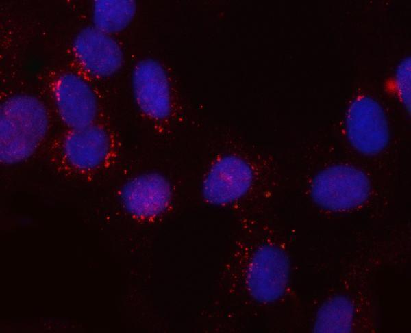
The IHC-validated antibody A06545 has IHC data from human paraffin-embedded ovarian cancer tissue and IF/ICC data from U20S cells (catalog IHC/IF captions).
A06545 will render with an IHC figure from a human paraffin-embedded ovarian cancer section (catalog IHC caption). It also has an IF/ICC figure from U20S cells; the catalog lists human, mouse, and rat reactivity (catalog IF caption; catalog reactivity).
Which to pick: Choose A06545 for paraffin-section IHC: its IHC caption shows EDTA pH 8.0 retrieval and 2 μg/ml primary antibody in human ovarian cancer tissue; the fixative is unreported (catalog IHC caption). The same SKU is listed for IF/ICC and has a U20S-cell IF figure (catalog applications; catalog IF caption). For mouse or rat samples, A06545 is the listed option based on reactivity, but those species are not shown in its IHC or IF captions; clonality is unreported (catalog reactivity; catalog IHC/IF captions; catalog clone field).