SON / Protein SON · IHC design guide

Design Immunohistochemistry for SON

Plan chromogenic SON IHC around the granular nuclear pattern seen in tissue (HPA tissue IHC). The catalog antibody's paraffin-section range is 2–5 μg/ml (datasheet A00288-1); esophageal squamous cells show high staining, while adipocytes are not detected (HPA tissue IHC).

Evidence assembled Oct 2026 · For research use; verify linked source records and product datasheet before use
Immunohistochemistry protocol sheet for SON (IHC for SON): expected localisation Granular nuclear staining in tissue (HPA tissue IHC), antibody A00288-1, validated IHC image, and IHC protocol steps
Printable SON IHC protocol sheet — expected localisation Granular nuclear staining in tissue (HPA tissue IHC), antibody A00288-1, controls and protocol steps. Open the full SON IHC guide →

SON Immunohistochemistry Experimental Design Guide

Expected localisation, validated protocols, controls and antibodies — the at-a-glance facts below, then the full design guide.

Must know before staining
Expected localisation Granular nuclear staining in tissue (HPA tissue IHC)
Staining pattern Widespread granular staining in nuclei (HPA tissue IHC)
Antigen retrieval EDTA pH 8.0 HIER, heat-mediated (datasheet A00288-1)
Positive control ⓘ Adrenal gland+4 more · see all
Negative control ⓘ Adipose tissue
Important caveats
Reasons your staining may differ from the expected pattern.
Fixation Keep fixation consistent across paraffin sections (standard IHC practice; not target-specific)
Caveat Adipocytes may be unstained despite widespread nuclear staining (HPA tissue IHC)
Regulation Leukocyte and heart expression is higher (UniProt)
Isoform / epitope 10 isoforms; check epitope coverage (UniProt)
Section 1

Recommended SON IHC & IF Protocols

The catalog antibody’s IHC-P protocol (datasheet A00288-1) is accompanied by one published Protein SON protocol using pancreatic tumor sections (PMC3576306).

Recommended immunohistochemistry (IHC-P) protocol parameters
SampleParaffin-embedded human breast cancer tissue; fixative not specified (datasheet A00288-1)
FixationImage fixative and duration unreported (datasheet A00288-1); verify before use.
Sectioning4–5 µm sections on charged slides (standard)
DeparaffinisationXylene, graded ethanol series to water (standard)
Antigen retrievalHeat retrieval: EDTA pH 8.0 (datasheet A00288-1); 20 min, 95–100 °C (standard)
Peroxidase block3% H2O2, 10 min, room temperature (standard)
Blocking10% goat serum (datasheet A00288-1)
Primary antibodyRabbit anti-SON, 2-5 μg/ml (datasheet A00288-1)
Primary incubationOvernight at 4 °C (datasheet A00288-1)
DetectionHRP-conjugated secondary, DAB chromogen (datasheet A00288-1)
CounterstainHematoxylin, blue, dehydrate and mount (standard)
Expected resultSON-positive staining in glandular cells of adrenal gland (HPA tissue IHC: High). HPA tissue profile: Ubiquitous nuclear expression with a granular pattern. No signal in the no-primary control.
💡Decision noteStart with heat-mediated EDTA pH 8.0 retrieval (datasheet A00288-1); the published protocol does not specify retrieval (PMC3576306).
Section 2

What Is the Expected SON Staining Pattern?

SON should show granular nuclear staining in many cell types, including glandular, epithelial, glial and neuronal cells (HPA: ubiquitous nuclear expression; High in the listed cells). Nuclear speckles are the expected compartment (UniProt P18583: nucleus speckle). SON has no transmembrane segment (UniProt P18583 topology). Treat intensity as tissue and cell dependent: HPA rates its tissue IHC evidence Supported, with medium consistency between staining and RNA expression (HPA: tissue IHC reliability).

What am I looking at on my slide?
Granular nuclear staining in appendix glandular cells or hippocampal neurons (HPA: High in both).This fits the reported cell distribution and speckled nuclear profile (HPA: tissue IHC profile; UniProt P18583: nucleus speckle). Compare nuclei within the same section; a granular pattern matters as well as signal strength.
Predominantly cytoplasmic or cell-surface chromogen, with little nuclear signal.This conflicts with SON's reported nuclear-speckle location and lack of a transmembrane segment (UniProt P18583). Check staining specificity and slide controls before scoring the signal as SON; chromogen outside nuclei alone is insufficient evidence.
Strong staining confined to adipocytes while nearby expected-positive cells lack nuclear signal.HPA reports SON as Not detected in adipocytes (HPA: adipose tissue IHC). Consider off-target staining or endogenous detection activity; neither can be identified from appearance alone (general IHC practice).
Uniform haze across nuclei, cytoplasm and extracellular spaces.The distribution obscures the expected granular nuclear pattern (HPA: tissue IHC profile). Assess no-primary and detection controls for nonspecific background, and judge positivity only where nuclear detail can be resolved (general IHC practice).
No nuclear signal in a section containing expected-positive appendix glandular cells.An absent signal conflicts with the reported High staining in those cells (HPA: appendix IHC), but does not establish SON absence. Review section quality, assay controls and detection steps before interpreting a negative result (general IHC practice).
💡Expected SON appearanceCall a result positive when identifiable glandular, epithelial, glial or neuronal cells show clear granular nuclear staining, often High in the listed HPA examples; diffuse cytoplasmic, surface or extracellular color without that nuclear pattern is suspect (HPA: tissue IHC; UniProt P18583: nucleus speckle).
How each factor affects the staining
Cell and tissue context (HPA: tissue IHC).HPA reports High staining in selected glandular, epithelial, glial and neuronal cells, Low in smooth muscle cells and chondrocytes, and Not detected in adipocytes. Its overall description is ubiquitous nuclear expression; use the named cell type when comparing intensity.
Compartment and texture (HPA: tissue IHC; UniProt P18583).The reported IHC profile is granular and nuclear, consistent with SON at nuclear speckles (HPA: tissue IHC; UniProt P18583: nucleus speckle). A uniformly filled nucleus is less characteristic; morphology and controls are needed before calling it artefact.
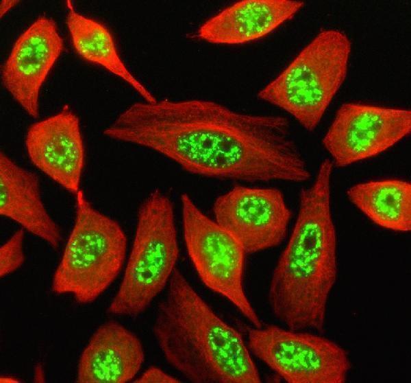
Antibody and IF/ICC evidence (HPA: antibody validation; subcellular).HPA023535 has Supported IHC status; its ICC status is Enhanced (HPA: antibody validation). Q: What should IF/ICC show? A: Nuclear speckles; HPA reports an enhanced speckle location and lists A-431, U-251MG and U2OS images (HPA: subcellular).
Isoforms and epitope interpretation (UniProt P18583).UniProt lists 10 SON isoforms, F through J (UniProt P18583: isoforms). If staining differs between samples, the supplied evidence cannot assign that difference to an isoform: the antibody epitope and isoform recognition are unspecified.
Protein architecture (UniProt P18583).SON has no transmembrane segment or signal peptide, and the listed protein chain spans residues 2–2426 (UniProt P18583: topology and processing). These annotations support a nuclear interpretation; they supply no evidence for a membrane or shed-protein staining pattern.
Why is my staining missing, weak or wrong?
SituationLikely causeNext action
Expected-positive cells lack nuclear staining.Possible assay failure or weak detection; HPA reports High staining in appendix glandular cells (HPA: appendix IHC). A negative slide alone cannot distinguish technical failure from sample variation.Confirm the expected cell type is present; compare a documented positive section and inspect primary-antibody, detection and counterstain steps (general IHC practice). Record any remaining discordance with the HPA reference.
Nuclei are faint and granules cannot be resolved.Low contrast or excessive counterstain may hide nuclear detail (general IHC practice). HPA's expected pattern is granular nuclear staining (HPA: tissue IHC profile).Inspect an appropriate positive control and adjust counterstain or detection conditions within the assay's established limits (general IHC practice). Score only nuclei whose signal and morphology remain distinguishable.
Widespread diffuse brown background masks cell boundaries.Nonspecific binding, incomplete blocking or detection background are possibilities (general IHC practice); HPA's reported SON signal is granular and nuclear (HPA: tissue IHC profile).Compare no-primary and detection controls, then review blocking, washing and chromogen development (general IHC practice). Do not count diffuse haze as SON-positive cells.
Cytoplasm or tissue edges stain more strongly than nuclei.The dominant location conflicts with nuclear speckles (UniProt P18583; HPA: subcellular). Edge-associated color can also complicate chromogenic interpretation (general IHC practice).Check whether intact internal cells retain granular nuclear signal; inspect controls for nonspecific color and review the detection workflow before assigning SON positivity (general IHC practice).
Adipocytes appear strongly positive.This differs from HPA's Not detected adipocyte result (HPA: adipose tissue IHC). Off-target binding or endogenous detection activity is possible, but appearance does not identify which (general IHC practice).Compare adipocytes with expected-positive cells on the section and with no-primary and detection controls (general IHC practice). Report the discrepancy rather than treating adipocytes as a validated positive reference.
Staining intensity varies between cell types in one section.Variation may reflect the reported cell distribution: HPA lists High staining in several cell types and Low staining in smooth muscle cells and chondrocytes (HPA: tissue IHC).Identify each cell type before scoring, compare its nuclear pattern with the matching HPA observation, and retain HPA's Supported, medium-consistency reliability caveat (HPA: tissue IHC).

Sample controls for SON IHC & IF

🧪Run adrenal gland first and look for SON staining in glandular cells (HPA: High in adrenal gland glandular cells); use adipose tissue adipocytes as the negative tissue (HPA: Not detected in adipocytes). Within the adrenal section, non-glandular stromal cells that show only background signal may serve as internal comparators, but they are not established SON-negative cells (UniProt: widely expressed).
Positive control tissue: Adrenal gland (Glandular cells, HPA High)
Negative control tissue: Adipose tissue (HPA Not detected)
ICC-IF cell lines (HPA subcellular resource): HPA ICC-IF images show SON in A-431, U-251MG, U2OS, with annotated localisation: Nuclear speckles (enhanced) (HPA subcellular).
Technical controls: Include a no-primary, secondary-only slide and a concentration-matched rabbit isotype control (selected-SKU IHC caption: rabbit primary antibody); confirm target specificity with SON knockout material or peptide blocking if the immunizing peptide is available (standard IHC practice). For adrenal DAB sections, quench endogenous peroxidase and check intrinsic brown pigment against the no-primary slide (standard IHC practice).
⚠️Feasibility: A target-specific fixation window or fixation effect is unreported, and the selected A00288-1 paraffin-section caption does not state a fixative (selected-SKU IHC caption: fixative not stated). Heat retrieval in EDTA at pH 8.0 was used, but a requirement for retrieval was not established (selected-SKU IHC caption: EDTA retrieval). The supplied evidence does not establish whether frozen sections or IF are easier; ICC-IF provides a nuclear-speckle readout, while intrinsic adrenal pigment should be checked when interpreting chromogenic staining (HPA subcellular: nuclear speckles; standard IHC practice).

HPA tissue IHC evidence for SON

Comprehensive Human Protein Atlas IHC scoring per tissue (reliability: Supported — Medium consistency between antibody staining and RNA expression data.). Rows are taken directly from the HPA tissue chart — click any row's HPA link to view the source.

Positive expression · recommended positive controls

TissueCell typeLevelEvidenceSource
Adrenal gland Glandular cells High Protein (IHC) HPA →
Appendix Glandular cells High Protein (IHC) HPA →
Cerebral cortex Glial cells High Protein (IHC) HPA →
Cervix Glandular cells High Protein (IHC) HPA →
Esophagus Squamous epithelial cells High Protein (IHC) HPA →

Undetected expression · recommended negative controls

TissueCell typeLevelEvidenceSource
Adipose tissue Adipocytes Not detected Protein (IHC) HPA →
Section 3

Advanced SON IHC Tips

Troubleshoot SON staining by checking nuclear speckle localisation, tissue context, and the documented paraffin-section workflow (UniProt P18583; HPA tissue IHC; datasheet A00288-1).

What should I try when SON staining is weak after antigen retrieval?
Start with heat-mediated retrieval in EDTA at pH 8.0 for paraffin sections (datasheet A00288-1). Keep section thickness, heating, cooling, and staining conditions consistent while comparing retrieval runs; assess whether nuclear granular staining improves without a matching rise in diffuse background (standard IHC practice; HPA tissue IHC). The documented breast cancer section was incubated with 2 μg/ml catalog antibody overnight at 4°C after retrieval, giving a defined starting point for troubleshooting (datasheet A00288-1). If staining remains weak, test an alternative retrieval buffer on adjacent sections as a fallback, with the EDTA condition retained as the reference (standard IHC practice; datasheet A00288-1).
How should I troubleshoot inconsistent SON staining between paraffin blocks?
SON-specific sensitivity to fixation is unknown from the supplied evidence: the selected paraffin-section caption does not state a fixative (datasheet A00288-1). Record each block’s fixative, fixation interval, processing history, and section age before attributing differences to SON biology (standard IHC practice). Compare blocks using the same EDTA pH 8.0 heat retrieval, antibody concentration, incubation, and DAB development conditions, then inspect whether the expected granular nuclear pattern is preserved (datasheet A00288-1; HPA tissue IHC; standard IHC practice). Include a comparable reference section in each run and treat unexplained between-block differences as a technical uncertainty when reporting staining (standard IHC practice).
Where should convincing SON chromogenic staining appear?
Look for granular staining within nuclei: SON localises to nuclear speckles, and tissue IHC describes a ubiquitous granular nuclear pattern (UniProt P18583 subcellular; HPA tissue IHC). Compare staining with the nuclear counterstain at high magnification so that discrete nuclear signal is distinguishable from pigment, precipitate, or diffuse cytoplasmic colour (standard IHC practice). SON has no transmembrane segment, so a predominantly membranous pattern is discordant with the supplied localisation evidence (UniProt P18583 topology; UniProt P18583 subcellular). Because SON is widely expressed, evaluate nuclear pattern and cell identity together instead of treating staining intensity alone as proof of specificity (UniProt P18583 tissue specificity; standard IHC practice).
Could SON isoforms or epitope accessibility explain variable staining?
SON has 10 listed isoforms, and the supplied product caption does not identify the antibody epitope or establish which isoforms it detects (UniProt P18583 isoforms; datasheet A00288-1). Check the catalog antibody’s immunogen and epitope documentation before claiming that a tissue pattern represents every SON isoform (standard IHC practice). The protein includes a G-patch domain at residues 2305–2351, a DRBM at 2371–2426, and reported modified residues, but those annotations do not establish an effect on this antibody’s binding (UniProt P18583 domains; UniProt P18583 modified residues). If interpretation depends on isoform recognition, compare an independently mapped antibody on adjacent sections and report the remaining epitope uncertainty (standard IHC practice).
How can I compare SON IHC with a multiplex IF experiment?
For the separate IF/ICC workflow, expect SON signal in nuclear speckles and consider SRSF2 as a colocalisation reference because the proteins colocalise (UniProt P18583 subcellular; HPA subcellular). Pair SON with a marker for the cell type under study, and check each single-marker channel before interpreting merged images (standard IF practice). Select fluorophores after measuring tissue autofluorescence, favouring a channel with clear signal separation for the weaker marker; include single-stain controls when channels may bleed into one another (standard IF practice). SON has no transmembrane segment, so use permeabilisation suitable for access to a nuclear epitope, while optimising it for the IF specimen and antibody (UniProt P18583 topology; UniProt P18583 subcellular; standard IF practice).
How do I distinguish SON signal from brown background?
First check whether brown deposits follow the expected granular nuclear pattern rather than tissue edges, extracellular material, or the entire section (HPA tissue IHC; standard IHC practice). The documented paraffin-section workflow used 10% goat serum, peroxidase-conjugated secondary detection, and DAB, providing a reference for reproducing its blocking and detection conditions (datasheet A00288-1). Include a no-primary control and an appropriate peroxidase block to assess detection-related background, then compare DAB development across sections processed together (standard IHC practice). If background persists, review washing and antibody concentration; the caption’s starting concentration was 2 μg/ml, with overnight incubation at 4°C (datasheet A00288-1; standard IHC practice).
How should I score SON staining across tissue samples? ⚠ ANSWER MARKED FOR VERIFICATION
Define positive cells by granular nuclear staining before scoring, using the same counterstain and image review rules across samples (HPA tissue IHC; standard IHC practice). Report the percentage of positive nuclei and, if intensity is reproducible, a nuclear H-score with the stated intensity categories; cell density per mm² can supplement these measures when cell abundance differs (standard IHC practice). Normalise positive counts to the number of evaluable nuclei in the same annotated cell population, and exclude folds, necrosis, and section edges by a prespecified rule (standard IHC practice). Keep retrieval, DAB development, and imaging settings comparable because technical variation can shift apparent intensity (standard IHC practice).
When is apparent SON positivity likely to be an artefact?
A convincing result places granular signal in nuclei of identifiable cells, consistent with SON’s nuclear speckle localisation and the tissue IHC profile (UniProt P18583 subcellular; HPA tissue IHC). Check suspect positivity against the counterstain and tissue morphology: edge-only colour, necrotic deposits, or strong diffuse cytoplasmic staining warrants technical review (standard IHC practice; HPA tissue IHC). Assess staining in the intended cell population rather than assuming that a positive nucleus belongs to a neighbouring cell; HPA reports high glandular-cell staining in the adrenal gland and no detection in adipocytes (HPA tissue IHC; standard IHC practice). Use a no-primary control to evaluate detection background, including residual endogenous peroxidase, before assigning faint DAB signal to SON (standard IHC practice).
Boster reagents

Best SON / Protein SON IHC Antibodies

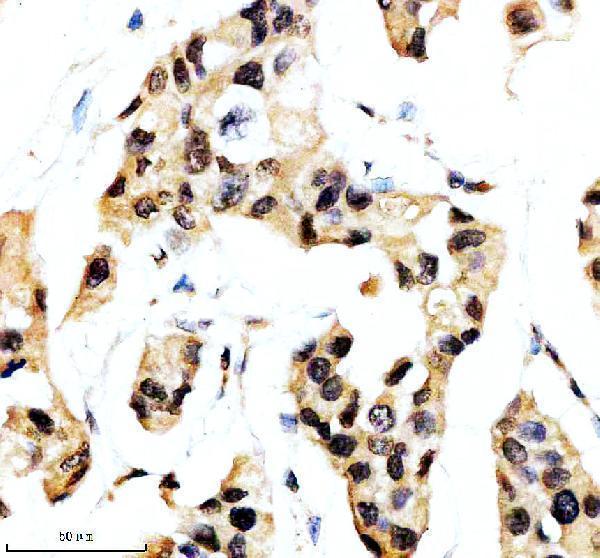
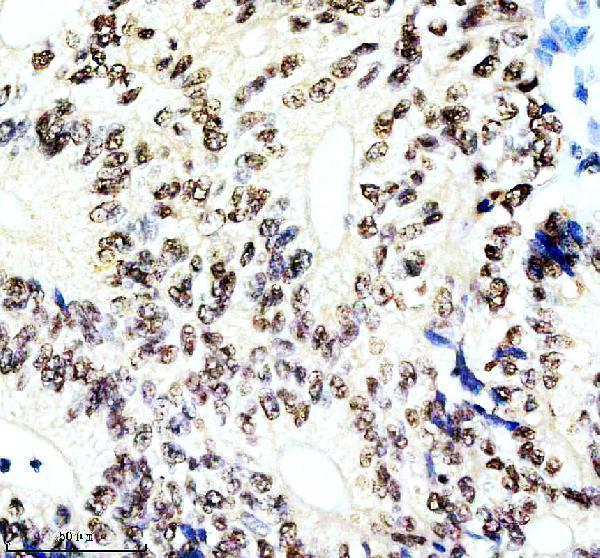
A00288-1 has IHC images from paraffin-embedded human breast and colon cancer and mouse brain tissue, plus IF images from Siha cells and human liver cancer tissue (catalog image captions).

Real IHC data IHC analysis of SON using anti-SON antibody (A00288-1). SON was detected in a paraffin-embedded section of human breast cancer tissue. Heat mediated antigen retrieval was performed in EDTA buffer (pH 8.0, epitope retrieval solution). The tissue section was blocked with 10% goat serum. The tissue section was then incubated with 2 μg/ml rabbit anti-SON Antibody (A00288-1) overnight at 4°C. Peroxidase Conjugated Goat Anti-rabbit IgG was used as secondary antibody and incubated for 30 minutes at 37°C. The tissue section was developed using HRP Conjugated Rabbit IgG Super Vision Assay Kit (Catalog # SV0002) with DAB as the chromogen.
Anti-SON Antibody ®
Cat # A00288-1

A00288-1 has IHC images from paraffin-embedded human breast and colon cancer and mouse brain tissue (catalog IHC image captions). Its IF images show Siha cells and paraffin-embedded human liver cancer tissue; IF and ICC are listed applications (catalog IF image captions; catalog applications).

Which to pick: Choose A00288-1 for paraffin-section IHC: its IHC captions show staining in human breast and colon cancer and mouse brain sections (catalog IHC image captions). The same SKU supports IF/ICC selection through its listed applications and IF images, and human/mouse work through its listed reactivity and IHC images (catalog applications; catalog IF image captions; catalog reactivity; catalog IHC image captions). The IHC captions do not report the fixative, and the catalog does not report clonality (catalog IHC image captions; catalog clone field).

Each figure is that product's own IHC / IF validation image from its datasheet.

References

  1. UniProt Consortium. UniProt entry P18583 (SON_HUMAN, Protein SON).
  2. Human Protein Atlas. SON tissue IHC expression (reliability: Supported).
  3. Human Protein Atlas. SON subcellular location (ICC-IF): Localized to the nuclear speckles..
  4. Human Protein Atlas. SON antibody validation summary (3 antibodies).
  5. Inhibitory inputs to avian ITD circuits. Trends in hearing 2026 — PMC13223351.
  6. Targeting of MAPK-associated molecules identifies SON as a prime target to attenuate the proliferation and tumorigenicity of pancreatic cancer cells. Molecular cancer 2012 — PMC3576306.
  7. Selective up-regulation of JunD transcript and protein expression in vasopressinergic supraoptic nucleus neurones in water-deprived rats. Journal of neuroendocrinology 2012 — PMC3499652.
  8. Mechanisms involved in dual vasopressin/apelin neuron dysfunction during aging. PloS one 2014 — PMC3914823.
  9. PubMed PMID:11707072 — UniProt-cited evidence.
  10. PubMed PMID:11306577 — UniProt-cited evidence.
  11. PubMed PMID:10470851 — UniProt-cited evidence.