SORBS3 / Vinexin · IHC design guide

Design Immunohistochemistry for SORBS3

Plan chromogenic IHC on paraffin sections using the catalog antibody's conditions (datasheet A05794). Compare staining with the cytoplasmic tissue pattern and high signal in cardiomyocytes and skeletal myocytes, while accounting for low antibody–RNA consistency (HPA tissue IHC).

Evidence assembled Oct 2026 · For research use; verify linked source records and product datasheet before use
Immunohistochemistry protocol sheet for SORBS3 (IHC for SORBS3): expected localisation Cytoplasmic tissue staining (HPA tissue IHC), antibody A05794, validated IHC image, and IHC protocol steps
Printable SORBS3 IHC protocol sheet — expected localisation Cytoplasmic tissue staining (HPA tissue IHC), antibody A05794, controls and protocol steps. Open the full SORBS3 IHC guide →

SORBS3 Immunohistochemistry Experimental Design Guide

Expected localisation, validated protocols, controls and antibodies — the at-a-glance facts below, then the full design guide.

Must know before staining
Expected localisation Cytoplasmic tissue staining (HPA tissue IHC)
Staining pattern Cytoplasmic signal in cardiomyocytes and skeletal myocytes (HPA tissue IHC)
Antigen retrieval EDTA pH 8.0 HIER, heat-mediated (datasheet A05794)
Positive control ⓘ Heart muscle+4 more · see all
Negative control ⓘ Adipose tissue+4 more · see all
Important caveats
Reasons your staining may differ from the expected pattern.
Fixation Keep fixation consistent between sections (standard IHC practice; not target-specific)
Caveat Low antibody–RNA consistency complicates tissue comparisons (HPA tissue IHC)
Regulation Isoform β is especially found in liver (UniProt)
Isoform / epitope 2 isoforms, alpha and beta; check epitope coverage (UniProt)
Section 1

Recommended SORBS3 IHC & IF Protocols

The catalog antibody has a datasheet IHC-P protocol (datasheet: A05794); one published SORBS3 IHC protocol provides additional conditions (PMC10084591).

Recommended immunohistochemistry (IHC-P) protocol parameters
SampleParaffin-embedded human liver cancer tissue; fixative not specified (datasheet A05794)
FixationImage fixative and duration unreported (datasheet A05794); verify before use.
Sectioning4–5 µm sections on charged slides (standard)
DeparaffinisationXylene, graded ethanol series to water (standard)
Antigen retrievalHeat retrieval: EDTA pH 8.0 (datasheet A05794); 20 min, 95–100 °C (standard)
Peroxidase block3% H2O2, 10 min, room temperature (standard)
Blocking10% goat serum (datasheet A05794)
Primary antibodyRabbit anti-SORBS3, 0.5-1μg/ml (datasheet A05794)
Primary incubationOvernight at 4 °C (datasheet A05794)
DetectionStreptavidin-biotin complex (SABC), DAB chromogen (datasheet A05794)
CounterstainHematoxylin, blue, dehydrate and mount (standard)
Expected resultSORBS3-positive staining in cardiomyocytes of heart muscle (HPA tissue IHC: High). HPA tissue profile: Cytoplasmic expression in several tissues. No signal in the no-primary control.
💡Decision noteStart with heat-mediated EDTA retrieval at pH 8.0 (datasheet: A05794); the published protocol reports antigen retrieval without specifying its conditions (PMC10084591).
Section 2

What Is the Expected SORBS3 Staining Pattern?

SORBS3 is associated with cell junctions and the cytoskeleton and can also occur in nuclei; it has no transmembrane segment (UniProt O60504). In paraffin IHC, expect chiefly cytoplasmic staining, strongest in cardiomyocytes and skeletal myocytes (HPA tissue IHC: High). HPA rates the IHC pattern Approved but reports low consistency with RNA expression, so judge each cell population and control separately (HPA tissue IHC).

What am I looking at on my slide?
Strong cytoplasmic staining in heart cardiomyocytes or skeletal muscle myocytes.This matches the clearest reported IHC positives: both cell populations score High (HPA tissue IHC). Junction-associated staining can be compatible with SORBS3 biology (UniProt O60504), but HPA's paraffin IHC summary describes cytoplasmic expression; do not require a sharply outlined junction pattern to call a section positive.
An exclusively luminal or extracellular deposit, with no convincing intracellular staining.This is discordant with the reported cytoplasmic IHC pattern (HPA tissue IHC) and with a protein lacking a signal peptide or transmembrane segment (UniProt O60504). Check tissue deposits and detection background before assigning it to SORBS3 (standard IHC practice). Nuclear staining alone needs caution: UniProt also lists a nuclear location (UniProt O60504).
Strong staining in adipocytes or bone marrow hematopoietic cells.Those specific populations are reported as Not detected, so this pattern needs confirmation (HPA tissue IHC). Review morphology and a no-primary control for cross-reactivity or endogenous detection activity (standard IHC practice). HPA's Approved rating does not remove this concern because antibody staining and RNA data show low consistency (HPA tissue IHC).
Widespread, nearly uniform color over cells, stroma and blank areas.A signal that disregards cell boundaries and expected positive populations is more consistent with background than with HPA's cell-specific pattern (HPA tissue IHC; standard IHC practice). Compare a no-primary control and inspect blocking, washes and chromogen development before scoring cells (standard IHC practice).
No cellular signal in a well-preserved heart or skeletal muscle section.Target-specific fixation effects are not established by the supplied assay evidence. Verify with a matched IHC source before attributing a result to fixation.
💡Expected SORBS3 appearanceCall a section positive when cardiomyocytes or skeletal myocytes show convincing, predominantly cytoplasmic staining at the reported High level (HPA tissue IHC); isolated extracellular color or staining confined to a no-primary control is a false-positive warning (UniProt O60504 topology; standard IHC practice).
How each factor affects the staining
Cell population and tissueHeart cardiomyocytes and skeletal myocytes are High; several glandular and endothelial populations are Medium, while listed adipocytes and marrow hematopoietic cells are Not detected (HPA tissue IHC). Score the named cell type within each section: a tissue-level label does not imply that every cell in that tissue should stain (HPA tissue IHC).
Subcellular locationHPA summarizes tissue IHC as cytoplasmic (HPA tissue IHC). UniProt lists junctions, cytoskeleton and nucleus (UniProt O60504). HPA's ICC-IF junction and plasma-membrane assignments are marked uncertain; they provide context, not an additional paraffin IHC pass criterion (HPA subcellular ICC-IF).
Isoforms and antibody coverageUniProt lists alpha and beta isoforms and notes beta especially in liver (UniProt O60504). Epitope coverage and isoform specificity are not supplied here, so these facts cannot predict relative IHC intensity or turn low cholangiocyte staining into an assay failure (HPA tissue IHC: Low in liver cholangiocytes).
IHC evidence strengthHPA marks the tissue IHC pattern Approved while flagging low consistency between staining and RNA expression; the listed IHC antibody is HPA015849 (HPA tissue IHC; HPA antibodies). Treat the observed pattern as a guide for controls, and investigate unexpected cell staining rather than treating approval as proof of specificity.
Retrieval and detection settingsRetrieval, blocking and chromogen development can affect IHC background and interpretability in general (standard IHC practice). No SORBS3-specific retrieval setting, fixation effect or dilution is supplied, so use the IHC-validated antibody's documented conditions and optimize against positive and no-primary controls (standard IHC practice).
Why is my staining missing, weak or wrong?
SituationLikely causeNext action
Heart or skeletal muscle positive control has no convincing staining.The result conflicts with High staining in the named myocytes (HPA tissue IHC); tissue handling, assay setup or detection may have failed (standard IHC practice).Confirm cell identity and preservation, inspect the detection control, and follow the IHC-validated antibody's documented retrieval and dilution before changing one condition at a time (standard IHC practice).
Color appears broadly across the section, including blank regions.Cell-independent color suggests nonspecific background or excess chromogen development (standard IHC practice), unlike the reported cell-specific tissue pattern (HPA tissue IHC).Run a no-primary control; review blocking, washes, antibody concentration and development time, then reassess cellular localization (standard IHC practice).
A reported Not detected cell population appears strongly positive.Adipocytes and bone marrow hematopoietic cells are listed as Not detected (HPA tissue IHC); cross-reactivity or endogenous detection activity is possible (standard IHC practice).Confirm the cell type on the counterstained section, compare a no-primary control and repeat with appropriate detection controls before reporting SORBS3 positivity (standard IHC practice).
Only a nuclear signal is visible.UniProt includes nuclear localization, but HPA summarizes tissue IHC as cytoplasmic (UniProt O60504; HPA tissue IHC). Nuclear-only IHC therefore remains ambiguous rather than automatically positive.Check whether known-positive myocytes also show the expected cytoplasmic signal; compare controls and report nuclear-only staining separately until independently supported (HPA tissue IHC; standard IHC practice).
The section lacks a crisp junctional outline despite cellular staining.HPA describes paraffin tissue IHC as cytoplasmic, while its junctional ICC-IF assignment is uncertain (HPA tissue IHC; HPA subcellular ICC-IF).Judge the IHC section by cell identity, cytoplasmic signal and controls; use the separate IF/ICC guide for junction imaging rather than changing the paraffin IHC acceptance rule (HPA tissue IHC; standard IHC practice).
Liver cholangiocytes stain weakly compared with muscle controls.HPA reports Low staining in cholangiocytes and High staining in heart cardiomyocytes and skeletal myocytes (HPA tissue IHC). UniProt's note that beta is especially found in liver does not establish cholangiocyte IHC intensity (UniProt O60504).Compare the named cell populations and their controls; avoid treating weak cholangiocyte staining alone as evidence that the assay failed (HPA tissue IHC; standard IHC practice).

Sample controls for SORBS3 IHC & IF

🧪Run heart muscle first and expect strong staining in cardiomyocytes (HPA: High in cardiomyocytes). Run adipose tissue as a negative comparator and assess adipocytes specifically (HPA: Not detected in adipocytes); on the heart slide, internal negative cells should show background staining, but no non-cardiomyocyte population is established as negative by the supplied HPA rows (HPA: High in cardiomyocytes).
Positive control tissue: Heart muscle (Cardiomyocytes, HPA High)
Negative control tissue: Adipose tissue (HPA Not detected)
ICC-IF cell lines (HPA subcellular resource): HPA ICC-IF images show SORBS3 in A-549, U2OS, hTERT-RPE1, with annotated localisation: Cell Junctions (uncertain) (HPA subcellular).
Technical controls: Include a no-primary (secondary-only) control, a species- and immunoglobulin-matched rabbit IgG isotype control for the captioned rabbit primary (caption: rabbit anti-SORBS3), and SORBS3-knockout tissue or cells as a biological negative (standard IHC practice). Block endogenous peroxidase and check for endogenous biotin background in heart sections because the captioned detection uses a streptavidin–biotin complex with DAB (caption: SABC/DAB; standard IHC practice).
⚠️Feasibility: A target-specific fixation window or fixation effect is unreported, and the selected A05794 tissue-IHC caption does not state a fixative (caption: fixative not stated). The caption uses heat retrieval in EDTA at pH 8.0, but does not establish that retrieval is required under other conditions (caption: EDTA pH 8.0); neither frozen sections nor IF can be judged easier from the supplied evidence (HPA: ICC-IF images in A-549, U2OS and hTERT-RPE1). Check heart sections for pigment and blood-associated chromogenic background when interpreting cardiomyocyte staining (standard IHC practice).

HPA tissue IHC evidence for SORBS3

Comprehensive Human Protein Atlas IHC scoring per tissue (reliability: Approved — Low consistency between antibody staining and RNA expression data.). Rows are taken directly from the HPA tissue chart — click any row's HPA link to view the source.

Positive expression · recommended positive controls

TissueCell typeLevelEvidenceSource
Heart muscle Cardiomyocytes High Protein (IHC) HPA →
Skeletal muscle Myocytes High Protein (IHC) HPA →
Appendix Glandular cells Medium Protein (IHC) HPA →
Breast Glandular cells Medium Protein (IHC) HPA →
Cerebral cortex Endothelial cells Medium Protein (IHC) HPA →

Undetected expression · recommended negative controls

TissueCell typeLevelEvidenceSource
Adipose tissue Adipocytes Not detected Protein (IHC) HPA →
Adrenal gland Glandular cells Not detected Protein (IHC) HPA →
Bone marrow Hematopoietic cells Not detected Protein (IHC) HPA →
Caudate Glial cells Not detected Protein (IHC) HPA →
Cerebellum Cells in granular layer Not detected Protein (IHC) HPA →
Section 3

Advanced SORBS3 IHC Tips

Troubleshoot SORBS3 staining in paraffin sections by checking retrieval, cell type, subcellular pattern and detection controls before interpreting signal.

Which retrieval conditions should I start with for SORBS3 IHC?
Start with heat-mediated retrieval in EDTA at pH 8.0 for paraffin sections (datasheet A05794). The selected tissue image used this retrieval before overnight incubation at 4°C with 1 μg/mL catalog antibody (datasheet A05794). If staining is weak, vary heating duration on matched sections while keeping antibody concentration and detection conditions constant (standard IHC practice). Include a positive tissue section in each comparison so tissue loss or uneven heating does not look like retrieval failure (standard IHC practice). Assess cytoplasmic and junction-associated signal in the expected cells, since SORBS3 has no transmembrane segment (UniProt O60504 topology and localisation).
How should I troubleshoot possible fixation effects on SORBS3 staining?
The selected paraffin-section caption does not report a fixative, so target-specific fixation sensitivity is unknown (datasheet A05794). Record the fixative and fixation duration for every specimen, then compare similarly processed sections before attributing intensity differences to SORBS3 abundance (standard IHC practice). If signal varies across a batch, inspect tissue preservation and compare retrieval and staining on sections processed together (standard IHC practice). Keep the documented EDTA pH 8.0 retrieval as the starting condition while investigating those differences (datasheet A05794). Neither the reported tissue distribution nor SORBS3 topology establishes a preferred fixative (HPA tissue IHC; UniProt O60504 topology).
Where should convincing SORBS3 staining appear in tissue?
Expect predominantly cytoplasmic tissue staining, while evaluating junction-associated signal where cell boundaries can be resolved (HPA tissue IHC; UniProt O60504 localisation). Both isoforms have been localized to focal adhesion and cell–cell adhesion sites, and UniProt also lists cytoskeletal and nuclear locations (UniProt O60504 localisation). HPA reports cell-junction and additional plasma-membrane localisation from IF images, but marks both assignments uncertain (HPA subcellular). Check whether a proposed boundary pattern follows intact cells across the section and persists away from cut edges (standard IHC practice). Treat isolated strong nuclear staining cautiously unless its cell distribution and controls support it (UniProt O60504 localisation; standard IHC practice).
Can this antibody distinguish SORBS3 isoforms or phosphorylation states in IHC?
The supplied caption gives no epitope or isoform specificity, so staining with this catalog antibody cannot identify alpha versus beta by itself (datasheet A05794). SORBS3 has alpha and beta isoforms, and beta is especially reported in liver (UniProt O60504 isoforms and tissue specificity). The protein contains a SoHo domain and three SH3 domains, but their positions do not establish where this antibody binds (UniProt O60504 domains; datasheet A05794). Multiple phosphoserines are recorded, including residues 348 and 395, without evidence that they alter this antibody’s IHC recognition (UniProt O60504 modified residues; datasheet A05794). Require an epitope-defined or isoform-specific reagent before assigning an isoform or phosphorylation state (standard IHC practice).
How can I assess SORBS3 by multiplex IF alongside tissue IHC?
For the secondary IF application, pair SORBS3 with a marker that identifies the cell population being assessed, such as a cardiomyocyte marker in heart sections (HPA: high in cardiomyocytes; standard IF practice). Select a spectrally separated, preferably far-red detection channel after checking unstained tissue for autofluorescence (standard IF practice). SORBS3 has no transmembrane segment and includes cytoplasmic and junction-associated locations, so test permeabilised sections when assessing intracellular signal (UniProt O60504 topology and localisation; standard IF practice). Because the antibody’s epitope is unspecified, compare permeabilised and nonpermeabilised controls before interpreting a missing junction signal (datasheet A05794; standard IF practice). Do not transfer the paraffin IHC caption into an IF fixation claim (datasheet A05794).
How do I separate SORBS3 signal from chromogenic background?
The selected liver cancer section used a biotinylated secondary antibody, streptavidin–biotin detection and DAB, so inspect each detection step when diffuse brown staining appears (datasheet A05794). Run a no-primary control to reveal secondary or detection-system background, and apply peroxidase blocking before DAB development (standard IHC practice). Check for endogenous biotin contribution when using the documented biotin-based system, particularly if background tracks tissue structures regardless of primary antibody (datasheet A05794; standard IHC practice). Compare suspicious staining with intact-cell cytoplasmic and junction-associated patterns instead of scoring pigment, debris or damaged edges as positive cells (HPA tissue IHC; UniProt O60504 localisation; standard IHC practice).
What is a defensible way to score SORBS3 IHC? ⚠ ANSWER MARKED FOR VERIFICATION
Define the cell population and compartment before scoring, because HPA reports cytoplasmic expression across several tissues and UniProt also lists junction-associated localisation (HPA tissue IHC; UniProt O60504 localisation). For cellular staining, record percent-positive cells and intensity, or calculate a cytoplasmic H-score on a 0–300 scale (standard IHC practice). If junctional staining is the endpoint, measure positive boundary length or area against the evaluable cell-boundary area rather than total section area (standard image-analysis practice). Normalize cell counts to the relevant viable-cell population, or report positive-cell density per mm² of viable tissue (standard IHC practice). Keep retrieval, exposure or DAB development, and scoring thresholds consistent across comparisons (standard IHC practice).
When is apparent SORBS3 positivity likely to be artefactual?
Give more weight to reproducible cytoplasmic or junction-associated staining in intact cells than to signal restricted to tissue edges or necrotic regions (HPA tissue IHC; UniProt O60504 localisation; standard IHC practice). Check cell identity: HPA reports high staining in cardiomyocytes and skeletal myocytes, but no detected staining in several other listed cell populations (HPA tissue IHC). Brown signal that persists without primary antibody suggests detection background or endogenous enzyme activity rather than SORBS3 (standard IHC practice). Treat a sharply nuclear-only pattern cautiously because the tissue IHC profile is cytoplasmic, although UniProt includes nuclear localisation (HPA tissue IHC; UniProt O60504 localisation). HPA rates its tissue antibody data Approved while reporting low consistency with RNA expression, so corroborate unexpected patterns independently (HPA tissue IHC).
Boster reagents

Best SORBS3 / Vinexin IHC Antibodies

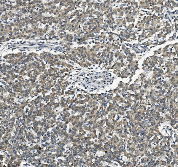
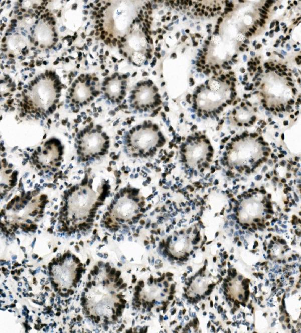
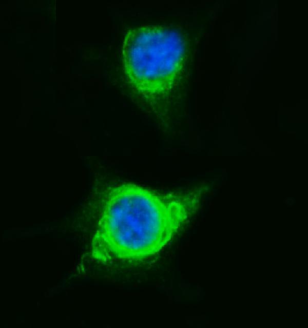
A05794 has IHC images from human liver cancer, mouse intestine and rat intestinal lymph node paraffin sections, plus an IF image from A549 cells (A05794 image captions).

Real IHC data IHC analysis of SORBS3 using anti-SORBS3 antibody (A05794). SORBS3 was detected in paraffin-embedded section of human liver cancer tissue. Heat mediated antigen retrieval was performed in EDTA buffer (pH8.0, epitope retrieval solution). The tissue section was blocked with 10% goat serum. The tissue section was then incubated with 1μg/ml rabbit anti-SORBS3 Antibody (A05794) overnight at 4°C. Biotinylated goat anti-rabbit IgG was used as secondary antibody and incubated for 30 minutes at 37°C. The tissue section was developed using Strepavidin-Biotin-Complex (SABC) (Catalog # SA1022) with DAB as the chromogen.
Anti-Vinexin/SORBS3 Antibody ®
Cat # A05794

A05794 will render with its human liver cancer paraffin-section IHC figure; additional IHC captions show mouse intestine and rat intestinal lymph node paraffin sections (A05794 IHC captions). Its IF caption shows A549 cells, and the catalog lists IHC, ICC and IF applications with human, mouse and rat reactivity (A05794 IF caption; A05794 catalog).

Which to pick: Choose A05794 for tissue IHC in paraffin sections, starting at 0.5–1 μg/ml; its IHC captions document EDTA retrieval at pH 8.0, while the fixative is unreported (A05794 catalog; A05794 IHC captions). Choose the same SKU for IF/ICC at 2 μg/ml, supported by its A549 cell IF image (A05794 catalog; A05794 IF caption). For cross-species IHC, its captions show human, mouse and rat sections; the antibody is rabbit-hosted, and clonality is unreported (A05794 IHC captions; A05794 catalog).

Each figure is that product's own IHC / IF validation image from its datasheet.

References

  1. UniProt Consortium. UniProt entry O60504 (VINEX_HUMAN, Vinexin).
  2. Human Protein Atlas. SORBS3 tissue IHC expression (reliability: Approved).
  3. Human Protein Atlas. SORBS3 subcellular location (ICC-IF): Mainly localized to the cell junctions. In addition localized to the plasma membrane..
  4. Human Protein Atlas. SORBS3 antibody validation summary (2 antibodies).
  5. SORBS3-β suppresses lymph node metastasis in cervical cancer by promoting the ubiquitination of β-catenin. Journal of translational medicine 2025 — PMC11978191.
  6. Elevated ZNF704 expression is associated with poor prognosis of uveal melanoma and promotes cancer cell growth by regulating AKT/mTOR signaling. Biomarker research 2023 — PMC10084591.
  7. PubMed PMID:9885244 — UniProt-cited evidence.
  8. PubMed PMID:16421571 — UniProt-cited evidence.
  9. PubMed PMID:15489334 — UniProt-cited evidence.