SORL1 / Sortilin-related receptor · IHC design guide

Design Immunohistochemistry for SORL1

Plan paraffin-section SORL1 IHC using cerebellar Purkinje cells as a high-staining reference (HPA tissue IHC). Interpret staining with the predominantly intracellular localization and possible ectodomain shedding in mind (UniProt).

Evidence assembled Oct 2026 · For research use; verify linked source records and product datasheet before use
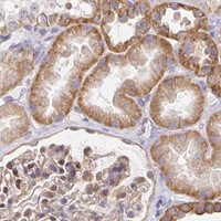
Immunohistochemistry protocol sheet for SORL1 (IHC for SORL1): expected localisation TGN/endosomal localization expected; tissue pattern uncertain (UniProt), antibody M01887-2, validated IHC image, and IHC protocol steps
Printable SORL1 IHC protocol sheet — expected localisation TGN/endosomal localization expected; tissue pattern uncertain (UniProt), antibody M01887-2, controls and protocol steps. Open the full SORL1 IHC guide →

SORL1 Immunohistochemistry Experimental Design Guide

Expected localisation, validated protocols, controls and antibodies — the at-a-glance facts below, then the full design guide.

Must know before staining
Expected localisation TGN/endosomal localization expected; tissue pattern uncertain (UniProt)
Staining pattern Purkinje cells: high; compartment unreported (HPA tissue IHC)
Antigen retrieval EDTA pH 8.0 HIER, heat-mediated (datasheet M01887-2)
Positive control ⓘ Cerebellum+4 more · see all
Negative control ⓘ Adipose tissue+4 more · see all
Important caveats
Reasons your staining may differ from the expected pattern.
Fixation Keep fixation consistent across sections. (standard IHC practice; not target-specific)
Caveat Shedding can release the extracellular domain (UniProt)
Regulation Reduced in Alzheimer frontal cortex (UniProt)
Isoform / epitope No isoforms listed; epitope side matters after shedding (UniProt)
Section 1

Recommended SORL1 IHC & IF Protocols

The catalog antibody protocol uses EDTA pH 8.0 retrieval (datasheet: M01887-2). Published IHC protocols cover human brain (PMC5465543) and interspinous ligament (PMC9019158).

Recommended immunohistochemistry (IHC-P) protocol parameters
SampleParaffin-embedded mouse brain tissue; fixative not specified (datasheet M01887-2)
FixationImage fixative and duration unreported (datasheet M01887-2); verify before use.
Sectioning4–5 µm sections on charged slides (standard)
DeparaffinisationXylene, graded ethanol series to water (standard)
Antigen retrievalHeat retrieval: EDTA pH 8.0 (datasheet M01887-2); 20 min, 95–100 °C (standard)
Peroxidase block3% H2O2, 10 min, room temperature (standard)
Blocking10% goat serum (datasheet M01887-2)
Primary antibodyRabbit monoclonal (clone 22S13) anti-SORL1, 1:50 (datasheet M01887-2)
Primary incubationOvernight at 4 °C (datasheet M01887-2)
DetectionHRP-conjugated secondary, DAB chromogen (datasheet M01887-2)
CounterstainHematoxylin, blue, dehydrate and mount (standard)
Expected resultSORL1-positive staining in purkinje cells of cerebellum (HPA tissue IHC: High). HPA tissue profile: plasma protein. No signal in the no-primary control.
💡Decision noteStart with heat-mediated EDTA pH 8.0 retrieval (datasheet: M01887-2); the published ligament protocol reports antigen repair without conditions (PMC9019158).
Section 2

What Is the Expected SORL1 Staining Pattern?

SORL1 should appear mainly intracellular, consistent with its trans-Golgi and endosomal localisation and single transmembrane segment (UniProt Q92673 subcellular location and topology). In paraffin-section IHC, cerebellar Purkinje cells are the clearest supplied positive example, with high staining; cerebral cortex neuropil shows medium staining (HPA: tissue IHC). Treat these as provisional reference patterns because HPA rates the tissue IHC evidence Uncertain (HPA: reliability).

What am I looking at on my slide?
Purkinje cells stain strongly, with predominantly intracellular signal; cortex neuropil may stain less strongly (HPA: High in Purkinje cells; Medium in cortex neuropil; UniProt Q92673 subcellular location).This fits the supplied tissue pattern and the receptor’s mainly trans-Golgi and endosomal distribution. Score the named cells or structures rather than treating an entire brain section as uniformly positive (HPA: tissue IHC; UniProt Q92673 subcellular location).
A dominant nuclear stain replaces the expected intracellular pattern (UniProt Q92673 subcellular location).Question the result and check antibody specificity and controls. UniProt reports that a cleaved cytosolic fragment can enter the nucleus, so nuclear signal alone cannot identify full-length SORL1; interpretation depends on the antibody’s epitope (UniProt Q92673 processing and topology).
Cells scored as negative by HPA stain as strongly as Purkinje cells, such as adipocytes or spleen red-pulp cells (HPA: Not detected in those cells; High in Purkinje cells).Investigate cross-reactivity or endogenous detection activity before assigning SORL1 positivity. HPA’s negative calls apply to those specified cell types, and its overall IHC reliability is Uncertain (HPA: tissue IHC and reliability; general IHC practice).
Chromogen is diffuse across the section, including areas without a clear cellular pattern (general IHC practice).This can reflect nonspecific background or detection activity; extracellular staining also requires care because the SORL1 ectodomain can be shed. Use controls and morphology to distinguish these possibilities (general IHC practice; UniProt Q92673 shedding).
Purkinje cells show no convincing signal in an otherwise assessable cerebellar section (HPA: High in Purkinje cells).Check the run and tissue before calling SORL1 absent. The HPA pattern is a useful reference, but its Uncertain reliability means a negative result needs independent support (HPA: tissue IHC and reliability; general IHC practice).
💡Expected SORL1 appearanceCall a result provisionally positive when Purkinje cells show high, predominantly intracellular staining, with possible medium cortex-neuropil staining; indiscriminate diffuse chromogen or equally strong staining in HPA-negative cell types warrants investigation (HPA: tissue IHC, Uncertain reliability; UniProt Q92673 subcellular location; general IHC practice).
How each factor affects the staining
Subcellular distribution (UniProt Q92673 subcellular location)SORL1 is mainly in the trans-Golgi network and endosomes, with lower plasma-membrane abundance. Judge intracellular and surface signals in that context; a membrane-only pattern needs scrutiny (UniProt Q92673 subcellular location).
Epitope and processing (UniProt Q92673 topology and processing)Most of the receptor is lumenal, followed by a transmembrane segment and short cytoplasmic tail. Ectodomain shedding and subsequent cleavage can separate signals from different regions; antibody epitope information is needed to interpret them (UniProt Q92673 topology and processing).
Cell-level reference patterns (HPA: tissue IHC)Use the named cell or structure: Purkinje cells are High, cortex neuropil Medium, and adipocytes Not detected. These entries do not imply uniform staining across their tissues (HPA: tissue IHC).
Strength of IHC evidence (HPA: reliability and antibody validation)HPA calls the tissue IHC profile Uncertain and lists IHC validation as Uncertain for HPA031321 and CAB011500. Treat agreement with its pattern as supportive rather than conclusive (HPA: reliability and antibody validation).
Chromogenic detection background (general IHC practice)Endogenous enzyme activity and nonspecific detection can mimic a positive deposit. Evaluate reagent and tissue controls when staining ignores cell boundaries or appears in an unexpected compartment (general IHC practice).
IF/ICC pattern? (HPA: subcellular ICC-IF)HPA summarises SORL1 as Membrane, Secreted but provides no main location or cell-line ICC-IF images here. This IHC pattern cannot establish an IF/ICC image pattern; consult the separate IF/ICC guide (HPA: subcellular ICC-IF).
Why is my staining missing, weak or wrong?
SituationLikely causeNext action
Purkinje cells are blank (HPA: High in Purkinje cells).The run may have insufficient detectable signal, or this section may not reproduce HPA’s Uncertain reference pattern (general IHC practice; HPA: reliability).Verify that the named cells are present, inspect the positive control, then review antigen retrieval, antibody dilution and detection performance using the catalog antibody’s IHC-P instructions (general IHC practice; HPA: tissue IHC).
Adipocytes or spleen red-pulp cells show strong deposit (HPA: Not detected in those cells).Cross-reactivity or endogenous detection activity is possible; a cell-level HPA negative is not proof of complete tissue absence (general IHC practice; HPA: tissue IHC).Compare a no-primary control and the positive reference cells, then reassess antibody specificity before scoring those cells as SORL1-positive (general IHC practice; HPA: tissue IHC).
The whole section has a diffuse brown haze (general IHC practice).Nonspecific binding, incomplete blocking or detection background can obscure a cellular pattern; extracellular SORL1 ectodomain is also biologically possible (general IHC practice; UniProt Q92673 shedding).Inspect no-primary and tissue controls, review blocking and wash steps, and score only interpretable cell-associated deposits (general IHC practice).
Staining is mainly at cell borders (UniProt Q92673 subcellular location).Some surface SORL1 is plausible, but UniProt describes the receptor as predominantly intracellular (UniProt Q92673 subcellular location).Compare intracellular signal and the expected Purkinje-cell pattern; check controls and the antibody epitope before interpreting a membrane-only result (UniProt Q92673 subcellular location; HPA: tissue IHC; general IHC practice).
Nuclei stain prominently (UniProt Q92673 processing).A released C-terminal fragment can localise to nuclei, while nonspecific nuclear staining remains possible (UniProt Q92673 processing; general IHC practice).Check whether the antibody recognises the cytoplasmic C-terminus, review no-primary controls, and avoid calling nuclear deposit full-length receptor without further validation (UniProt Q92673 topology and processing; general IHC practice).
A result conflicts with the HPA tissue pattern (HPA: tissue IHC).HPA rates this IHC profile Uncertain and notes that secreted variants complicate RNA–protein agreement (HPA: reliability description).Record the exact cell type and compartment, compare matched controls, and seek independent support before treating either the new stain or the HPA pattern as definitive (HPA: reliability; general IHC practice).

Sample controls for SORL1 IHC & IF

🧪Run cerebellum first: Purkinje cells should stain (HPA: High in Purkinje cells). Use adipose tissue as the negative tissue, focusing on adipocytes (HPA: Not detected in adipocytes); on the cerebellum slide, treat neighboring cells without specific staining as an internal negative reference only if observed, since the supplied HPA row assigns a level only to Purkinje cells (HPA: High in Purkinje cells).
Positive control tissue: Cerebellum (Purkinje cells, HPA High)
Negative control tissue: Adipose tissue (HPA Not detected)
ICC-IF cell lines (HPA subcellular resource): HPA carries no ICC-IF cell line for SORL1; derive a cell-line control from the positive tissue's cell type (Purkinje cells) and confirm it by RNA or western blot first.
Technical controls: Include a no-primary (secondary-only) control, a matched rabbit IgG isotype control, and SORL1 knockout tissue as a biological negative (caption: rabbit primary antibody). Quench endogenous peroxidase in cerebellum sections before HRP/DAB detection (caption: HRP/DAB detection in mouse brain tissue).
⚠️Feasibility: A target-specific fixation window and fixation effect are unreported, and the selected M01887-2 tissue-IHC caption does not state a fixative (selected-SKU caption: fixative not stated). Its paraffin-section example uses heat retrieval in EDTA at pH 8.0 and primary antibody at 1:50 overnight at 4°C; use these as a starting condition, since retrieval dependence has not been established by a comparison (selected-SKU caption). The supplied evidence does not establish whether frozen sections or IF are easier; in brain sections, distinguish specific staining from endogenous pigment during chromogenic scoring and account for autofluorescence if adapting to IF (UniProt Q92673: brain expression; selected-SKU caption: mouse brain HRP/DAB IHC).

HPA tissue IHC evidence for SORL1

Comprehensive Human Protein Atlas IHC scoring per tissue (reliability: Uncertain — At least one protein variant secreted, tissue location of RNA and protein might differ and correlation is complex. Pending external verification.). Rows are taken directly from the HPA tissue chart — click any row's HPA link to view the source.

Positive expression · recommended positive controls

TissueCell typeLevelEvidenceSource
Cerebellum Purkinje cells High Protein (IHC) HPA →
Adrenal gland Glandular cells Medium Protein (IHC) HPA →
Appendix Glandular cells Medium Protein (IHC) HPA →
Breast Glandular cells Medium Protein (IHC) HPA →
Bronchus Respiratory epithelial cells Medium Protein (IHC) HPA →

Undetected expression · recommended negative controls

TissueCell typeLevelEvidenceSource
Adipose tissue Adipocytes Not detected Protein (IHC) HPA →
Bone marrow Hematopoietic cells Not detected Protein (IHC) HPA →
Liver Cholangiocytes Not detected Protein (IHC) HPA →
Placenta Decidual cells Not detected Protein (IHC) HPA →
Soft tissue Chondrocytes Not detected Protein (IHC) HPA →
Section 3

Advanced SORL1 IHC Tips

Troubleshoot SORL1 staining in paraffin sections by checking retrieval, compartment, cell type, and controls before comparing signal intensity (UniProt Q92673; HPA tissue IHC).

Which retrieval conditions should I try first for weak SORL1 staining?
Start with heat-mediated antigen retrieval in EDTA at pH 8.0 for paraffin sections (datasheet M01887-2). The catalog antibody produced a mouse brain IHC image after this retrieval, followed by 10% goat serum blocking and a 1:50 primary incubation overnight at 4°C (datasheet M01887-2). If staining is weak, adjust heating duration within a controlled series while keeping section thickness, antibody incubation, and detection consistent (standard IHC practice). Compare each condition with a no-primary control and an identically processed positive section, because stronger DAB signal alone cannot establish specificity (standard IHC practice). Interpret improvement against the expected predominantly intracellular SORL1 distribution (UniProt Q92673 subcellular location).
How should I troubleshoot fixation when SORL1 staining is inconsistent?
The selected IHC caption identifies a paraffin-embedded mouse brain section but does not state its fixative, so SORL1-specific fixation sensitivity is unknown (datasheet M01887-2). Record fixative, fixation duration, processing history, and section age for each specimen before comparing staining across batches (standard IHC practice). Keep retrieval in EDTA at pH 8.0 and the primary incubation at 1:50 overnight at 4°C when benchmarking against the caption (datasheet M01887-2). If signal varies, compare matched sections processed together and include no-primary controls to assess processing and detection effects (standard IHC practice). Do not assign a fixation effect from tissue staining patterns alone (standard IHC practice).
Where should convincing SORL1 staining appear in a paraffin section?
Look first for intracellular, often perinuclear or punctate staining consistent with the trans-Golgi network and endosomes, where SORL1 is predominantly found (UniProt Q92673 subcellular location). Limited cell-surface signal can also be plausible, because only low levels normally reside at the plasma membrane (UniProt Q92673 subcellular location). In cerebellum, Purkinje cells provide a reported high-staining cell population, although the HPA tissue-IHC reliability assessment is uncertain (HPA tissue IHC: High in Purkinje cells; reliability Uncertain). Compare that pattern with neighboring cells and a no-primary section under the same DAB development conditions (standard IHC practice). Diffuse extracellular staining needs separate scrutiny because SORL1 can release a soluble ectodomain (UniProt Q92673 subcellular location).
How does antibody epitope position change interpretation of SORL1 staining?
Determine whether the antibody recognizes the lumenal ectodomain, spanning residues 82–2137, or the cytoplasmic tail, spanning 2159–2214 (UniProt Q92673 topology). The supplied caption does not identify the epitope, so these alternatives cannot be assigned to catalog antibody M01887-2 from its IHC image (datasheet M01887-2). Ectodomain staining could include membrane-associated receptor and shed material, whereas tail-directed staining would assess a different portion of the receptor (UniProt Q92673 processing and subcellular location). Numerous annotated glycosylation sites lie in the ectodomain and may matter when evaluating epitope accessibility, but no retrieval sensitivity is established here (UniProt Q92673 glycosylation; datasheet M01887-2). The record lists 0 isoforms, so do not explain discordant patterns by an undocumented isoform (UniProt Q92673 isoforms).
How can I adapt this SORL1 question to a multiplex IF experiment?
Treat the paraffin-section DAB image as IHC evidence; it does not establish an IF/ICC staining condition for catalog antibody M01887-2 (datasheet M01887-2). Pair SORL1 with a validated marker for the cell population being examined, and use matched single-stain controls to distinguish co-localisation from spectral bleed-through (standard IF practice). Choose fluorophores after inspecting unstained tissue autofluorescence, favoring channels with clearer separation from that background (standard IF practice). For an intracellular epitope, optimise permeabilisation; for an extracellular epitope, compare conditions that preserve surface staining, because SORL1 spans the membrane with distinct lumenal and cytoplasmic regions (UniProt Q92673 topology; standard IF practice). Verify any apparent perinuclear or punctate signal against no-primary controls before interpreting it as SORL1 trafficking (UniProt Q92673 subcellular location; standard IF practice).
What should I change when SORL1 DAB staining is widespread?
Inspect a no-primary section first to assess signal from secondary detection, endogenous peroxidase, or DAB development (standard IHC practice). Include a peroxidase-blocking step in the chromogenic workflow and keep its duration consistent across sections; the selected caption reports peroxidase-based detection but does not specify a blocking step (datasheet M01887-2; standard IHC practice). The caption used 10% goat serum and a 1:50 primary dilution, which provide documented starting conditions for this antibody (datasheet M01887-2). If background persists, titrate primary concentration and detection time using matched sections while retaining the EDTA pH 8.0 retrieval condition (datasheet M01887-2; standard IHC practice). Assess diffuse extracellular signal cautiously because SORL1 can shed its ectodomain (UniProt Q92673 subcellular location).
How should I score SORL1 signal across stained sections? ⚠ ANSWER MARKED FOR VERIFICATION
Define the cell population and compartment before scoring, since SORL1 is predominantly intracellular and can also appear at the cell surface or extracellularly after shedding (UniProt Q92673 subcellular location). For cell-based comparisons, record the percentage of positive cells and an intensity-based H-score using a fixed threshold and scoring rubric (standard IHC practice). For punctate or region-based staining, report positive area or puncta density per mm² of analyzable tissue, excluding folds and damaged edges consistently (standard IHC practice). Normalise to the same cell population or tissue area and compare sections processed with identical retrieval and DAB development (standard IHC practice). Keep Purkinje-cell and neuropil measurements separate when relevant, because their reported staining levels differ (HPA tissue IHC: High in cerebellar Purkinje cells; Medium in cerebral-cortex neuropil).
How do I distinguish true SORL1 staining from artefact?
Favor a reproducible intracellular, often perinuclear or punctate pattern in appropriate cells, consistent with SORL1 enrichment in the trans-Golgi network and endosomes (UniProt Q92673 subcellular location). High staining in cerebellar Purkinje cells is a useful comparison, but the HPA tissue-IHC reliability rating is uncertain and requires independent verification (HPA tissue IHC: High in Purkinje cells; reliability Uncertain). Question signal concentrated at section edges, folds, or necrotic regions, and compare it with a no-primary control for endogenous enzyme or detection background (standard IHC practice). Interpret broad extracellular signal carefully because the receptor ectodomain can be shed (UniProt Q92673 subcellular location). Confirm a disputed pattern with an independently validated epitope or orthogonal evidence before attributing a biological change to SORL1 (standard IHC practice).
Boster reagents

Best SORL1 / Sortilin-related receptor IHC Antibodies

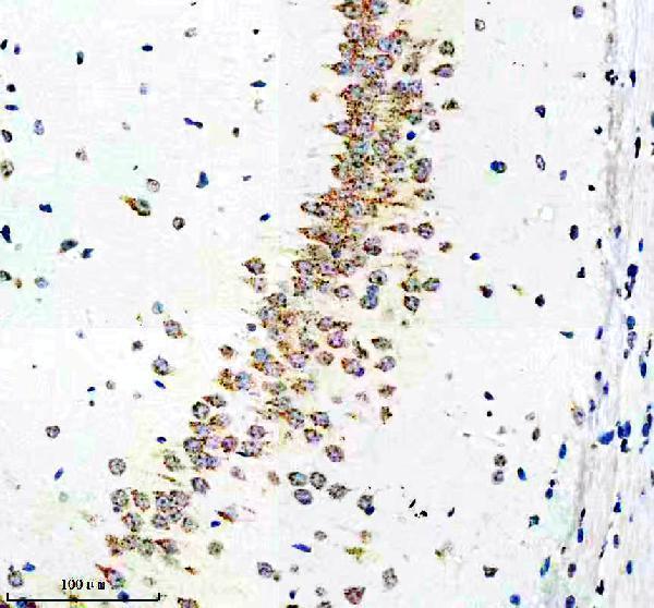
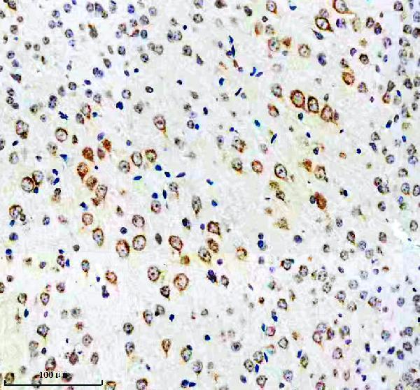
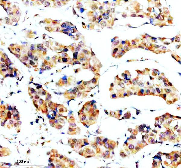
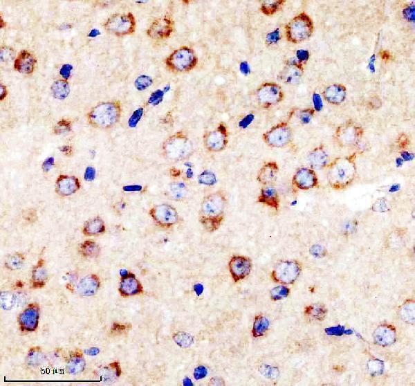
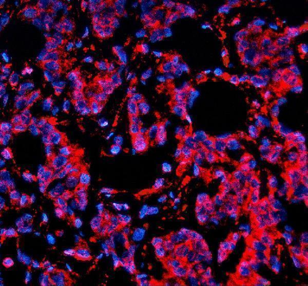
Two anti-SORL1 antibodies have IHC images from paraffin sections: mouse and rat brain for both, plus human breast cancer tissue for A01887-1 (catalog IHC captions). A01887-1 also has a human tissue IF image (catalog IF caption).

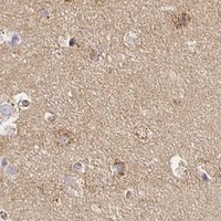
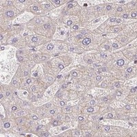
Real IHC data IHC analysis of SORLA using anti-SORLA antibody (M01887-2). SORLA was detected in a paraffin-embedded section of mouse brain tissue. Heat mediated antigen retrieval was performed in EDTA buffer (pH 8.0, epitope retrieval solution). The tissue section was blocked with 10% goat serum. The tissue section was then incubated with 1:50 rabbit anti-SORLA Antibody (M01887-2) overnight at 4°C. Peroxidase Conjugated Goat Anti-rabbit IgG was used as secondary antibody and incubated for 30 minutes at 37°C. The tissue section was developed using HRP Conjugated Rabbit IgG Super Vision Assay Kit (Catalog # SV0002) with DAB as the chromogen.
Anti-SorLA Rabbit Monoclonal Antibody
Cat # M01887-2
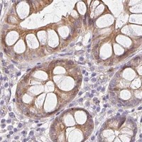
Real IHC data IHC analysis of SORLA/SORL1 using anti-SORLA/SORL1 antibody (A01887-1). SORLA/SORL1 was detected in a paraffin-embedded section of human breast cancer tissue. Heat mediated antigen retrieval was performed in EDTA buffer (pH 8.0, epitope retrieval solution). The tissue section was blocked with 10% goat serum. The tissue section was then incubated with 2 μg/ml rabbit anti-SORLA/SORL1 Antibody (A01887-1) overnight at 4°C. Peroxidase Conjugated Goat Anti-rabbit IgG was used as secondary antibody and incubated for 30 minutes at 37°C. The tissue section was developed using HRP Conjugated Rabbit IgG Super Vision Assay Kit (Catalog # SV0002) with DAB as the chromogen.
Anti-SORLA/SORL1 Antibody ®
Cat # A01887-1

M01887-2 is listed for IHC in human, mouse and rat; its images show paraffin sections of mouse and rat brain (catalog applications, reactivity and IHC captions). A01887-1 is listed for IHC and IF in human, mouse and rat; its images show IHC in human breast cancer tissue and mouse and rat brain, plus IF in human breast cancer tissue (catalog applications, reactivity and image captions).

Which to pick: For brain tissue IHC, choose M01887-2 if a rabbit monoclonal is preferred; its own captions show paraffin sections of mouse and rat brain, while human reactivity is listed without a human IHC image (catalog clone, reactivity and M01887-2 IHC captions). For IF, choose A01887-1: its IF caption shows a paraffin section of human breast cancer tissue; ICC validation is unreported (catalog applications and A01887-1 IF caption). For IHC across the three listed species, A01887-1 has images for each; both antibodies’ IHC captions describe paraffin sections and leave the fixative unreported (catalog reactivity and IHC captions).

Each figure is that product's own IHC / IF validation image from its datasheet.

References

  1. UniProt Consortium. UniProt entry Q92673 (SORL_HUMAN, Sortilin-related receptor).
  2. Human Protein Atlas. SORL1 tissue IHC expression (reliability: Uncertain).
  3. Human Protein Atlas. SORL1 subcellular location (ICC-IF): Membrane, Secreted.
  4. Human Protein Atlas. SORL1 antibody validation summary (2 antibodies).
  5. Identification and description of three families with familial Alzheimer disease that segregate variants in the SORL1 gene. Acta neuropathologica communications 2017 — PMC5465543.
  6. Upregulated of ANXA3, SORL1, and Neutrophils May Be Key Factors in the Progressionof Ankylosing Spondylitis. Frontiers in immunology 2022 — PMC9019158.
  7. A genetically modified minipig model for Alzheimer's disease with SORL1 haploinsufficiency. Cell reports. Medicine 2022 — PMC9512670.
  8. The transcriptomes of hypothalamic micropunches reveal sex differences in regulatory processes across hibernation in the Arctic ground squirrel. Physiological genomics 2025 — PMC12679999.
  9. PubMed PMID:9157966 — UniProt-cited evidence.
  10. PubMed PMID:8940146 — UniProt-cited evidence.
  11. PubMed PMID:16554811 — UniProt-cited evidence.