SOS1 / Son of sevenless homolog 1 · IHC design guide

Design Immunohistochemistry for SOS1

Plan SOS1 chromogenic IHC in paraffin sections using the catalog antibody’s validated protocol (datasheet A00837-1). Compare cytoplasmic staining across cell types while accounting for the tissue evidence’s uncertain reliability (HPA tissue IHC).

Evidence assembled Oct 2026 · For research use; verify linked source records and product datasheet before use
Immunohistochemistry protocol sheet for SOS1 (IHC for SOS1): expected localisation Cytoplasmic staining in tissue sections (HPA tissue IHC), antibody A00837-1, validated IHC image, and IHC protocol steps
Printable SOS1 IHC protocol sheet — expected localisation Cytoplasmic staining in tissue sections (HPA tissue IHC), antibody A00837-1, controls and protocol steps. Open the full SOS1 IHC guide →

SOS1 Immunohistochemistry Experimental Design Guide

Expected localisation, validated protocols, controls and antibodies — the at-a-glance facts below, then the full design guide.

Must know before staining
Expected localisation Cytoplasmic staining in tissue sections (HPA tissue IHC)
Staining pattern Variable cytoplasmic staining across cell types (HPA tissue IHC)
Antigen retrieval EDTA pH 8.0 HIER, heat-mediated (datasheet A00837-1)
Positive control ⓘ Cerebral cortex+4 more · see all
Negative control ⓘ Caudate+4 more · see all
Important caveats
Reasons your staining may differ from the expected pattern.
Fixation Keep fixation consistent across sections. (standard IHC practice; not target-specific)
Caveat Tissue staining awaits external verification (HPA tissue IHC)
Regulation Intensity varies by cell type (HPA tissue IHC)
Isoform / epitope 2 isoforms; epitope coverage is unspecified (UniProt)
Section 1

Recommended SOS1 IHC & IF Protocols

The catalog antibody’s IHC-P protocol (datasheet A00837-1) is accompanied by published SOS1 IHC methods for prostate tissue (PMC9974262), ovarian tissue (PMC3059077), and organoids (PMC10113742).

Recommended immunohistochemistry (IHC-P) protocol parameters
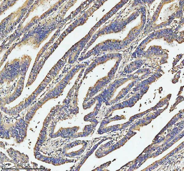
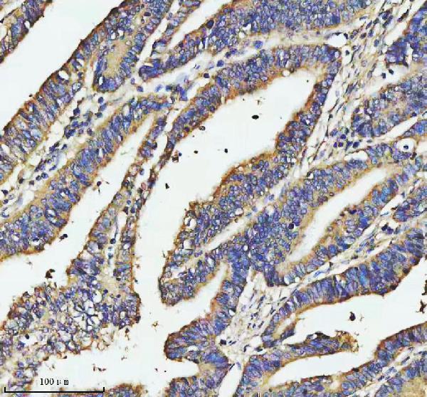
SampleParaffin-embedded human colon cancer tissue; fixative not specified (datasheet A00837-1)
FixationImage fixative and duration unreported (datasheet A00837-1); verify before use.
Sectioning4–5 µm sections on charged slides (standard)
DeparaffinisationXylene, graded ethanol series to water (standard)
Antigen retrievalHeat retrieval: EDTA pH 8.0 (datasheet A00837-1); 20 min, 95–100 °C (standard)
Peroxidase block3% H2O2, 10 min, room temperature (standard)
Blocking10% goat serum (datasheet A00837-1)
Primary antibodyRabbit anti-SOS1, 2-5 μg/ml (datasheet A00837-1)
Primary incubationOvernight at 4 °C (datasheet A00837-1)
DetectionHRP-conjugated secondary, DAB chromogen (datasheet A00837-1)
CounterstainHematoxylin, blue, dehydrate and mount (standard)
Expected resultSOS1-positive staining in neuronal cells of cerebral cortex (HPA tissue IHC: High). HPA tissue profile: Cytoplasmic expression at variable levels in several different cell types. No signal in the no-primary control.
💡Decision noteStart with heat-mediated EDTA retrieval at pH 8.0 for the catalog antibody (datasheet A00837-1). Use the published retrieval conditions when reproducing the organoid method (PMC10113742).
Section 2

What Is the Expected SOS1 Staining Pattern?

In paraffin-section IHC, expect predominantly cytoplasmic SOS1 staining at variable levels across cell types (HPA tissue IHC: Uncertain, pending external verification). HPA reports high staining in cerebral cortex and hippocampal neuronal cells, ovarian follicle cells, placental trophoblastic cells, and cells in seminiferous ducts (HPA tissue IHC). SOS1 has no transmembrane segment; UniProt does not annotate a subcellular location (UniProt Q07889 topology; UniProt Q07889 subcellular record).

What am I looking at on my slide?
Cytoplasmic chromogen in placental trophoblastic cells or ovarian follicle cells.This matches cell types scored High by HPA, but the tissue IHC profile remains Uncertain pending external verification (HPA tissue IHC). Compare stained cells with their morphology and a counterstain (general IHC practice).
Predominantly nuclear staining, with little cytoplasmic signal.That distribution conflicts with HPA's cytoplasmic tissue profile and approved cytosolic ICC-IF location (HPA tissue IHC; HPA ICC-IF). Check staining controls and morphology before assigning the nuclear signal to SOS1 (general IHC practice).
Strong chromogen in oral-mucosa squamous cells while expected positive cells are unstained.HPA reports SOS1 as Not detected in those squamous cells (HPA tissue IHC). Consider antibody cross-reactivity or endogenous detection activity, then compare a no-primary control; HPA's Uncertain IHC profile makes this a discrepancy to investigate, not proof of a false positive (HPA tissue IHC; general IHC practice).
Diffuse chromogen obscures cell borders and counterstained nuclei.The distribution cannot support cell-level localisation or intensity scoring (general IHC practice). Assess background with a no-primary control and compare it with HPA's reported cytoplasmic pattern (general IHC practice; HPA tissue IHC).
No signal in placental trophoblastic cells selected as a positive reference.HPA scores these cells High, so review whether they are present and whether the staining controls worked (HPA tissue IHC; general IHC practice). A negative section alone cannot establish absent SOS1 because the HPA IHC profile is Uncertain (HPA tissue IHC).
💡Expected SOS1 appearanceA plausible positive is variable-intensity cytoplasmic chromogen in correctly identified cells, such as HPA-High placental trophoblastic cells; isolated nuclear staining or widespread cell-independent colour warrants investigation (HPA tissue IHC: Uncertain; general IHC practice).
How each factor affects the staining
IHC evidence and antibody validationThe tissue pattern is Uncertain, pending external verification, and CAB005396 has Uncertain IHC status (HPA tissue IHC; HPA antibodies). Treat agreement with HPA as supportive, not definitive.
Protein topologySOS1 has no transmembrane segment and no UniProt subcellular annotation (UniProt Q07889 topology; UniProt Q07889 subcellular record). Interpret localisation against HPA's observed cytoplasmic tissue pattern (HPA tissue IHC).
Isoforms and epitopeUniProt lists 2 SOS1 isoforms (UniProt Q07889 isoforms). The supplied records do not map the catalog antibody's epitope, so isoform-specific staining cannot be inferred.
Modified residuesUniProt records phosphorylated residues, including Ser-1078 and Ser-1082 (UniProt Q07889 modified residues). The supplied sources do not establish whether these modifications alter this antibody's IHC staining.
IF/ICC Q: Where should SOS1 appear?A: Primarily in the cytosol (HPA ICC-IF: approved). This is an IF/ICC localisation reference; CAB005396 has Approved ICC status and Uncertain IHC status (HPA antibodies).
Why is my staining missing, weak or wrong?
SituationLikely causeNext action
Positive-reference section has no visible chromogen.The expected cells may be absent from the section, or an IHC step may have failed (general IHC practice).Confirm the cell type with morphology, then check retrieval, primary antibody, detection and controls using the catalog IHC-P instructions (general IHC practice).
Signal is nuclear rather than cytoplasmic.The result conflicts with HPA tissue and ICC-IF localisation; its cause is unresolved (HPA tissue IHC; HPA ICC-IF).Inspect a no-primary control and counterstained morphology; repeat interpretation only after the cytoplasmic reference pattern is assessable (general IHC practice).
Colour appears in HPA-Not-detected cell types.Possible cross-reactivity or endogenous detection activity; HPA's Uncertain IHC profile limits certainty (HPA tissue IHC; general IHC practice).Compare no-primary and positive-reference sections, and score cell types separately rather than treating all tissue colour as SOS1 (general IHC practice).
Diffuse background masks cytoplasm.Nonspecific staining or detection background may prevent localisation assessment (general IHC practice).Review blocking, washes, primary dilution and detection controls under the chosen IHC workflow; reassess cell-level staining after background falls (general IHC practice).
Only a sharp membrane rim is scored as positive.A rim alone does not match HPA's reported cytoplasmic tissue profile (HPA tissue IHC).Inspect cytoplasm and cell borders at higher magnification and compare staining controls before calling the rim SOS1 (general IHC practice).
Staining differs between sections or cell populations.HPA reports variable cytoplasmic expression and low tissue specificity, with Uncertain IHC reliability (HPA tissue IHC).Record the tissue, identified cell type, compartment and intensity for each section; use the same positive reference and controls when comparing runs (general IHC practice).

Sample controls for SOS1 IHC & IF

🧪Run placenta first: trophoblastic cells should stain strongly (HPA: High in trophoblastic cells). Use cervix glandular cells as a negative tissue control (HPA: Not detected in glandular cells); unstained non-trophoblastic cells on the placenta slide can indicate local background, but their SOS1-negative status is unverified.
Positive control tissue: Cerebral cortex (Neuronal cells, HPA High)
Negative control tissue: Caudate (HPA Not detected)
ICC-IF cell lines (HPA subcellular resource): HPA ICC-IF images show SOS1 in A-431, U-251MG, U2OS, with annotated localisation: Cytosol (approved) (HPA subcellular).
Technical controls: Include no-primary (secondary-only), a rabbit IgG isotype control matched to the primary antibody’s clonality, and SOS1-knockout material or a validated peptide-block control (hero caption: rabbit primary antibody). Check endogenous peroxidase in the placenta section before interpreting HRP/DAB staining, particularly in blood-containing areas (hero caption: HRP/DAB detection; standard IHC practice).
⚠️Feasibility: A target-specific fixation window or fixation effect is unreported, and the selected A00837-1 paraffin-section caption does not state a fixative (hero caption: fixative unreported). That caption uses heat-mediated retrieval in EDTA at pH 8.0, but does not establish whether retrieval is essential (hero caption: EDTA retrieval). Whether frozen sections or IF are easier is unestablished; placental blood can complicate chromogenic interpretation through endogenous peroxidase activity (standard IHC practice).

HPA tissue IHC evidence for SOS1

Comprehensive Human Protein Atlas IHC scoring per tissue (reliability: Uncertain — Pending external verification.). Rows are taken directly from the HPA tissue chart — click any row's HPA link to view the source.

Positive expression · recommended positive controls

TissueCell typeLevelEvidenceSource
Cerebral cortex Neuronal cells High Protein (IHC) HPA →
Hippocampus Neuronal cells High Protein (IHC) HPA →
Ovary Follicle cells High Protein (IHC) HPA →
Placenta Trophoblastic cells High Protein (IHC) HPA →
Testis Cells in seminiferous ducts High Protein (IHC) HPA →

Undetected expression · recommended negative controls

TissueCell typeLevelEvidenceSource
Caudate Glial cells Not detected Protein (IHC) HPA →
Cervix Glandular cells Not detected Protein (IHC) HPA →
Oral mucosa Squamous epithelial cells Not detected Protein (IHC) HPA →
Prostate Glandular cells Not detected Protein (IHC) HPA →
Skeletal muscle Myocytes Not detected Protein (IHC) HPA →
Section 3

Advanced SOS1 IHC Tips

Troubleshoot SOS1 staining in paraffin sections by checking retrieval, antibody controls, cellular distribution and scoring before interpreting differences between specimens.

Which antigen retrieval should I try first for SOS1 IHC?
Start with heat-mediated retrieval in EDTA pH 8.0 for this catalog antibody (datasheet A00837-1). Its paraffin-section example used that retrieval before 2 μg/ml primary antibody overnight at 4°C, so use the example as a starting condition rather than a guarantee for every specimen (datasheet A00837-1). If staining is weak, adjust heating duration in small steps while keeping buffer and pH fixed, and process a reference section alongside each trial (standard IHC practice). Compare signal in glandular cells with section background; HPA reports medium colon glandular-cell staining but marks its tissue-IHC reliability uncertain (HPA: Colon; HPA: reliability Uncertain).
Could fixation explain weak or uneven SOS1 staining?
Target-specific sensitivity to fixation is unknown because the catalog paraffin-section caption does not state a fixative (datasheet A00837-1). Record the fixative, fixation duration and tissue thickness for each specimen, then compare sections processed together before changing antibody concentration (standard IHC practice). Include a reference section retrieved in EDTA pH 8.0 and stained with the same 2 μg/ml primary-antibody starting condition (datasheet A00837-1). If weak staining tracks a processing batch, examine morphology and retrieval consistency before attributing it to SOS1 abundance (standard IHC practice). HPA tissue patterns cannot establish how fixation affects this epitope (HPA: tissue IHC; datasheet A00837-1).
Where should SOS1 staining appear, and how should membrane-adjacent signal be judged?
Expect predominantly cytoplasmic staining in tissue sections (HPA: cytoplasmic expression) and cytosolic localisation in cell imaging (HPA: Cytosol, approved). SOS1 has no annotated transmembrane segment, so a sharp continuous cell-surface outline alone needs additional validation (UniProt Q07889 topology). Membrane-adjacent enrichment may be biologically plausible because SOS1 interacts through its C-terminus with GRB2, but this does not establish a fixed IHC pattern (UniProt Q07889: subunit). Examine high-power views against neighbouring cells, and compare serial sections stained at 2 μg/ml with a no-primary control (datasheet A00837-1; standard IHC practice). Treat isolated nuclear or luminal deposits cautiously when cytoplasmic signal is absent (HPA: tissue IHC profile; standard IHC practice).
How can isoforms or epitope placement affect SOS1 IHC interpretation?
SOS1 has 2 annotated isoforms, but the supplied catalog caption does not identify the antibody epitope or establish isoform coverage (UniProt Q07889: isoforms; datasheet A00837-1). Request epitope information before interpreting different staining patterns as isoform-specific; a shared-domain antibody may not distinguish the isoforms (standard IHC practice). Relevant mapped regions include the DH domain at residues 200–390 and Ras-GEF domain at 780–1019 (UniProt Q07889: domains). Several C-terminal serines are phosphorylated, including 1078 and 1082, but their effect on this antibody’s IHC binding is unreported (UniProt Q07889: modified residues; datasheet A00837-1). Validate any proposed epitope-dependent difference with an independently mapped antibody or orthogonal assay (standard IHC practice).
How should I adapt SOS1 localisation checks to multiplex IF?
Use IF to assess the expected cytosolic distribution, ideally alongside a marker that identifies the cell population being scored (HPA: Cytosol, approved; standard IF practice). For neuronal tissue, select a neuronal-cell marker because HPA reports high SOS1 staining in cerebral-cortex and hippocampal neuronal cells, while its tissue-IHC reliability remains uncertain (HPA: Cerebral cortex; HPA: Hippocampus; HPA: reliability Uncertain). Choose a spectrally separated, preferably far-red SOS1 fluorophore after checking unstained-tissue autofluorescence and single-label bleed-through (standard IF practice). SOS1 lacks a transmembrane segment, so optimise permeabilisation for access to an intracellular epitope rather than assuming extracellular accessibility (UniProt Q07889 topology; standard IF practice). The supplied IHC caption gives no IF dilution or IF fixation condition (datasheet A00837-1).
What should I check when DAB background obscures SOS1 staining?
First compare a no-primary section with the stained section to identify signal from the detection system or tissue itself (standard IHC practice). The catalog example used 10% goat-serum blocking, a peroxidase-conjugated secondary and DAB development, but it does not report a peroxidase-block step (datasheet A00837-1). Add a peroxidase block where appropriate, then review wash quality, secondary-antibody specificity and DAB development time as general chromogenic controls (standard IHC practice). If background rises with primary concentration, titrate around the documented 2 μg/ml starting condition while keeping retrieval at EDTA pH 8.0 (datasheet A00837-1). Judge improvement by clearer cellular cytoplasm, consistent with the reported tissue pattern (HPA: tissue IHC profile).
How should SOS1 IHC be scored across heterogeneous tissue sections? ⚠ ANSWER MARKED FOR VERIFICATION
Define the cell population and compartment before scoring, because HPA describes variable cytoplasmic staining among cell types (HPA: tissue IHC profile). For each region, record the percentage of positive cells and staining intensity, then calculate an H-score using 0–3 intensity categories if that scheme fits the study design (standard IHC practice). Normalise positive counts to eligible, morphologically intact cells, or report positive-cell density per mm² of viable analysed tissue (standard IHC practice). Keep retrieval, imaging and threshold settings consistent across slides, and score reference sections within the same batch (standard IHC practice). Report the HPA tissue-IHC uncertainty when using its patterns to choose comparison regions (HPA: reliability Uncertain).
How can I distinguish plausible SOS1 positivity from staining artefact?
A plausible result shows intracellular, chiefly cytoplasmic signal in identifiable cells rather than staining confined to section edges, necrotic debris or luminal material (HPA: tissue IHC profile; standard IHC practice). Compare the cell type as well as intensity: HPA reports high staining in placental trophoblastic cells and no detected staining in prostate glandular cells, with uncertain overall tissue-IHC reliability (HPA: Placenta; HPA: Prostate; HPA: reliability Uncertain). Check a no-primary section for endogenous enzyme or detection background, particularly before calling diffuse DAB deposition positive (standard IHC practice). The catalog colon-cancer image demonstrates staining after EDTA pH 8.0 retrieval, but one image does not establish diagnostic specificity (datasheet A00837-1). Confirm consequential findings with an independent antibody or orthogonal evidence (standard IHC practice).
Boster reagents

Best SOS1 / Son of sevenless homolog 1 IHC Antibodies

A00837-1 has a human colon cancer paraffin-section IHC image (catalog image caption). Both antibodies list Human, Mouse and Rat reactivity; only M00837 lists ICC/IF (catalog applications/reactivity).

Real IHC data IHC analysis of SOS1 using anti-SOS1 antibody (A00837-1). SOS1 was detected in a paraffin-embedded section of human colon cancer tissue. Heat mediated antigen retrieval was performed in EDTA buffer (pH 8.0, epitope retrieval solution). The tissue section was blocked with 10% goat serum. The tissue section was then incubated with 2 μg/ml rabbit anti-SOS1 Antibody (A00837-1) overnight at 4°C. Peroxidase Conjugated Goat Anti-rabbit IgG was used as secondary antibody and incubated for 30 minutes at 37°C. The tissue section was developed using HRP Conjugated Rabbit IgG Super Vision Assay Kit (Catalog # SV0002) with DAB as the chromogen.
Anti-SOS1 Antibody ®
Cat # A00837-1

A00837-1 will render with an IHC image from a human colon cancer paraffin section; its listed reactivity is Human, Mouse and Rat (A00837-1 image caption; catalog reactivity). M00837 will render with IHC and ICC/IF listed for Human, Mouse and Rat, but no IHC or IF image is supplied (catalog applications/reactivity; image payload).

Which to pick: For tissue IHC, choose A00837-1: its own caption documents staining in a human colon cancer paraffin section with EDTA retrieval; the fixative is unreported (A00837-1 image caption). For IF/ICC, choose M00837 because this rabbit monoclonal lists ICC/IF, though no IF image is supplied (catalog host/clone/applications; image payload). For cross-species work, both list Human, Mouse and Rat reactivity, so choose by application and verify staining in the intended species (catalog applications/reactivity; standard IHC practice).

Each figure is that product's own IHC / IF validation image from its datasheet.

References

  1. UniProt Consortium. UniProt entry Q07889 (SOS1_HUMAN, Son of sevenless homolog 1).
  2. Human Protein Atlas. SOS1 tissue IHC expression (reliability: Uncertain).
  3. Human Protein Atlas. SOS1 subcellular location (ICC-IF): Localized to the cytosol..
  4. Human Protein Atlas. SOS1 antibody validation summary (1 antibodies).
  5. Identification and Validation of FGF-Related Prognostic Signatures in Prostate Cancer. Disease markers 2023 — PMC9974262.
  6. Integrity of SOS1/EPS8/ABI1 tri-complex determines ovarian cancer metastasis. Cancer research 2010 — PMC3059077.
  7. Translational relevance of SOS1 targeting for KRAS-mutant colorectal cancer. Molecular carcinogenesis 2023 — PMC10330439.
  8. Development of SOS1 Inhibitor-Based Degraders to Target KRAS-Mutant Colorectal Cancer. Journal of medicinal chemistry 2022 — PMC10113742.
  9. PubMed PMID:8493579 — UniProt-cited evidence.
  10. PubMed PMID:14702039 — UniProt-cited evidence.
  11. PubMed PMID:15815621 — UniProt-cited evidence.