This website uses cookies to ensure you get the best experience on our website.


- Table of Contents
Plan SPAG5 paraffin IHC with the catalog antibody at 2–5 µg/mL (datasheet A07062-2). Compare cytoplasmic staining in testis and kidney tubules with a negative-control section, while accounting for low consistency between antibody staining and RNA expression (HPA tissue IHC).

Expected localisation, validated protocols, controls and antibodies — the at-a-glance facts below, then the full design guide.
| Expected localisation | Cytoplasmic staining in most tissues (HPA tissue IHC) | |
| Staining pattern | Variable cytoplasmic staining across most tissues, including testis (HPA tissue IHC) | |
| Antigen retrieval | EDTA pH 8.0 HIER, heat-mediated (datasheet A07062-2) | |
| Positive control | Bone marrow+4 more · see all | |
| Negative control | Heart muscle+4 more · see all |
| Fixation | Keep paraffin-section fixation consistent across samples. (standard IHC practice; not target-specific) | |
| Caveat | Antibody staining has low consistency with RNA expression (HPA tissue IHC) | |
| Regulation | Tissue enhanced RNA: marrow, kidney, testis (HPA tissue IHC: RNA specificity) | |
| Isoform / epitope | No annotated isoforms or processing variants (UniProt) |
The catalog antibody’s IHC-P protocol (datasheet: A07062-2) is accompanied by three published SPAG5 IHC protocols (PMC9116224; PMC11657553; PMC7007403).
| Sample | Paraffin-embedded human renal clear cell carcinoma tissue; fixative not specified (datasheet A07062-2) |
| Fixation | Image fixative and duration unreported (datasheet A07062-2); verify before use. |
| Sectioning | 4–5 µm sections on charged slides (standard) |
| Deparaffinisation | Xylene, graded ethanol series to water (standard) |
| Antigen retrieval | Heat retrieval: EDTA pH 8.0 (datasheet A07062-2); 20 min, 95–100 °C (standard) |
| Peroxidase block | 3% H2O2, 10 min, room temperature (standard) |
| Blocking | 10% goat serum (datasheet A07062-2) |
| Primary antibody | Rabbit anti-SPAG5, 2-5μg/ml (datasheet A07062-2) |
| Primary incubation | Overnight at 4 °C (datasheet A07062-2) |
| Detection | Streptavidin-biotin complex (SABC), DAB chromogen (datasheet A07062-2) |
| Counterstain | Hematoxylin, blue, dehydrate and mount (standard) |
| Expected result | SPAG5-positive staining in hematopoietic cells of bone marrow (HPA tissue IHC: High). HPA tissue profile: Cytoplasmic expression at variable levels in most tissues, including testis. No signal in the no-primary control. |
SPAG5 is a non-membrane protein associated with cytoplasm, microtubules and the mitotic spindle (UniProt Q96R06 topology and subcellular location). In paraffin sections, expect cytoplasmic staining in cell populations reported positive by HPA, particularly seminiferous duct cells, kidney tubule cells and bone marrow hematopoietic cells (HPA tissue IHC: High). Interpret intensity cautiously: HPA rates its tissue IHC staining Approved but reports low consistency with RNA expression (HPA tissue IHC reliability).
| Distinct cytoplasmic staining in seminiferous duct cells, kidney tubules or bone marrow hematopoietic cells. | This fits the reported High staining in those cell populations (HPA tissue IHC). Compare the named cells with adjacent cells; HPA's levels describe observed staining, not an intensity threshold for every cell (HPA tissue IHC). |
| Signal concentrated around mitotic figures, with less conspicuous staining in neighboring cells. | A spindle-associated pattern is biologically plausible: SPAG5 occurs at spindle poles, across the spindle and later at midzone microtubules as mitosis progresses (UniProt Q96R06 subcellular location). A chromogenic section may not resolve each structure (general IHC practice). |
| Predominantly nuclear-only staining across many cells, without a convincing cytoplasmic pattern. | Reassess compartment assignment, counterstain and detection controls before calling it SPAG5 (general IHC practice). HPA describes cytoplasmic tissue IHC, although its separate ICC-IF data support nuclear bodies; nuclear signal alone is therefore not a universal artefact (HPA tissue IHC; HPA subcellular ICC-IF). |
| Strong staining in cardiomyocytes or pancreatic exocrine glandular cells. | Those specific cell populations were not detected in HPA tissue IHC. Check morphology and controls for possible cross-reactivity or endogenous detection activity before treating the signal as SPAG5 (HPA tissue IHC; general IHC practice). |
| Diffuse deposit obscures cell boundaries, or no signal appears in a known-positive comparison section. | Diffuse background prevents a reliable compartment call; a blank positive comparison leaves assay performance unresolved (general IHC practice). Neither result alone establishes SPAG5 absence in the test section, especially given the reported variability in tissue staining (HPA tissue IHC). |
| Cell population and tissue | HPA reports High staining in bone marrow hematopoietic cells, kidney tubules and testis seminiferous ducts, but not detected in cardiomyocytes (HPA tissue IHC). Score the identified cells, not the entire section. |
| Cell-cycle state | Interphase SPAG5 can be punctate; mitotic localization shifts from spindle poles to spindle and midzone structures (UniProt Q96R06 subcellular location). The visible pattern can therefore vary among cells in one section. |
| Strength of tissue evidence | HPA labels tissue IHC Approved while noting low staining–RNA consistency; two listed antibodies have IHC Approved status (HPA tissue IHC; HPA antibodies). Treat an unexpected pattern as a finding to verify. |
| Topology and processing | UniProt lists no transmembrane segment or signal peptide and annotates one full-length chain (UniProt Q96R06 topology and processing). These annotations support an intracellular expectation; they do not predict retrieval or fixation sensitivity. |
| IF/ICC Q: Should its pattern match tissue IHC? | HPA ICC-IF supports nuclear bodies and mitotic spindle, with additional cytosol, whereas tissue IHC describes variable cytoplasmic staining (HPA subcellular ICC-IF; HPA tissue IHC). Interpret each application with its own evidence. |
| Situation | Likely cause | Next action |
|---|---|---|
| No staining in the test section or the positive comparison. | Assay failure remains possible; HPA reports High staining in the named testis, kidney and marrow cell populations (HPA tissue IHC). | Repeat with a documented positive tissue and verify the IHC-P retrieval, antibody and chromogenic detection steps used in the run (general IHC practice). |
| The positive comparison stains, but the test section does not. | Expression may differ by cell population; HPA also reports several specific cell types as not detected (HPA tissue IHC). | Identify the cell type on the counterstain and compare its reported HPA level before interpreting the absence (HPA tissue IHC; general IHC practice). |
| A HPA-negative cell population stains strongly. | Cross-reactivity or endogenous detection activity is possible; the discrepancy alone cannot identify its cause (HPA tissue IHC; general IHC practice). | Inspect antibody-omission and detection controls, then reassess morphology and the affected cell population (general IHC practice). |
| Diffuse chromogen obscures intracellular detail. | Background prevents a dependable cytoplasmic or spindle-associated interpretation (general IHC practice). | Review blocking, antibody dilution, washing and detection controls using the established IHC-P workflow; reassess only after cell boundaries are readable (general IHC practice). |
| Signal appears nuclear-only in tissue IHC. | It differs from HPA's cytoplasmic tissue description, although nuclear bodies are supported in ICC-IF (HPA tissue IHC; HPA subcellular ICC-IF). | Check counterstain alignment and controls, then report the application and compartment explicitly instead of equating the two HPA patterns (general IHC practice; HPA subcellular ICC-IF). |
| A positive section shows uneven intensity among cells. | HPA describes variable cytoplasmic tissue staining, and UniProt describes cell-cycle-dependent localization (HPA tissue IHC; UniProt Q96R06 subcellular location). | Score defined cell populations and compartments separately; compare sections using the same detection and scoring conditions (general IHC practice). |


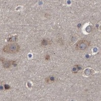


Comprehensive Human Protein Atlas IHC scoring per tissue (reliability: Approved — Low consistency between antibody staining and RNA expression data.). Rows are taken directly from the HPA tissue chart — click any row's HPA link to view the source.
| Tissue | Cell type | Level | Evidence | Source |
|---|---|---|---|---|
| Bone marrow | Hematopoietic cells | High | Protein (IHC) | HPA → |
| Kidney | Cells in tubules | High | Protein (IHC) | HPA → |
| Testis | Cells in seminiferous ducts | High | Protein (IHC) | HPA → |
| Tonsil | Non-germinal center cells | High | Protein (IHC) | HPA → |
| Adrenal gland | Glandular cells | Medium | Protein (IHC) | HPA → |
| Tissue | Cell type | Level | Evidence | Source |
|---|---|---|---|---|
| Heart muscle | Cardiomyocytes | Not detected | Protein (IHC) | HPA → |
| Oral mucosa | Squamous epithelial cells | Not detected | Protein (IHC) | HPA → |
| Pancreas | Exocrine glandular cells | Not detected | Protein (IHC) | HPA → |
| Salivary gland | Glandular cells | Not detected | Protein (IHC) | HPA → |
| Skeletal muscle | Myocytes | Not detected | Protein (IHC) | HPA → |
Use cell type, compartment and staining controls to assess SPAG5 in paraffin section IHC (HPA tissue IHC; UniProt Q96R06).
A07062-2 has human paraffin-section IHC images and a U20S-cell IF image (catalog images); its listed reactivity is human (catalog: reactivity).
A07062-2 has IHC images from human renal clear cell carcinoma, liver cancer, lung cancer, and rectal cancer paraffin sections (A07062-2 IHC captions). It also has an IF image from U20S cells (A07062-2 IF caption).
Which to pick: Choose A07062-2 for human paraffin-section IHC: its renal clear cell carcinoma image documents EDTA retrieval at pH 8.0 and 2 μg/ml primary antibody (A07062-2 IHC caption); the fixative is unreported (A07062-2 IHC captions). The same SKU lists IF/ICC and shows IF in U20S cells at 5 μg/ml (A07062-2 applications and IF caption). No cross-species choice is supported because the listed reactivity is human only; clonality is unreported (A07062-2 catalog).