This website uses cookies to ensure you get the best experience on our website.


- Table of Contents
Plan chromogenic IHC on paraffin sections using the catalog antibody's reported conditions (datasheet A09906-1) and ciliated-cell staining as a tissue reference (HPA tissue IHC). Interpret staining with the reported off-target binding caveat (HPA tissue IHC) and stress-dependent mitochondrial relocalization in mind (UniProt).

Expected localisation, validated protocols, controls and antibodies — the at-a-glance facts below, then the full design guide.
| Expected localisation | Ciliary rootlets, cell bodies and cytoplasm (HPA tissue IHC) | |
| Staining pattern | Ciliated-cell rootlets/body; cytoplasm in seminiferous ducts (HPA tissue IHC) | |
| Antigen retrieval | EDTA pH 8.0 HIER, heat-mediated (datasheet A09906-1) | |
| Positive control | Bronchus+4 more · see all | |
| Negative control | Adipose tissue+4 more · see all |
| Fixation | Keep fixation consistent across sections (standard IHC practice; not target-specific); Matched tissue-IHC evidence does not establish the fixation claim. Validate the specimen-specific method before use. (selected-SKU IHC image A09906-1) | |
| Caveat | Presumed off-target binding may confound staining (HPA tissue IHC) | |
| Regulation | Stress triggers mitochondrial relocalization (UniProt) | |
| Isoform / epitope | 2 isoforms; epitope differences unspecified (UniProt) |
The catalog antibody’s IHC-P protocol is paired with published SPATA18 methods for colorectal cancer, clear cell renal cell carcinoma, and mouse paraffin sections (PMC8910917; PMC9924025; PMC10845071).
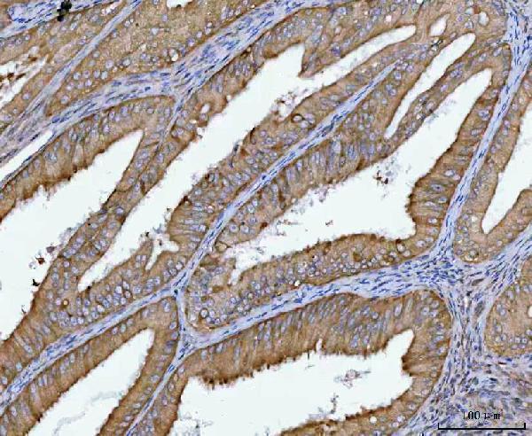
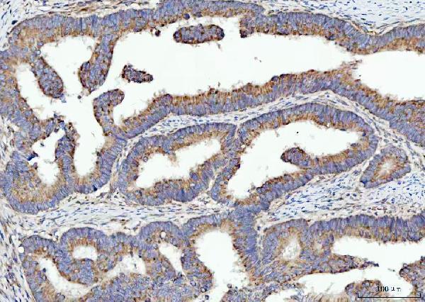
| Sample | Paraffin-embedded human endometrial carcinoma tissue; fixative not specified (datasheet A09906-1) |
| Fixation | Image fixative and duration unreported (datasheet A09906-1); verify before use. |
| Sectioning | 4–5 µm sections on charged slides (standard) |
| Deparaffinisation | Xylene, graded ethanol series to water (standard) |
| Antigen retrieval | Heat retrieval: EDTA pH 8.0 (datasheet A09906-1); 20 min, 95–100 °C (standard) |
| Peroxidase block | 3% H2O2, 10 min, room temperature (standard) |
| Blocking | 10% goat serum (datasheet A09906-1) |
| Primary antibody | Rabbit anti-SPATA18, 2-5 μg/ml (datasheet A09906-1) |
| Primary incubation | Overnight at 4 °C (datasheet A09906-1) |
| Detection | HRP-conjugated secondary, DAB chromogen (datasheet A09906-1) |
| Counterstain | Hematoxylin, blue, dehydrate and mount (standard) |
| Expected result | SPATA18-positive staining in ciliated cells (ciliary rootlets) of bronchus (HPA tissue IHC: High). HPA tissue profile: Expression mainly in ciliated cells of respiratory epithelium, fallopian tube and endometrium. Cytoplasmic expression in cells in seminiferous ducts and choroid plexus. No signal in the no-primary control. |
SPATA18 staining should be assessed by cell type and compartment: bronchial and fallopian tube ciliated cells and late spermatids show high IHC signal (HPA: tissue IHC). Cytoplasmic staining is plausible because SPATA18 is cytosolic under normal conditions and can relocate to mitochondria after stress (UniProt Q8TC71: subcellular location; no transmembrane segment). HPA rates tissue IHC reliability as Enhanced, while noting medium RNA agreement and disregarded presumed off-target binding (HPA: tissue IHC).
| Strong chromogenic signal at bronchial or fallopian tube ciliary rootlets, or in late spermatids. | This matches the reported high-staining cells; nasopharyngeal ciliated cell bodies are another high-signal reference (HPA: tissue IHC). Score the identified cells and structures rather than treating an entire tissue section as uniformly positive. |
| Predominantly cytoplasmic staining in the expected cells, with a less sharply defined intracellular distribution. | A cytoplasmic pattern is compatible with normal cytosolic localization (UniProt Q8TC71: subcellular location). Mitochondrial relocation after stress is also reported, so a single IHC section cannot establish the cell's stress state from staining distribution alone (UniProt Q8TC71: subcellular location). |
| Isolated nuclear-only or extracellular staining dominates the chromogenic section. | Question its specificity against the expected tissue pattern (HPA: tissue IHC) and cytosolic or mitochondrial localization (UniProt Q8TC71: subcellular location). Nuclear-only IHC should not be declared biological solely from this image; HPA's additional nucleoplasmic localization comes from ICC-IF (HPA: subcellular). |
| Strong staining appears in adipocytes or bone marrow hematopoietic cells. | These cells are reported as not detected (HPA: tissue IHC). Check for cross-reactivity and endogenous detection activity using appropriate controls (general IHC practice); HPA also notes presumed off-target binding that was disregarded in its tissue assessment (HPA: tissue IHC). |
| No staining appears in an otherwise interpretable bronchus or fallopian tube section. | Those tissues contain high-staining ciliated rootlets in the HPA profile (HPA: tissue IHC). Confirm that the relevant ciliated cells are present, then assess antibody and detection performance with controls; absence of signal alone does not establish biological absence (general IHC practice). |
| Cell and tissue selection | HPA reports high signal in bronchial and fallopian tube ciliary rootlets, nasopharyngeal ciliated cell bodies, and late spermatids (HPA: tissue IHC). Its negative calls apply to specified cells, such as adipocytes and bone marrow hematopoietic cells, rather than every cell in those tissues (HPA: tissue IHC). |
| Compartment and cellular state | SPATA18 is cytosolic under normal conditions and relocates to the mitochondrial outer membrane after cellular stress; matrix condensates are proposed (UniProt Q8TC71: subcellular location). HPA's supported main mitochondrial location comes from ICC-IF (HPA: subcellular), so do not require IHC chromogen to resolve individual mitochondria. |
| Antibody validation and interpretation | Both listed antibodies have Enhanced IHC status, meaning the pattern was reproduced by independent antibodies or orthogonal data (HPA: antibodies). HPA nevertheless reports medium agreement with RNA and disregarded presumed off-target binding (HPA: tissue IHC); validation supports the reported pattern without making every stained structure specific. |
| Isoforms and unreported epitope coverage | Two isoforms are annotated, with one 1–538 chain and no annotated signal peptide or propeptide (UniProt Q8TC71: isoforms and processing). The supplied record does not map the IHC antibody epitope, so staining cannot be assigned to a particular isoform or interpreted as cleavage-dependent from these data. |
| Situation | Likely cause | Next action |
|---|---|---|
| Known-positive ciliated tissue has no chromogenic signal. | The sampled field may lack the relevant ciliated cells, or the IHC workflow may have failed; HPA reports high staining at bronchial and fallopian tube rootlets (HPA: tissue IHC). | Verify the cell type and slide integrity, then check primary antibody, antigen retrieval, detection reagents, and a concurrently processed positive control (general IHC practice). Do not infer target-specific retrieval or fixation sensitivity from HPA's staining levels. |
| Adipocytes or bone marrow hematopoietic cells stain strongly. | Those cell types are listed as not detected (HPA: tissue IHC); nonspecific antibody binding or endogenous detection activity can produce misleading chromogen (general IHC practice). | Review a no-primary control and the detection-system controls, then compare localization with an expected positive cell type (general IHC practice; HPA: tissue IHC). Treat discordant staining cautiously because HPA notes disregarded presumed off-target binding (HPA: tissue IHC). |
| Brown haze covers cells, stroma, and empty areas. | Diffuse background can result from inadequate blocking or washing, excessive antibody concentration, or detection background (general IHC practice). It does not resemble the cell-restricted high-signal profile (HPA: tissue IHC). | Inspect no-primary and reagent controls; optimize blocking, washing, antibody concentration, and detection conditions according to the assay instructions (general IHC practice). Judge any remaining signal by its location in the expected cells (HPA: tissue IHC). |
| A section shows mainly nuclear-only signal. | The expected tissue IHC description emphasizes ciliated structures and cytoplasmic expression (HPA: tissue IHC). Additional nucleoplasmic localization is approved in ICC-IF, but that evidence does not establish nuclear-only chromogenic IHC as the expected pattern (HPA: subcellular). | Compare an expected positive tissue and a no-primary control, and assess whether cytoplasmic or rootlet staining is also present (general IHC practice; HPA: tissue IHC). Keep the nuclear finding provisional unless supported independently. |
| Bronchial rootlets stain, but adjacent nonciliated cells do not. | A cell-restricted result is consistent with the high bronchial ciliary-rootlet finding (HPA: tissue IHC). The HPA profile does not assign a single intensity to every cell in the bronchus. | Score ciliated and nonciliated populations separately, recording the stained structure and intensity (general IHC practice). Use the HPA cell-level pattern, rather than whole-section uniformity, for interpretation (HPA: tissue IHC). |
| Q: Should an ICC-IF image show exactly the same pattern as chromogenic IHC? | A: HPA's supported main ICC-IF location is mitochondrial, with additional approved cytosolic and nucleoplasmic locations (HPA: subcellular); tissue IHC instead reports staining in specific cells and structures (HPA: tissue IHC). | Interpret ICC-IF localization in its dedicated guide; here, use the tissue IHC cell pattern to assess paraffin sections (HPA: tissue IHC and subcellular). Do not equate apparent mitochondrial detail in fluorescence with what chromogenic IHC must resolve. |


Comprehensive Human Protein Atlas IHC scoring per tissue (reliability: Enhanced — Medium consistency between antibody staining and RNA expression data. Presumed off target binding observed and disregarded. Protein expression in respiratory epithelia can be correlated with lung RNA-seq data.). Rows are taken directly from the HPA tissue chart — click any row's HPA link to view the source.
| Tissue | Cell type | Level | Evidence | Source |
|---|---|---|---|---|
| Bronchus | Ciliated cells (ciliary rootlets) | High | Protein (IHC) | HPA → |
| Fallopian tube | Ciliated cells (ciliary rootlets) | High | Protein (IHC) | HPA → |
| Nasopharynx | Ciliated cells (cell body) | High | Protein (IHC) | HPA → |
| Testis | Elongated or late spermatids | High | Protein (IHC) | HPA → |
| Adrenal gland | Glandular cells | Medium | Protein (IHC) | HPA → |
| Tissue | Cell type | Level | Evidence | Source |
|---|---|---|---|---|
| Adipose tissue | Adipocytes | Not detected | Protein (IHC) | HPA → |
| Appendix | Glandular cells | Not detected | Protein (IHC) | HPA → |
| Bone marrow | Hematopoietic cells | Not detected | Protein (IHC) | HPA → |
| Caudate | Glial cells | Not detected | Protein (IHC) | HPA → |
| Cerebellum | Cells in granular layer | Not detected | Protein (IHC) | HPA → |
Troubleshoot SPATA18 staining in paraffin sections by checking retrieval, cell type, compartment, controls and scoring against the documented IHC evidence.
A09906-1 has real IHC data from human paraffin-embedded tumor sections (A09906-1 IHC captions). The catalog reports no IF/ICC validation or nonhuman reactivity (catalog applications/reactivity).
A09906-1 is listed for human IHC and has images from paraffin-embedded endometrial carcinoma, rectum adenocarcinoma, and testicular germ cell tumor sections (catalog applications/reactivity; A09906-1 IHC captions). It is the only SKU supplied, and no IF image is supplied (catalog payload).
Which to pick: Choose A09906-1 for human paraffin-section IHC; its rabbit antibody is listed for IHC, and its own captions document staining in the three human tumor section types (catalog host/applications/reactivity; A09906-1 IHC captions). The captions do not report the fixative, and the catalog does not report a clone (A09906-1 IHC captions; catalog clone). No SKU in the payload is validated for IF/ICC or listed as reactive with another species (catalog applications/reactivity).