SPINK4 / Serine protease inhibitor Kazal-type 4 · IHC design guide

Design Immunohistochemistry for SPINK4

Plan SPINK4 IHC in paraffin sections using the selective cytoplasmic goblet-cell pattern in gastrointestinal mucosa as a reference (HPA tissue IHC). The guide covers fixation, staining interpretation and the possibility of signal beyond producing cells for a secreted protein (UniProt).

Evidence assembled Oct 2026 · For research use; verify linked source records and product datasheet before use
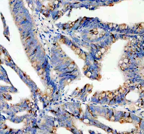
Immunohistochemistry protocol sheet for SPINK4 (IHC for SPINK4): expected localisation Goblet-cell cytoplasm in gastrointestinal mucosa (HPA tissue IHC), antibody A17177, validated IHC image, and IHC protocol steps
Printable SPINK4 IHC protocol sheet — expected localisation Goblet-cell cytoplasm in gastrointestinal mucosa (HPA tissue IHC), antibody A17177, controls and protocol steps. Open the full SPINK4 IHC guide →

SPINK4 Immunohistochemistry Experimental Design Guide

Expected localisation, validated protocols, controls and antibodies — the at-a-glance facts below, then the full design guide.

Must know before staining
Expected localisation Goblet-cell cytoplasm in gastrointestinal mucosa (HPA tissue IHC)
Staining pattern Selective cytoplasmic staining in gastrointestinal goblet cells (HPA tissue IHC)
Antigen retrieval EDTA pH 8.0 HIER, heat-mediated (datasheet A17177)
Positive control ⓘ Appendix+4 more · see all
Negative control ⓘ Adipose tissue+4 more · see all
Important caveats
Reasons your staining may differ from the expected pattern.
Fixation Keep fixation consistent across sections (standard IHC practice; not target-specific). Selected-image fixative and duration unreported (datasheet A17177); verify before use.
Caveat Secreted protein may appear beyond producing cells (UniProt)
Regulation Intestine-enriched expression; regulation unreported (HPA tissue IHC)
Isoform / epitope No annotated isoforms; mature secreted chain spans residues 27–86 (UniProt)
Section 1

Recommended SPINK4 IHC & IF Protocols

Start with the catalog antibody’s IHC protocol (datasheet: A17177), then compare published SPINK4 staining of rectal and colorectal tumors (PMC8293060; PMC10961291; PMC6938003).

Recommended immunohistochemistry (IHC-P) protocol parameters
SampleParaffin-embedded human colon cancer tissue; fixative not specified (datasheet A17177)
FixationImage fixative and duration unreported (datasheet A17177); verify before use.
Sectioning4–5 µm sections on charged slides (standard)
DeparaffinisationXylene, graded ethanol series to water (standard)
Antigen retrievalHeat retrieval: EDTA pH 8.0 (datasheet A17177); 20 min, 95–100 °C (standard)
Peroxidase block3% H2O2, 10 min, room temperature (standard)
Blocking10% goat serum (datasheet A17177)
Primary antibodyRabbit anti-SPINK4, 2-5 μg/ml (datasheet A17177)
Primary incubationOvernight at 4 °C (datasheet A17177)
DetectionHRP-conjugated secondary, DAB chromogen (datasheet A17177)
CounterstainHematoxylin, blue, dehydrate and mount (standard)
Expected resultSPINK4-positive staining in goblet cells of appendix (HPA tissue IHC: High). HPA tissue profile: Selective cytoplasmic expression in goblet cells of gastrointestinal mucosa. No signal in the no-primary control.
💡Decision noteTry EDTA pH 8.0 heat retrieval first for the catalog antibody (datasheet: A17177). The published citrate method used a different primary antibody (PMC10961291).
Section 2

What Is the Expected SPINK4 Staining Pattern?

SPINK4 is secreted and has no transmembrane segment (UniProt O60575: topology). In paraffin-section IHC, expect selective cytoplasmic staining in gastrointestinal goblet cells (HPA: tissue IHC, Enhanced reliability); stomach glandular cells show a weaker reported signal (HPA: Medium). The HPA reliability assessment reflects consistency between staining and RNA data, while noting that secretion can complicate their spatial correlation (HPA: reliability description).

What am I looking at on my slide?
Strong cytoplasmic staining in goblet cells of appendix, colon, duodenum, rectum, or small intestine (HPA: High in goblet cells).This matches the reported tissue pattern (HPA: selective cytoplasmic expression in gastrointestinal goblet cells). Score the identified cell type and compartment, rather than calling the entire mucosa positive. Stomach glandular cells provide a separate, Medium reference pattern (HPA: stomach glandular cells).
Nuclear staining dominates a gastrointestinal section, with little goblet-cell cytoplasmic signal (HPA: expected tissue IHC pattern).That is discordant with the main tissue IHC pattern and warrants review of morphology, detection background, and controls (HPA: tissue IHC; general IHC practice). Nuclear signal alone does not prove an artefact: nucleoplasm is an additional approved ICC-IF location (HPA: subcellular).
Staining predominates in nongoblet cells of a tissue whose reported positive cells are goblet cells (HPA: tissue IHC).Check whether the cells were identified correctly, then compare appropriate detection and antibody controls (general IHC practice). Persistent unexpected staining could reflect cross-reactivity or endogenous detection activity; the supplied HPA tissue profile does not establish which explanation applies.
Brown signal spreads broadly across tissue or appears similarly in cells reported as negative (HPA: tissue IHC).Treat widespread colour as background until controls support specific staining (general chromogenic IHC practice). Adipocytes in adipose tissue and respiratory epithelial cells in bronchus are reported Not detected (HPA: tissue IHC), but their status does not make every cell in those tissues a negative control.
Goblet cells in a known-positive gastrointestinal section show no convincing cytoplasmic signal (HPA: High in listed intestinal sites).An absent signal conflicts with the HPA reference pattern but cannot, by itself, identify a technical cause (HPA: tissue IHC; general IHC practice). Review tissue identity, staining run, antibody and detection controls before interpreting an experimental sample as SPINK4-negative.
💡Expected SPINK4 appearanceCall a positive IHC result when goblet-cell cytoplasm stains prominently in the listed intestinal tissues (HPA: High, selective cytoplasmic); broad staining of unrelated cells is a suspect positive pattern (HPA: tissue IHC; general IHC practice).
How each factor affects the staining
Reference tissue and cell typeAppendix, colon, duodenum, rectum, and small intestine each have High goblet-cell staining (HPA: tissue IHC). Stomach has Medium glandular-cell staining (HPA: tissue IHC). Choose a reference that tests the intended cell-level readout.
Secretion and processingThe precursor is 86 aa, with a signal peptide at residues 1–26 and mature chain at 27–86 (UniProt O60575: processing). Secreted status and absence of a transmembrane segment make a fixed membrane outline an unsupported expectation (UniProt O60575: subcellular location, topology).
Antibody evidenceHPA records IHC as Enhanced for HPA007286 and ICC as Approved (HPA: antibody validation). These labels support using the reported patterns as references; they do not determine the cause of an unexpected stain in a particular run.
IF/ICC Q&A: What location is reported?Golgi apparatus and vesicles are approved main locations, with nucleoplasm an approved additional location (HPA: subcellular ICC-IF). That cell-image result can inform interpretation, but the primary paraffin IHC readout remains cytoplasmic goblet-cell staining (HPA: tissue IHC).
Fixation evidence boundaryTarget-specific fixation sensitivity and antigen-retrieval response are unreported in the supplied UniProt and HPA records. Retrieval can be checked against a known-positive section as general IHC practice; no particular retrieval condition or SPINK4-specific fixation effect follows from these sources.
Why is my staining missing, weak or wrong?
SituationLikely causeNext action
No signal in a listed intestinal positive tissue (HPA: High in goblet cells).Possible run failure, missed goblet cells, or inadequate detection; the image alone cannot distinguish these (general IHC practice).Confirm tissue morphology, inspect the positive-control section, and check antibody and detection steps before changing the biological interpretation (general IHC practice).
Only faint staining where the reference is High (HPA: intestinal goblet cells).Weak run performance or section variability may contribute (general IHC practice); HPA does not specify a SPINK4-specific fixation effect.Compare a known-positive section stained in the same run, then review the validated IHC procedure and detection performance (general IHC practice).
Colour covers both expected cells and adjacent tissue (HPA: selective goblet-cell pattern).Nonspecific binding or incomplete control of endogenous detection activity can produce broad chromogenic background (general IHC practice).Examine the no-primary and detection controls; review blocking, washes, and chromogen development while retaining the same positive reference (general IHC practice).
Unexpected cells stain strongly while goblet cells do not (HPA: tissue IHC pattern).Cell misidentification, cross-reactivity, or detection background are possibilities; the supplied sources cannot assign one cause.Recheck morphology and compare the suspect section with a listed goblet-cell-positive tissue and appropriate controls (HPA: tissue IHC; general IHC practice).
Nuclear staining is the dominant IHC finding (HPA: selective cytoplasmic tissue pattern).Its meaning is uncertain: HPA reports nucleoplasm as an additional ICC-IF location, while tissue IHC emphasizes goblet-cell cytoplasm (HPA: subcellular; tissue IHC).Evaluate nuclear colour against controls and the cytoplasmic positive reference; do not score nuclear staining alone as the expected IHC result (general IHC practice; HPA: tissue IHC).
A negative-reference cell appears positive, such as bronchial respiratory epithelium (HPA: Not detected).Background or an unexpected antibody reaction may explain the finding, but HPA's reference level cannot diagnose the mechanism (HPA: tissue IHC).Verify the exact cell type, inspect no-primary controls, and repeat comparison with a goblet-cell-positive section in the same run (general IHC practice; HPA: tissue IHC).

Sample controls for SPINK4 IHC & IF

🧪Run colon first and expect staining in goblet cells (HPA: High in colon goblet cells). Use bronchus as the negative tissue, with respiratory epithelial cells at background level (HPA: Not detected in bronchus respiratory epithelial cells); on the colon slide, assess adjacent non-goblet cells as internal background controls (HPA: High in colon goblet cells).
Positive control tissue: Appendix (Goblet cells, HPA High)
Negative control tissue: Adipose tissue (HPA Not detected)
ICC-IF cell lines (HPA subcellular resource): HPA ICC-IF images show SPINK4 in HeLa, SiHa, U2OS, with annotated localisation: Golgi apparatus (approved), Vesicles (approved) (HPA subcellular).
Technical controls: Include a no-primary, secondary-only control and a rabbit IgG isotype control matched to the primary antibody (selected-SKU tissue-IHC caption: rabbit anti-SPINK4); where available, confirm specificity with SPINK4 knockout material or peptide blocking. Quench endogenous peroxidase for chromogenic detection and check colon mucus for nonspecific DAB signal (selected-SKU tissue-IHC caption: HRP/DAB; HPA: High in colon goblet cells).
⚠️Feasibility: A target-specific fixation window or fixation effect is unreported, and the selected-SKU tissue-IHC caption does not state the fixative. The caption documents heat retrieval in EDTA at pH 8.0, but does not establish that retrieval is required (selected-SKU tissue-IHC caption). Whether frozen sections or IF are easier is unreported; because SPINK4 is secreted, interpret luminal signal cautiously alongside goblet-cell staining and mucus background (UniProt O60575: Secreted; HPA: High in colon goblet cells).

HPA tissue IHC evidence for SPINK4

Comprehensive Human Protein Atlas IHC scoring per tissue (reliability: Enhanced — High consistency between antibody staining and RNA expression data. At least one protein variant secreted, tissue location of RNA and protein might differ and correlation is complex.). Rows are taken directly from the HPA tissue chart — click any row's HPA link to view the source.

Positive expression · recommended positive controls

TissueCell typeLevelEvidenceSource
Appendix Goblet cells High Protein (IHC) HPA →
Colon Goblet cells High Protein (IHC) HPA →
Duodenum Goblet cells High Protein (IHC) HPA →
Rectum Goblet cells High Protein (IHC) HPA →
Small intestine Goblet cells High Protein (IHC) HPA →

Undetected expression · recommended negative controls

TissueCell typeLevelEvidenceSource
Adipose tissue Adipocytes Not detected Protein (IHC) HPA →
Adrenal gland Glandular cells Not detected Protein (IHC) HPA →
Bone marrow Hematopoietic cells Not detected Protein (IHC) HPA →
Breast Adipocytes Not detected Protein (IHC) HPA →
Bronchus Respiratory epithelial cells Not detected Protein (IHC) HPA →
Section 3

Advanced SPINK4 IHC Tips

Troubleshoot SPINK4 staining in paraffin sections by checking retrieval, cell type, compartment, and controls before comparing signal intensity.

Which retrieval condition should I start with for weak SPINK4 staining?
Start with heat-mediated retrieval in EDTA at pH 8.0 for the A17177 paraffin-section IHC assay (datasheet A17177). The reported example used 2 μg/ml primary antibody overnight at 4°C, so compare retrieval conditions while holding antibody concentration and incubation constant (datasheet A17177; standard IHC practice). Include a goblet-cell-rich section as a positive reference, because gastrointestinal goblet cells show high SPINK4 staining (HPA tissue IHC). If signal remains weak, test retrieval heating time on adjacent sections and inspect morphology alongside staining (standard IHC practice). Document any alternative buffer or pH as an optimization outside the reported A17177 condition (datasheet A17177; standard IHC practice).
Could fixation explain inconsistent SPINK4 staining between paraffin blocks?
The A17177 tissue caption identifies a paraffin section but does not state its fixative, so target-specific fixation sensitivity is unknown (datasheet A17177). Record each block’s fixative and fixation duration before comparing SPINK4 signal across cases (standard IHC practice). Run sections with different fixation histories through the same EDTA retrieval at pH 8.0 and the same 2 μg/ml primary-antibody condition to limit procedural variation (datasheet A17177; standard IHC practice). Compare preserved goblet-cell morphology and staining in a reference section processed alongside each batch (HPA tissue IHC; standard IHC practice). Treat a weak block as inconclusive until fixation history, tissue preservation, and assay controls have been reviewed (standard IHC practice).
Where should convincing SPINK4 staining appear in gastrointestinal sections?
In tissue IHC, expect selective cytoplasmic staining of gastrointestinal goblet cells, with high staining reported in colon, rectum, appendix, duodenum, and small intestine (HPA tissue IHC). SPINK4 has a signal peptide at residues 1–26, a mature chain at 27–86, and no transmembrane segment; it is annotated as secreted (UniProt O60575). These features make cell-associated secretory staining biologically plausible, while extracellular signal alone does not identify its cell of origin (UniProt O60575; standard IHC interpretation). Compare staining with goblet-cell morphology and an appropriate reference section before assigning a positive cell population (HPA tissue IHC; standard IHC practice). Evaluate diffuse luminal staining separately from intracellular staining (standard IHC practice).
How should processing and an unknown epitope affect SPINK4 IHC interpretation?
SPINK4 has no annotated isoforms, and its precursor comprises 86 residues with a signal peptide at 1–26 and a mature chain at 27–86 (UniProt O60575). The supplied A17177 caption does not identify its recognized epitope, so staining cannot establish whether the antibody detects precursor, mature protein, or both (datasheet A17177; UniProt O60575). The mature region contains a Kazal-like domain at residues 31–86, which is relevant when reviewing any subsequently obtained immunogen information (UniProt O60575). Compare staining across sections processed under the same EDTA pH 8.0 retrieval condition before attributing differences to processing (datasheet A17177; standard IHC practice). Avoid assigning a cleavage state from chromogenic localisation alone (standard IHC interpretation).
How can IF help investigate a disputed SPINK4 IHC pattern?
Use IF/ICC as a separate localisation check, pairing SPINK4 with a marker that identifies the expected goblet-cell population when examining gastrointestinal tissue (HPA tissue IHC; standard IF practice). Choose spectrally separated fluorophores and assess an unstained section so tissue autofluorescence is distinguishable from specific signal (standard IF practice). For an intracellular epitope, optimize permeabilisation to reach the secretory pathway; for a proposed extracellular epitope, compare staining without permeabilisation, because SPINK4 is secreted and lacks a transmembrane segment (UniProt O60575; standard IF practice). HPA reports Golgi and vesicle localisation, with additional nucleoplasmic signal, in subcellular imaging; those observations can guide an IF comparison but do not establish the compartment of every chromogenic tissue signal (HPA subcellular; standard IHC interpretation).
How do I reduce diffuse brown signal without losing goblet-cell staining?
Begin with a no-primary control to assess detection-system staining, and check endogenous peroxidase blocking before interpreting DAB deposits (standard IHC practice). The A17177 example used 10% goat serum blocking, 2 μg/ml primary antibody overnight at 4°C, and a peroxidase-conjugated secondary for 30 minutes at 37°C (datasheet A17177). If background rises, compare shorter primary incubations or lower primary concentration on matched sections while preserving the reported EDTA pH 8.0 retrieval as the starting condition (datasheet A17177; standard IHC practice). Inspect whether brown signal follows damaged edges, necrotic material, or the detection control rather than goblet-cell cytoplasm (HPA tissue IHC; standard IHC interpretation).
What should I score when SPINK4 positivity is patchy? ⚠ ANSWER MARKED FOR VERIFICATION
Define the analysis region and score viable goblet-cell cytoplasm separately from luminal material, necrosis, and section edges (HPA tissue IHC; standard IHC practice). Report the percentage of positive goblet cells and, when intensity matters, an H-score calculated from the percentages at intensity levels 0–3 (standard IHC practice). Normalize cell-based scores to the number of evaluable goblet cells or the relevant epithelial area; if reporting positive-cell density, specify cells per mm² of that area (standard IHC practice). Apply one threshold and the same imaging and DAB-development conditions across all sections (standard IHC practice). Keep glandular-cell scoring separate if stomach sections are included, because HPA reports medium staining there (HPA tissue IHC).
When is an unexpected SPINK4-positive area likely to be artefactual?
Give greatest weight to cytoplasmic signal in morphologically intact gastrointestinal goblet cells, the selective tissue pattern reported for SPINK4 (HPA tissue IHC). SPINK4 is secreted, so luminal material may contain antigen, but luminal staining alone cannot demonstrate expression by nearby cells (UniProt O60575; standard IHC interpretation). Flag isolated nuclear-only or broadly uniform staining for review against the expected goblet-cell pattern; subcellular imaging also reports additional nucleoplasmic localisation, so nuclear signal should be judged with controls rather than dismissed automatically (HPA tissue IHC; HPA subcellular; standard IHC practice). Check edge effects, necrotic areas, and no-primary controls, including endogenous peroxidase activity, before calling an unexpected population positive (standard IHC practice).
Boster reagents

Best SPINK4 / Serine protease inhibitor Kazal-type 4 IHC Antibodies

A17177 has real chromogenic IHC data from a human paraffin-embedded colon cancer section (catalog A17177 IHC image caption).

Real IHC data IHC analysis of SPINK4 using anti-SPINK4 antibody (A17177). SPINK4 was detected in a paraffin-embedded section of human colon cancer tissue. Heat mediated antigen retrieval was performed in EDTA buffer (pH 8.0, epitope retrieval solution). The tissue section was blocked with 10% goat serum. The tissue section was then incubated with 2 μg/ml rabbit anti-SPINK4 Antibody (A17177) overnight at 4°C. Peroxidase Conjugated Goat Anti-rabbit IgG was used as secondary antibody and incubated for 30 minutes at 37°C. The tissue section was developed using HRP Conjugated Rabbit IgG Super Vision Assay Kit (Catalog # SV0002) with DAB as the chromogen.
Anti-SPINK4 Antibody
Cat # A17177

A17177 is listed for human IHC and shown staining a paraffin-embedded human colon cancer section (catalog applications, reactivity, and A17177 IHC image caption). Its caption reports EDTA retrieval at pH 8.0 and DAB detection (A17177 IHC image caption).

Which to pick: Choose A17177 for human paraffin-section IHC: it is a rabbit polyclonal antibody listed for human IHC, with supporting paraffin-section data; the fixative is unreported (catalog A17177 applications and reactivity; A17177 IHC image caption). There is no IF/ICC-validated SKU or IF figure in this payload (catalog applications and IF image alts). There is no supported cross-species choice because A17177 lists human reactivity only (catalog A17177 reactivity).

Each figure is that product's own IHC / IF validation image from its datasheet.

References

  1. UniProt Consortium. UniProt entry O60575 (ISK4_HUMAN, Serine protease inhibitor Kazal-type 4).
  2. Human Protein Atlas. SPINK4 tissue IHC expression (reliability: Enhanced).
  3. Human Protein Atlas. SPINK4 subcellular location (ICC-IF): Mainly localized to the Golgi apparatus and vesicles. In addition localized to the nucleoplasm..
  4. Human Protein Atlas. SPINK4 antibody validation summary (1 antibodies).
  5. High SPINK4 Expression Predicts Poor Outcomes among Rectal Cancer Patients Receiving CCRT. Current oncology (Toronto, Ont.) 2021 — PMC8293060.
  6. The association of tumor-expressed REG4, SPINK4 and alpha-1 antitrypsin with cancer-associated thrombosis in colorectal cancer. Journal of thrombosis and thrombolysis 2024 — PMC10961291.
  7. Crosstalk of SPINK4 Expression With Patient Mortality, Immunotherapy and Metastasis in Pan-Cancer Based on Integrated Multi-Omics Analyses. OncoTargets and therapy 2025 — PMC11806753.
  8. Downregulated SPINK4 is associated with poor survival in colorectal cancer. BMC cancer 2019 — PMC6938003.
  9. PubMed PMID:15164053 — UniProt-cited evidence.
  10. PubMed PMID:15489334 — UniProt-cited evidence.