This website uses cookies to ensure you get the best experience on our website.
- Table of Contents
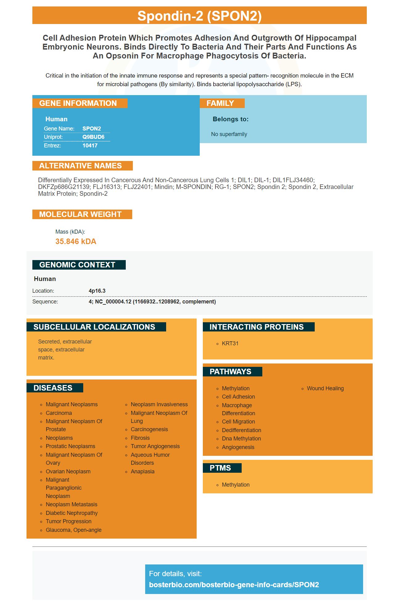
Facts about Spondin-2.
Critical in the initiation of the innate immune response and represents a special pattern- recognition molecule in the ECM for microbial pathogens (By similarity). Binds bacterial lipopolysaccharide (LPS).
Human | |
---|---|
Gene Name: | SPON2 |
Uniprot: | Q9BUD6 |
Entrez: | 10417 |
Belongs to: |
---|
No superfamily |
Differentially expressed in cancerous and non-cancerous lung cells 1; DIL1; DIL-1; DIL1FLJ34460; DKFZp686G21139; FLJ16313; FLJ22401; Mindin; M-SPONDIN; RG-1; SPON2; Spondin 2; spondin 2, extracellular matrix protein; spondin-2
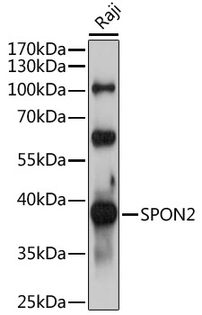
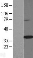
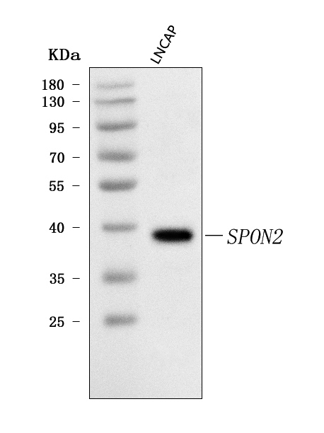
Mass (kDA):
35.846 kDA
Human | |
---|---|
Location: | 4p16.3 |
Sequence: | 4; NC_000004.12 (1166932..1208962, complement) |
Secreted, extracellular space, extracellular matrix.
Optimizing your Boster Bio flow protocols can improve your ability to analyze the data from your experiments. Flow procedures are the core of any experimental design. This article will discuss FITC and ELISA and SPON2 Marker. This guide will help you optimize your experiments and answer common questions. When you want to control the amount SPON2 in your samples, FITC is the best option.
Whether you're looking for a high-affinity antibody to detect SPON2 or a broad range of proteins, Boster has the right product for you. Its primary antibodies have been well cited for use in Western Blotting, Immunohistochemistry, and ELISA. With more than 25 years of experience, you can trust the quality of these antibodies. For more information, visit bosterbio.com/spon2 mark.
Human lung tumors are highly affected by the SPON2 gene. We used this gene to identify lung tumor cells in the present study. We found a significant relationship between SPON2 protein levels and ADC-cell size. This gene is found to be differentially expressed in normal and cancerous lung tissue. It is not clear if SPON2 plays a role in tumor progression or metastasis. We used tissue samples taken from patients with and without lung adenocarcinoma to validate the SPON2 marker.
There are many ways to detect the SPON2 gene. It is highly sensitive in diagnosing BA and is associated FEV1/FVC%, and serum neutrophils. Its positive correlations with IL-12/IL-4/IL-13 may be beneficial in diagnosing and monitoring a range diseases. However, it is not as reliable in urine samples and tissues. However, it is a promising indicator for diagnosing autoimmune and inflammatory conditions.
The SPON2 marker can be used in many research applications. Its positive correlation with tumor cells was confirmed by immunofluorescence staining of TAM markers. SPON2 expression is associated with poor prognosis in patients suffering from colorectal or gastric cancers. It is still being tested in clinical trials. It is critical to recognize the SPON2 mutation in a tumor for cancer detection.
The SPON2 gene can be detected by using qRT-PCR. It can also be used to detect tumor cells and molecular diagnosis. It can also be used for the assessment of the a5b1integrin-receptor. The SPON2 protein is abundant in human tissues. This gene can be found on human skin tumor cells. qPCR can detect gene expression levels.
SPON2, which is a strong chemoattractant can be used to attract macrophages to tumors and monocytes. Its ability to attract these cells is further supported by studies that show a strong correlation between SPON2 and HCC. The SPON2 gene has the ability to act as a chemical attractant and an inhibitor of hepatitis virus. Furthermore, the SPON2-abundant HCC will see an accumulation of macrophages with M1-like characteristics. These cells will exhibit strong antitumor immune responses.
Besides being a valuable prognostic biomarker for colorectal cancer, SPON2 has also been investigated as a serum and histologic diagnostic biomarker for ovarian cancer. Multiple studies have found conflicting results. It is known that it interacts with MACC1 as well as the a5b1 receptor of the integrin, thereby inducing cell mobility. It could also be used as a biomarker for poor prognosis. SPON2 also blocks HCC cells' invasion and migration.
SPON2 expression can be found in all types of tumors. Furthermore, higher SPON2 expression in a patient's tumor was associated with poorer overall survival and TNM stage, and high SPON2 mRNA was associated with poorer prognosis (P = 0.0016).
FITC is a powerful and reliable SPON2 marker, which also serves as an integrin ligand. FITC can be used to study SPON2 in immunohistochemical experiments. Its high levels within the ccRCc tissues, cell lines, and in the blood have led to SPON2 being developed as an integrin binding ligand.
ELISA, a type of immunoassay, uses antibodies to measure certain analytes. ELISAs are a popular method to evaluate various research and diagnostic targets. They were first developed in the 1970s to replace radioimmunoassays. They are still widely used today. ELISAs can be used to measure multiple substances in one well. This increased sensitivity allows them be extremely sensitive and allows for direct cell based output.
SPON2 sandwich ELISA Kit measures SPON2 concentrations in serum, tissue homogenates, plasma, and urine. The SPON2 sandwich ELISA kits uses a sensitive antibody to detect the protein. This antibody can be used in multiple applications and is compatible to many immunoassay methods. This kit contains the necessary components for performing a high-quality ELISA. It also includes a standard buffer and curve.
For small molecules that are difficult to sandwich between two antibodies, a competitive ELISA test is the most common. In this method, a capture antibody coats the microplate, while a conjugated antigen completes the binding process with the antigen present in the sample. The binding power of conjugated antibodies will decrease the more antigen in the sample. The signal level is determined by the amount of protein in your sample.
Another type ELISA sandwich uses an antibody which binds to the antigen. These antibodies are monoclonal in their nature and are known to have high sensitivity and specificity. They also have a wide dynamic range. These ELISAs represent the most common type of sandwich ELISA. You can use either one of these types of antibodies. You should make sure that you have the right combination for detection and primary antibodies.
Flow Cytometry is an effective technique for identifying specific structures on cells. These bioparticles are easily detected by flow Cytometry, as they are fluorescent. A pulse of photon emission is formed when bacteria in the sample interacts with the source of light. This happens over a period. PMTs detect the pulse and convert it into an electrical pulse. The area of the voltage pulse is related to the fluorescence intensity in the cell. Therefore, the more intense the fluorescence, the more channels.
The SPON2 marker integrates with many flow-cytometry analysis software packages. FlowJo 10.3 has a wide range of features, including gate-based selection of cell populations. It can be used to determine if a group is a helper cell based on CD3+ and CD4+ expression, and to check for activation via activation markers or IFN-g.
Flow cytometry allows researchers to compare and analyze cells suspended in liquid. The technology is extremely sensitive, fast, and can analyze thousands if cells per second. In addition, flow cytometry allows researchers to determine cell type, phenotype, and function. The technology is also useful for sorting live cells in culture. This technology is versatile and will help you locate the right cell for research.
This tool is a great option for those who want to determine which cells are affected. Boster provides high-affinity antibodies to this assay. Moreover, they have been validated by Western Blotting, Immunohistochemistry, and ELISA methods. You can also combine this product with other flow cytometry products.
PMID: 10512675 by Manda R., et al. Identification of genes (SPON2 and C20orf2) differentially expressed between cancerous and noncancerous lung cells by mRNA differential display.
PMID: 19153605 by Li Y., et al. Structure of the F-spondin domain of mindin, an integrin ligand and pattern recognition molecule.