SPRED1 / Sprouty-related, EVH1 domain-containing protein 1 · IHC design guide

Design Immunohistochemistry for SPRED1

Plan chromogenic SPRED1 IHC-P with the human-validated catalog antibody at 2.5 μg/mL (datasheet: A03613). Expect mainly cytoplasmic tissue staining, but interpret intensity cautiously because antibody staining and RNA show low consistency (HPA tissue IHC).

Evidence assembled Oct 2026 · For research use; verify linked source records and product datasheet before use
Immunohistochemistry protocol sheet for SPRED1 (IHC for SPRED1): expected localisation Mainly cytoplasmic in tissue (HPA tissue IHC), antibody A03613, validated IHC image, and IHC protocol steps
Printable SPRED1 IHC protocol sheet — expected localisation Mainly cytoplasmic in tissue (HPA tissue IHC), antibody A03613, controls and protocol steps. Open the full SPRED1 IHC guide →

SPRED1 Immunohistochemistry Experimental Design Guide

Expected localisation, validated protocols, controls and antibodies — the at-a-glance facts below, then the full design guide.

Must know before staining
Expected localisation Mainly cytoplasmic in tissue (HPA tissue IHC)
Staining pattern Mainly cytoplasmic staining across most tissues (HPA tissue IHC)
Antigen retrieval Tris-EDTA pH 9.0 HIER, 95–98 °C, 20 min (rule: nuclear antigen)
Positive control ⓘ Adrenal gland+4 more · see all
Negative control ⓘ Adipose tissue+4 more · see all
Important caveats
Reasons your staining may differ from the expected pattern.
Fixation Keep fixation consistent between tissue samples (standard IHC practice; not target-specific)
Caveat Antibody staining and RNA show low consistency (HPA tissue IHC)
Regulation Expression regulation not established (UniProt)
Isoform / epitope No annotated isoforms or transmembrane segment (UniProt)
Section 1

Recommended SPRED1 IHC & IF Protocols

The catalog antibody’s IHC-P protocol (datasheet) is accompanied by two published chromogenic paraffin IHC protocols (PMC4959414; PMC13383870).

Recommended immunohistochemistry (IHC-P) protocol parameters
SampleTissue sections; selected-image fixative not specified (standard IHC workflow)
FixationImage fixative and duration unreported (datasheet A03613); verify before use.
Sectioning4–5 µm sections on charged slides (standard)
DeparaffinisationXylene, graded ethanol series to water (standard)
Antigen retrievalHeat-induced epitope retrieval in Tris-EDTA buffer, pH 9.0, 20 min at 95–98 °C (standard rule: nuclear antigen)
Peroxidase block3% H2O2, 10 min, room temperature (standard)
Blocking10% normal serum of the secondary host, 30 min, room temperature (standard)
Primary antibodyRabbit anti-SPRED1, 2.5 μg/mL (datasheet A03613)
Primary incubationOvernight at 4 °C (standard)
DetectionHRP-polymer secondary, DAB chromogen 5–10 min (standard)
CounterstainHematoxylin, blue, dehydrate and mount (standard)
Expected resultSPRED1-positive staining in glandular cells of adrenal gland (HPA tissue IHC: Medium). HPA tissue profile: Mainly cytoplasmic expression in most tissues. No signal in the no-primary control.
💡Decision noteStart with Tris-EDTA pH 9.0 at 95–98 °C for 20 min (page retrieval rule: nuclear antigen); compare citrate pH 6.0 (PMC4959414).
Section 2

What Is the Expected SPRED1 Staining Pattern?

SPRED1 is reported at the cell membrane, in caveolae, and in the nucleus; it has no transmembrane segment (UniProt Q7Z699). In tissue IHC, expect mainly cytoplasmic staining across many tissues, including medium staining in selected glandular, epithelial, hematopoietic, glial, and cerebellar granular-layer cells (HPA: tissue IHC). HPA rates its tissue staining Approved but reports low consistency between antibody staining and RNA expression (HPA: tissue IHC reliability).

What am I looking at on my slide?
Cytoplasmic staining in adrenal gland or endometrial glandular cells, bronchial respiratory epithelial cells, or bone marrow hematopoietic cells.This fits the reported medium staining in those cell populations and the mainly cytoplasmic tissue pattern (HPA: tissue IHC). Assess the named cells rather than treating every cell in a positive tissue as an expected positive (HPA: tissue IHC).
Signal confined to extracellular spaces, gland lumens, or section edges, with no convincing cellular staining.These locations do not fit the reported cellular IHC pattern (HPA: tissue IHC). Treat the signal as a possible artefact and check its distribution against a known positive cell population. Nuclear signal alone needs separate review: UniProt includes the nucleus among SPRED1 locations (UniProt Q7Z699).
Prominent staining of adipocytes, duodenal glandular cells, or lymph-node germinal center cells.These specific populations are reported as not detected (HPA: tissue IHC). Staining there raises concern for cross-reactivity or endogenous detection activity; inspect a reagent control and compare the pattern with a positive reference section (general IHC practice).
Diffuse chromogen covers several tissue compartments and obscures cell boundaries.The haze cannot establish SPRED1 localisation or cell-specific staining. The reported tissue pattern is mainly cytoplasmic (HPA: tissue IHC). Review the reagent control, blocking, washes, and chromogen development before scoring the section (general IHC practice).
No cellular signal in adrenal glandular cells or bone marrow hematopoietic cells.Both are reported at medium intensity (HPA: tissue IHC). A blank run warrants a check of section quality, antibody dilution, retrieval conditions, and detection controls (general IHC practice). HPA also reports low antibody–RNA consistency, so RNA alone cannot settle an ambiguous IHC result (HPA: tissue IHC reliability).
💡Expected SPRED1 appearanceCall a positive result when identifiable cells show predominantly cytoplasmic, approximately medium staining in an HPA-listed positive population; signal concentrated in extracellular spaces, lumens, or HPA-listed undetected cells is suspect (HPA: tissue IHC).
How each factor affects the staining
Tissue and cell choiceUse a named medium population, such as adrenal glandular cells or bronchial respiratory epithelial cells, to judge the run; compare with the named undetected cell population within any negative tissue (HPA: tissue IHC).
Membrane and nuclear localisationUniProt lists membrane, caveola, and nuclear localisation, with no transmembrane segment (UniProt Q7Z699). HPA describes tissue IHC as mainly cytoplasmic (HPA: tissue IHC). Do not require a continuous membrane outline to accept an otherwise appropriate cellular IHC pattern.
IHC antibody evidenceThe HPA042193 antibody has Approved IHC status, while HPA reports low consistency between antibody staining and RNA expression (HPA: antibody validation; HPA: tissue IHC reliability). These labels support use as a reference pattern but leave discrepant staining open to investigation.
Processing and epitope limitsUniProt lists no signal peptide or propeptide and annotates a 2–444 chain (UniProt Q7Z699). The supplied record does not locate the IHC antibody epitope or report SPRED1-specific retrieval or fixation sensitivity; choose retrieval from the catalog antibody's IHC-P instructions (general IHC practice).
IF/ICC Q&A: should its location match tissue IHC?HPA reports supported nucleoplasmic ICC-IF localisation, with images in PC-3 and U2OS, for HPA061805 (HPA: subcellular; HPA: antibody validation). Tissue IHC is mainly cytoplasmic and uses a separately IHC-approved antibody, HPA042193 (HPA: tissue IHC; HPA: antibody validation).
Why is my staining missing, weak or wrong?
SituationLikely causeNext action
Expected positive cells are blank.The section or detection run may have failed; the chosen cells should show medium staining in HPA tissue IHC (HPA: tissue IHC).Check an on-run positive section and detection controls, then review the catalog antibody's IHC-P dilution and retrieval instructions (general IHC practice).
Only strong nuclear staining appears in tissue IHC.HPA tissue IHC is mainly cytoplasmic, although nuclear SPRED1 localisation is reported by UniProt and nucleoplasmic ICC-IF is supported by HPA (HPA: tissue IHC; UniProt Q7Z699; HPA: subcellular).Compare cellular morphology and a positive reference section; investigate a nuclear-only IHC pattern before calling it SPRED1 (general IHC practice).
Negative-reference cells stain prominently.Adipocytes and duodenal glandular cells are reported as not detected; unexpected signal may reflect cross-reactivity or endogenous detection activity (HPA: tissue IHC; general IHC practice).Check a reagent control and the detection background, then compare the same run with an HPA-listed positive cell population (general IHC practice).
Broad brown haze prevents cell-level scoring.Diffuse background can obscure the mainly cytoplasmic cellular pattern reported for SPRED1 tissue IHC (HPA: tissue IHC; general IHC practice).Review blocking, washes, antibody concentration, and chromogen development; score only when cell boundaries and compartments are interpretable (general IHC practice).
A whole positive tissue looks patchy.HPA assigns intensity to specified cell populations, such as medium glial cells in cerebral cortex, rather than to every cell in the section (HPA: tissue IHC).Identify the HPA-listed population before judging the run; record staining by cell type and compartment (HPA: tissue IHC; general IHC practice).
IHC disagrees with RNA or an IF image.HPA notes low tissue antibody–RNA consistency; its supported nucleoplasmic ICC-IF result and mainly cytoplasmic tissue IHC result come from different antibody validations (HPA: tissue IHC reliability; HPA: subcellular; HPA: antibody validation).Interpret each assay against its own HPA pattern and validation status, then resolve the tissue IHC call using its cell-level staining and controls (HPA: tissue IHC; general IHC practice).

Sample controls for SPRED1 IHC & IF

🧪Run bone marrow first: hematopoietic cells should show SPRED1 staining (HPA: Medium in bone marrow hematopoietic cells). Use adipose tissue as the negative tissue (HPA: Not detected in adipocytes); on the bone marrow slide, unstained neighboring cells and acellular areas should show only background, without being assumed to be validated SPRED1-negative populations (standard IHC control practice).
Positive control tissue: Adrenal gland (Glandular cells, HPA Medium)
Negative control tissue: Adipose tissue (HPA Not detected)
ICC-IF cell lines (HPA subcellular resource): HPA ICC-IF images show SPRED1 in PC-3, U2OS, with annotated localisation: Nucleoplasm (supported) (HPA subcellular).
Technical controls: Include a no-primary (secondary-only) control, a host-species- and isotype-matched control antibody, and SPRED1-knockout material as a biological negative where available (standard IHC control practice). Block endogenous peroxidase in bone marrow before chromogenic detection because myeloid cells can contribute peroxidase background (standard IHC practice).
⚠️Feasibility: A SPRED1-specific fixation window and antigen-retrieval dependency are unreported in the supplied evidence; the selected A03613 brain IHC caption also does not report a fixative (caption: fixative not stated). The evidence does not establish whether frozen sections or IF are easier than paraffin IHC, although ICC-IF images exist for PC-3 and U2OS cells (HPA: subcellular images). In bone marrow, endogenous peroxidase can complicate chromogenic scoring, so interpret hematopoietic-cell staining against the peroxidase-blocked controls (standard IHC practice).

HPA tissue IHC evidence for SPRED1

Comprehensive Human Protein Atlas IHC scoring per tissue (reliability: Approved — Low consistency between antibody staining and RNA expression data.). Rows are taken directly from the HPA tissue chart — click any row's HPA link to view the source.

Positive expression · recommended positive controls

TissueCell typeLevelEvidenceSource
Adrenal gland Glandular cells Medium Protein (IHC) HPA →
Appendix Glandular cells Medium Protein (IHC) HPA →
Bone marrow Hematopoietic cells Medium Protein (IHC) HPA →
Bronchus Respiratory epithelial cells Medium Protein (IHC) HPA →
Cerebellum Cells in granular layer Medium Protein (IHC) HPA →

Undetected expression · recommended negative controls

TissueCell typeLevelEvidenceSource
Adipose tissue Adipocytes Not detected Protein (IHC) HPA →
Duodenum Glandular cells Not detected Protein (IHC) HPA →
Lymph node Germinal center cells Not detected Protein (IHC) HPA →
Oral mucosa Squamous epithelial cells Not detected Protein (IHC) HPA →
Ovary Ovarian stroma cells Not detected Protein (IHC) HPA →
Section 3

Advanced SPRED1 IHC Tips

Troubleshoot SPRED1 chromogenic IHC in paraffin sections using compartment, cell type, and control staining alongside signal intensity.

How should I optimize retrieval when SPRED1 staining is weak?
Start with Tris-EDTA, pH 9.0, heat-induced retrieval at 95–98 °C for 20 min (page retrieval rule: nuclear antigen). Cool sections in the retrieval buffer, then compare the same antibody dilution and detection conditions across runs (standard IHC practice). If staining remains weak, test a second retrieval condition on adjacent sections while monitoring tissue damage and background; the supplied evidence does not establish a product-specific retrieval optimum (supplied catalog evidence). Score cytoplasmic and nuclear signal separately because tissue staining is mainly cytoplasmic, while nucleoplasm is a supported subcellular location (HPA: tissue IHC profile; HPA: subcellular).
Could fixation explain weak or uneven SPRED1 staining?
SPRED1-specific fixation sensitivity is unknown: the selected brain IHC caption does not state a fixative (A03613 tissue-IHC caption). Record fixation duration and processing for each block before comparing staining intensity, since those variables can alter antigen accessibility in paraffin sections (standard IHC practice). Compare adjacent sections with matched retrieval and detection, and include a control section processed alongside the study samples (standard IHC practice). Do not attribute a weak nuclear or cytoplasmic result to a SPRED1-specific fixation effect without a controlled comparison; both compartments have supporting localization evidence, but neither establishes fixation behavior (HPA: tissue IHC profile; HPA: subcellular; UniProt Q7Z699: subcellular location).
Should SPRED1 stain cytoplasm, nuclei, or cell membranes?
Evaluate cytoplasmic staining first because the tissue IHC profile describes mainly cytoplasmic expression across most tissues (HPA: tissue IHC profile). Also record nuclear and peripheral staining separately: nucleoplasm is a supported IF location, and membrane, caveola, and nucleus are annotated locations (HPA: subcellular; UniProt Q7Z699: subcellular location). SPRED1 has no transmembrane segment, so a membrane-associated pattern need not form a continuous cell outline (UniProt Q7Z699: topology). Compare each compartment with the same section's cell morphology and negative controls; isolated diffuse staining across every compartment deserves investigation before being scored as SPRED1 (standard IHC practice).
How can epitope location change the apparent SPRED1 pattern?
Check the antibody's documented immunogen or epitope before interpreting a compartment-specific loss of staining; none is supplied here (supplied antibody evidence). The annotated protein contains WH1 at residues 6–123, KBD at 233–285, and SPR at 334–442, with no listed isoforms (UniProt Q7Z699: domains and isoforms). Annotated modifications include phosphoserine at 238 and 308, so epitope masking by modification is a hypothesis to test, not an established cause of IHC differences (UniProt Q7Z699: modified residues). Compare matched retrieval conditions and controls before assigning divergent nuclear or cytoplasmic staining to an epitope effect (standard IHC practice).
How should I cross-check SPRED1 IHC with multiplex IF?
Use IF as a separate cross-check and multiplex SPRED1 with a marker for the cell population being assessed, such as a glial marker when examining cortical glia (HPA: medium staining in cerebral cortex glial cells; standard IF practice). Select a fluorophore channel after inspecting unstained tissue autofluorescence, and include single-label controls to assess bleed-through (standard IF practice). Because SPRED1 lacks a transmembrane segment and has nuclear as well as membrane-associated annotations, test permeabilisation appropriate to the epitope's location once that epitope is known (UniProt Q7Z699: topology and subcellular location; standard IF practice). Treat IF and chromogenic IHC patterns as separate observations until antibody specificity and compartment agreement have been checked (standard IHC/IF practice).
What should I check when SPRED1 DAB staining appears widespread?
Run a no-primary control and inspect whether pigment, precipitate, or endogenous enzyme signal persists without the antibody (standard chromogenic IHC practice). Include a peroxidase block before DAB development, and shorten development if signal obscures cell boundaries; these are general workflow adjustments, not SPRED1-specific evidence (standard chromogenic IHC practice). Titrate the catalog antibody around the caption's 2.5 µg/mL brain-tissue use while holding retrieval and exposure conditions constant (A03613 tissue-IHC caption; standard IHC practice). Interpret faint widespread staining cautiously because the tissue profile is mainly cytoplasmic and its antibody staining has low consistency with RNA expression (HPA: tissue IHC profile and reliability description).
How can I compare SPRED1 staining across paraffin sections? ⚠ ANSWER MARKED FOR VERIFICATION
Predefine the scored cell population and compartment, then report percent positive cells and an intensity-weighted H-score for cytoplasmic staining (standard IHC scoring practice; HPA: mainly cytoplasmic tissue profile). If spatial distribution matters, report positive cells per mm² of viable, annotated tissue rather than per total image area (standard IHC scoring practice). Normalize comparisons to the same cell type, sampled tissue area, staining batch, and exposure or DAB development conditions (standard IHC practice). Report nuclear staining separately because nucleoplasm is supported in subcellular data, and avoid treating different compartment scores as interchangeable (HPA: subcellular; standard IHC scoring practice).
When is a SPRED1-positive cell convincing rather than artefactual?
Look for cell-bound staining in a plausible compartment and population: cortical glial cells and bone-marrow hematopoietic cells have medium tissue staining, while the overall profile is mainly cytoplasmic (HPA: tissue IHC profile). Nuclear signal can be plausible because nucleoplasm is a supported location, but it still needs appropriate controls and intact cell morphology (HPA: subcellular; standard IHC practice). Discount staining confined to section edges, necrotic regions, or pigment deposits, and investigate signal persisting in a no-primary control (standard chromogenic IHC practice). Keep conclusions qualified when staining conflicts with other evidence because antibody staining and RNA expression show low consistency (HPA: reliability description).
Boster reagents

Best SPRED1 / Sprouty-related, EVH1 domain-containing protein 1 IHC Antibodies

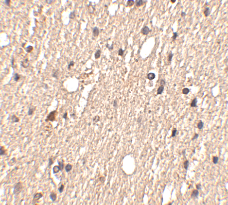
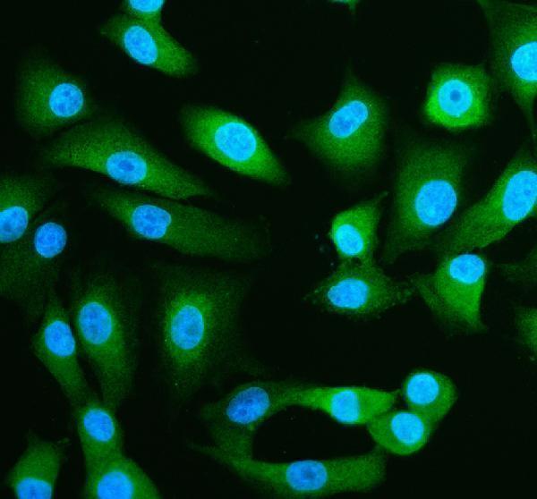
The catalog includes SPRED1 antibodies with IHC data from human brain tissue (A03613 image caption) and IF data from A549 cells (A03613-1 image caption); listed reactivity includes human, mouse, and rat (A03613 catalog).

Real IHC data Immunohistochemistry of Spred1 in human brain tissue with Spred1 antibody at 2.5 μg/mL.
Anti-Spred1 Antibody
Cat # A03613
Real IF data IF analysis of SPRED1 using anti-SPRED1 antibody (A03613-1). SPRED1 was detected in an immunocytochemical section of A549 cells. Enzyme antigen retrieval was performed using IHC enzyme antigen retrieval reagent (AR0022) for 15 mins. The cells were blocked with 10% goat serum. And then incubated with 5 μg/mL rabbit anti-SPRED1 Antibody (A03613-1) overnight at 4°C. DyLight®488 Conjugated Goat Anti-Rabbit IgG (BA1127) was used as secondary antibody at 1:500 dilution and incubated for 30 minutes at 37°C. The section was counterstained with DAPI. Visualize using a fluorescence microscope and filter sets appropriate for the label used.
Anti-SPRED1 Antibody ®
Cat # A03613-1

A03613 has an IHC-P application listing and a human brain tissue IHC image at 2.5 μg/mL (A03613 catalog; A03613 image caption). A03613-1 has IF/ICC application listings and an A549 cell IF image at 5 μg/mL (A03613-1 catalog; A03613-1 image caption).

Which to pick: Choose A03613 for paraffin-section tissue IHC: IHC-P is listed and its own image shows human brain tissue staining; the fixative is unreported (A03613 catalog; A03613 image caption). Choose A03613-1 for IF/ICC in human cells, supported by its A549 image; it has no listed IHC-P application (A03613-1 catalog; A03613-1 image caption). For mouse or rat samples, A03613 is the candidate based on listed reactivity, although its cited IHC image is from human tissue (A03613 catalog; A03613 image caption).

Each figure is that product's own IHC / IF validation image from its datasheet.

References

  1. UniProt Consortium. UniProt entry Q7Z699 (SPRE1_HUMAN, Sprouty-related, EVH1 domain-containing protein 1).
  2. Human Protein Atlas. SPRED1 tissue IHC expression (reliability: Approved).
  3. Human Protein Atlas. SPRED1 subcellular location (ICC-IF): Localized to the nucleoplasm..
  4. Human Protein Atlas. SPRED1 antibody validation summary (2 antibodies).
  5. Enhancing the sensitivity of lung adenocarcinoma to immune therapeutic agents through SPRED1. Translational lung cancer research 2025 — PMC12432670.
  6. Inhibition of microRNA-126 promotes the expression of Spred1 to inhibit angiogenesis in hepatocellular carcinoma after transcatheter arterial chemoembolization: in vivo study. OncoTargets and therapy 2016 — PMC4959414.
  7. Legius syndrome mutations in the Ras-regulator SPRED1 abolish its membrane localization and potentially cause neurodegeneration. The Journal of biological chemistry 2024 — PMC11648228.
  8. Downregulation of SPRED1 drives keratinocyte hyperproliferation in psoriasis via activation of the ERK-RSK1 signaling axis. iScience 2026 — PMC13383870.
  9. PubMed PMID:15683364 — UniProt-cited evidence.
  10. PubMed PMID:14702039 — UniProt-cited evidence.
  11. PubMed PMID:15489334 — UniProt-cited evidence.