SPTBN2 / Spectrin beta chain, non-erythrocytic 2 · IHC design guide

Design Immunohistochemistry for SPTBN2

Plan SPTBN2 chromogenic IHC in paraffin sections using the catalog antibody’s documented conditions (datasheet A05492). Compare cytoplasmic and membranous staining with the tissue patterns and controls reported by HPA (HPA tissue IHC).

Evidence assembled Oct 2026 · For research use; verify linked source records and product datasheet before use
Immunohistochemistry protocol sheet for SPTBN2 (IHC for SPTBN2): expected localisation Cytoplasmic and membranous staining (HPA tissue IHC), antibody A05492, validated IHC image, and IHC protocol steps
Printable SPTBN2 IHC protocol sheet — expected localisation Cytoplasmic and membranous staining (HPA tissue IHC), antibody A05492, controls and protocol steps. Open the full SPTBN2 IHC guide →

SPTBN2 Immunohistochemistry Experimental Design Guide

Expected localisation, validated protocols, controls and antibodies — the at-a-glance facts below, then the full design guide.

Must know before staining
Expected localisation Cytoplasmic and membranous staining (HPA tissue IHC)
Staining pattern Cytoplasm/membrane in CNS, distal tubules and squamous epithelia (HPA tissue IHC)
Antigen retrieval EDTA pH 8.0 HIER, heat-mediated (datasheet A05492)
Positive control ⓘ Caudate+4 more · see all
Negative control ⓘ Adipose tissue+4 more · see all
Important caveats
Reasons your staining may differ from the expected pattern.
Fixation Keep paraffin-section fixation consistent (standard IHC practice; not target-specific)
Caveat Staining and RNA expression show medium consistency (HPA tissue IHC)
Regulation Brain- and skin-enhanced RNA expression (HPA tissue RNA)
Isoform / epitope 2 isoforms; check epitope coverage across variants (UniProt)
Section 1

Recommended SPTBN2 IHC & IF Protocols

The catalog antibody’s IHC-P protocol (datasheet: A05492) is accompanied by a published lung adenocarcinoma IHC protocol (PMC8563792).

Recommended immunohistochemistry (IHC-P) protocol parameters
SampleParaffin-embedded mouse brain tissue; fixative not specified (datasheet A05492)
FixationImage fixative and duration unreported (datasheet A05492); verify before use.
Sectioning4–5 µm sections on charged slides (standard)
DeparaffinisationXylene, graded ethanol series to water (standard)
Antigen retrievalHeat retrieval: EDTA pH 8.0 (datasheet A05492); 20 min, 95–100 °C (standard)
Peroxidase block3% H2O2, 10 min, room temperature (standard)
Blocking10% goat serum (datasheet A05492)
Primary antibodyRabbit anti-SPTBN2, 2-5 μg/ml (datasheet A05492)
Primary incubationOvernight at 4 °C (datasheet A05492)
DetectionHRP-conjugated secondary, DAB chromogen (datasheet A05492)
CounterstainHematoxylin, blue, dehydrate and mount (standard)
Expected resultSPTBN2-positive staining in neuronal cells of caudate (HPA tissue IHC: High). HPA tissue profile: Cytoplasmic and membranous expression in most cell types, highly expressed in CNS, renal distal tubules and squamous epithelia. No signal in the no-primary control.
💡Decision noteStart with heat-mediated EDTA at pH 8.0 (datasheet: A05492); compare the published sodium citrate retrieval when optimizing lung adenocarcinoma sections (PMC8563792).
Section 2

What Is the Expected SPTBN2 Staining Pattern?

In paraffin-section IHC, expect SPTBN2 staining at the cytoplasm and cell periphery, consistent with its cytoskeletal and cell-cortex location and lack of a transmembrane segment (UniProt O15020). Strong examples include neuronal cells, renal collecting ducts, pancreatic exocrine cells and skin keratinocytes (HPA: High). HPA rates the tissue IHC profile Enhanced, with medium consistency between antibody staining and RNA expression (HPA: tissue IHC).

What am I looking at on my slide?
Strong cytoplasmic and peripheral staining in kidney collecting ducts, with little staining in nearby unstained cells.This fits a positive SPTBN2 pattern: HPA reports collecting ducts as High and describes cytoplasmic and membranous staining in most cell types (HPA: tissue IHC). Interpret the stained cell type and compartment together; intensity alone cannot establish specificity (general IHC practice).
Predominantly nuclear staining, without a convincing cytoplasmic or peripheral pattern.A mainly nuclear pattern lacks support from the supplied localisations: cytoskeleton and cell cortex (UniProt O15020), and cytoplasm and membrane in tissue IHC (HPA: tissue IHC). Treat it as suspect and compare it with controls before scoring it as SPTBN2 (general IHC practice).
Strong staining of a cell type recorded as undetected, such as adipocytes or heart cardiomyocytes.HPA reports SPTBN2 as Not detected in these specific cell types (HPA: adipocytes; heart cardiomyocytes). Unexpected colour may reflect antibody cross-reactivity or detection-system activity; confirm cell identity and examine control sections before assigning a cause (general IHC practice).
Colour spreads across the section without a clear cell boundary or reproducible cell-type pattern.Diffuse colour is difficult to reconcile with HPA's cell-resolved cytoplasmic and membranous profile (HPA: tissue IHC). Check background using a no-primary control, then review blocking, antibody concentration, washes and chromogen development (general IHC practice).
No staining in an included high-expression reference, such as skin keratinocytes.HPA records keratinocytes as High, so an unstained reference makes a technical failure plausible (HPA: skin). Check that the reference cells are present, then review antibody dilution, retrieval and detection controls (general IHC practice). One negative section alone does not establish absent SPTBN2.
💡Expected SPTBN2 appearanceCall a result positive when the expected cells show convincing cytoplasmic and cell-peripheral colour, with strong staining possible in renal collecting ducts or skin keratinocytes (HPA: High; HPA: tissue IHC); isolated nuclear colour or colour in an HPA-undetected cell type warrants control review before it is called specific (UniProt O15020; HPA: tissue IHC).
How each factor affects the staining
Compartment and topologySPTBN2 is assigned to the cytoskeleton and cell cortex and has no transmembrane segment (UniProt O15020). Peripheral staining can therefore fit its location without implying that it spans the plasma membrane.
Cell-type variation within tissuesHPA reports kidney collecting ducts as High but adipocytes and heart cardiomyocytes as Not detected (HPA: tissue IHC). Choose reference cells by their recorded cell type, rather than treating every cell in a section as equally positive.
Evidence strengthThe tissue IHC profile is Enhanced yet has medium staining-to-RNA consistency (HPA: tissue IHC). HPA039293 and CAB009844 have Enhanced IHC validation; HPA043529 is Supported (HPA: antibodies). Use this distinction when weighing an unexpected pattern.
Isoforms and processingTwo isoforms are listed, with no signal peptide or propeptide and a chain spanning residues 2–2390 (UniProt O15020). The supplied evidence gives no antibody epitope, so it cannot establish whether the catalog antibody distinguishes isoforms or processed material.
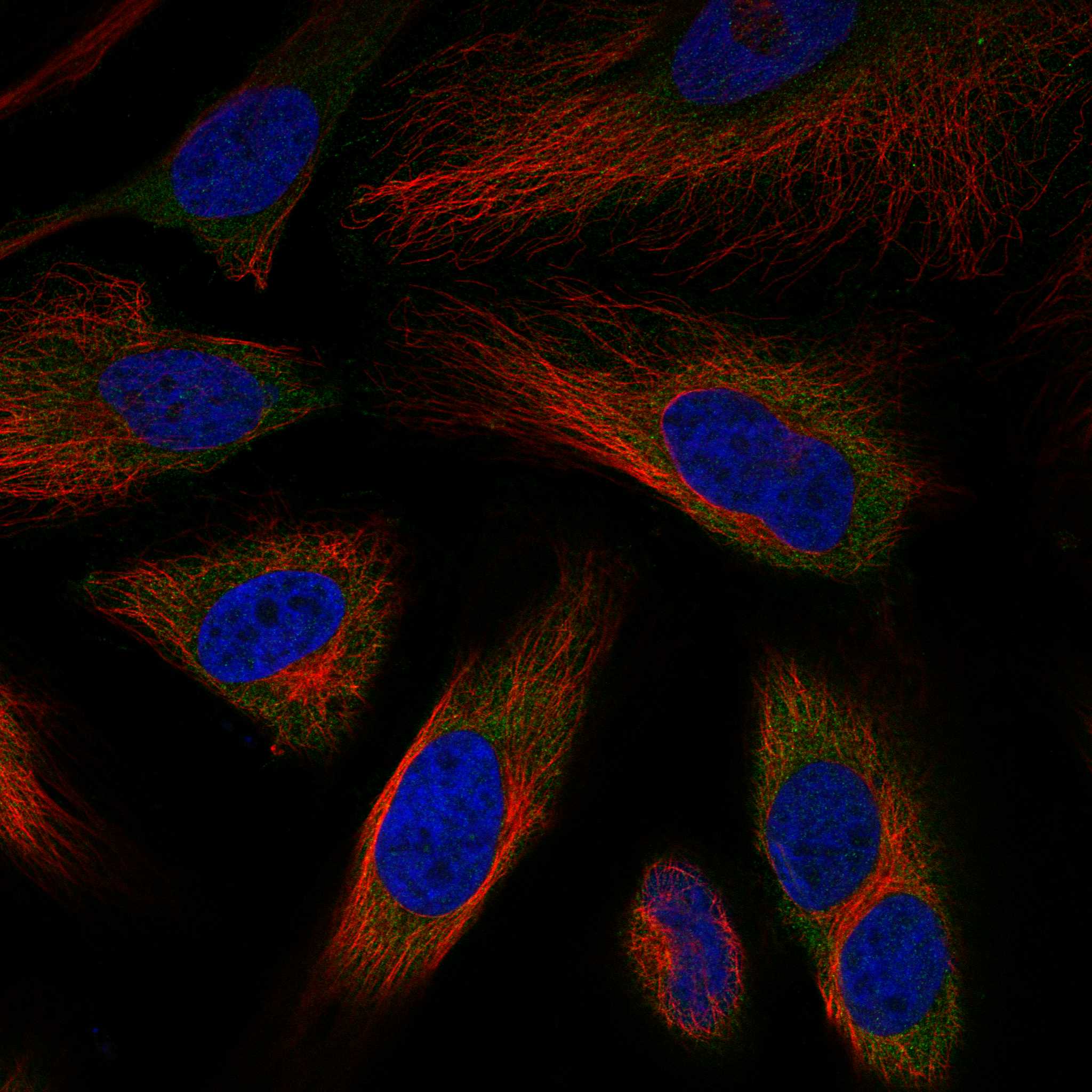
IF/ICC: where should signal appear?HPA places the main IF/ICC signal at the plasma membrane (approved) and cytosol (supported); vesicles are an additional uncertain location (HPA: subcellular). Treat this as an IF/ICC localisation reference, not as a paraffin-IHC protocol.
Why is my staining missing, weak or wrong?
SituationLikely causeNext action
A known-positive reference remains blank.A missing reference cell population or a failed IHC step may explain the blank section (general IHC practice); HPA lists skin keratinocytes as High (HPA: skin).Confirm the expected cells are present. Check reagent delivery, retrieval, dilution and detection with appropriate controls before interpreting the specimen as negative (general IHC practice).
Staining appears mainly in nuclei.The pattern does not match the reported cytoskeletal and cell-cortex localisation (UniProt O15020) or the tissue IHC compartment profile (HPA: tissue IHC).Compare with a high-staining reference and a no-primary control; review section morphology and detection background before scoring nuclear colour (general IHC practice).
Strong colour appears in an HPA-undetected cell type.Cross-reactivity or detection-system background is possible (general IHC practice); HPA reports adipocytes, for example, as Not detected (HPA: adipocytes).Verify the cell type and compare it with a high-staining reference. Use a no-primary control to test detection background; do not infer specificity from colour alone (general IHC practice).
The whole section has diffuse chromogenic background.Insufficient blocking or washing, excess antibody, or overdevelopment can raise background (general IHC practice). Diffuse colour obscures HPA's cell-resolved pattern (HPA: tissue IHC).Inspect the no-primary control, then adjust blocking, antibody dilution, washing or development time one variable at a time (general IHC practice).
Colour persists in a no-primary control.Primary-antibody binding cannot explain that control's colour. Detection reagents or endogenous activity may contribute, depending on the detection system (general IHC practice).Review the detection chemistry and its matching blocking step, including endogenous enzyme blocking if enzyme-based detection is used (general IHC practice).
A low-staining tissue is judged negative despite a working positive reference.HPA records low staining in lung macrophages and liver hepatocytes, while other listed cells are High (HPA: tissue IHC). A weak result needs a cell-specific interpretation.Score the specified cell population and compartment against the positive reference and background controls; report weak or undetected staining as observed, without assigning a target-specific fixation cause (HPA: tissue IHC; general IHC practice).

Sample controls for SPTBN2 IHC & IF

🧪Run caudate first and assess staining in neuronal cells (HPA: High in caudate neuronal cells). Use adipose tissue adipocytes as the negative tissue (HPA: Not detected in adipocytes); internal negative cells on the caudate slide should show counterstain without specific signal, but their target-negative status must be verified (standard IHC control practice).
Positive control tissue: Caudate (Neuronal cells, HPA High)
Negative control tissue: Adipose tissue (HPA Not detected)
ICC-IF cell lines (HPA subcellular resource): HPA ICC-IF images show SPTBN2 in HeLa, MCF-7, U2OS, with annotated localisation: Plasma membrane (approved), Cytosol (supported) (HPA subcellular).
Technical controls: Include a no-primary, secondary-only control and a host-, class-, and clonality-matched rabbit IgG isotype control; use SPTBN2 knockout tissue or validated peptide blocking as a biological specificity control (caption: rabbit primary antibody; standard IHC control practice). Quench endogenous peroxidase for DAB detection, and account for brain autofluorescence if evaluating IF (caption: HRP/DAB detection; standard IHC/IF practice).
⚠️Feasibility: A target-specific fixation window and fixation effect are unreported, and the selected A05492 paraffin-section caption does not state the fixative (caption: fixative unreported). The reported IHC example uses heat retrieval in EDTA at pH 8.0, but it does not establish whether retrieval is required for other specimens (caption: EDTA retrieval, pH 8.0). The supplied evidence does not establish whether frozen sections or IF are easier; brain autofluorescence can complicate IF interpretation (HPA: ICC-IF images available; standard IF practice).

HPA tissue IHC evidence for SPTBN2

Comprehensive Human Protein Atlas IHC scoring per tissue (reliability: Enhanced — Medium consistency between antibody staining and RNA expression data.). Rows are taken directly from the HPA tissue chart — click any row's HPA link to view the source.

Positive expression · recommended positive controls

TissueCell typeLevelEvidenceSource
Caudate Neuronal cells High Protein (IHC) HPA →
Cerebellum GLUC cells - cytoplasm/membrane High Protein (IHC) HPA →
Cerebral cortex Neuropil High Protein (IHC) HPA →
Cervix Glandular cells High Protein (IHC) HPA →
Kidney Collecting ducts High Protein (IHC) HPA →

Undetected expression · recommended negative controls

TissueCell typeLevelEvidenceSource
Adipose tissue Adipocytes Not detected Protein (IHC) HPA →
Appendix Glandular cells Not detected Protein (IHC) HPA →
Bone marrow Hematopoietic cells Not detected Protein (IHC) HPA →
Colon Endothelial cells Not detected Protein (IHC) HPA →
Duodenum Glandular cells Not detected Protein (IHC) HPA →
Section 3

Advanced SPTBN2 IHC Tips

Troubleshoot SPTBN2 staining in paraffin sections by checking retrieval, compartment, cell type and controls before comparing signal intensity.

How should I retrieve SPTBN2 in paraffin sections with A05492?
Start with heat-mediated retrieval in EDTA at pH 8.0 for paraffin sections (datasheet A05492). The reported mouse brain image used this condition, followed by 2 μg/mL primary antibody overnight at 4°C (caption A05492). If staining is weak, compare retrieval times on adjacent sections while keeping antibody concentration and detection constant; record tissue damage alongside signal (standard IHC practice). Include a no-primary control to separate retrieval-related background from specific staining (standard IHC practice). Judge improvement by cytoplasmic or cortical staining in the expected cells, rather than by darker DAB alone (UniProt O15020 localisation; HPA tissue IHC).
Could fixation explain weak SPTBN2 staining?
Target-specific fixation sensitivity is unknown: the selected paraffin-section caption does not report a fixative (caption A05492). Record the actual fixative and fixation duration for each specimen, then compare matched sections with the same EDTA pH 8.0 retrieval and detection conditions (datasheet A05492; standard IHC practice). If staining differs between processing batches, include a shared positive-control section in each run before attributing the difference to SPTBN2 abundance (standard IHC practice). Assess morphology and background with the signal, because poor preservation or excessive retrieval can make chromogenic staining hard to interpret (standard IHC practice). Do not assign a SPTBN2-specific fixation effect from these observations alone.
Where should convincing SPTBN2 DAB signal appear?
Expect a cytoplasmic or cell-cortical pattern, consistent with SPTBN2's annotated cytoskeletal and cortical localisation (UniProt O15020 localisation). HPA reports cytoplasmic and membranous tissue staining, including high signal in cerebellar GLUC cells and cerebral-cortex neuropil (HPA tissue IHC). Use those patterns to guide region selection, while checking cellular outlines and adjacent morphology before calling a membrane-associated DAB rim specific (HPA tissue IHC; standard IHC practice). SPTBN2 has no annotated transmembrane segment, so a surface-like pattern alone does not establish membrane insertion (UniProt O15020 topology). Score diffuse nuclear-only colour or staining outside preserved cells cautiously and review no-primary controls (standard IHC practice).
How do I interpret variable staining across SPTBN2 isoforms?
SPTBN2 has 2 annotated isoforms, but the supplied A05492 caption does not identify its epitope or establish isoform recognition (UniProt O15020 isoforms; caption A05492). Confirm the catalog antibody's immunogen and mapped epitope before claiming isoform-specific staining, and compare that sequence with each isoform (standard antibody-validation practice). The protein contains CH domains at residues 57–161 and 176–281, plus a PH domain at 2218–2328 (UniProt O15020 domains). Annotated phosphorylation and N-terminal acetylation may matter only if an antibody epitope overlaps an affected residue; no such overlap is supplied here (UniProt O15020 modified residues). Treat regional intensity differences as staining differences until epitope specificity is independently established (standard IHC practice).
How can IF help verify a puzzling IHC pattern?
Use IF as a separate validation experiment and compare its cellular pattern with the chromogenic section, without assuming identical processing or antibody performance (standard IHC/IF practice). Multiplex SPTBN2 with a neuronal marker when examining brain regions where neuronal or neuropil staining is expected (HPA tissue IHC; standard IF practice). Choose a far-red fluorophore and measure an unstained section to assess tissue autofluorescence before setting exposure and thresholds (standard IF practice). Because SPTBN2 is cytosolic or cortical and lacks a transmembrane segment, optimise permeabilisation for access to intracellular epitopes while monitoring morphology (UniProt O15020 localisation and topology; standard IF practice). No target-specific IF fixation or permeabilisation condition is supplied here.
How can I reduce diffuse or patchy DAB background?
Start with a no-primary section and review whether colour persists in the same regions; persistent signal points to detection or tissue background (standard IHC practice). The A05492 image used 10% goat-serum blocking and 2 μg/mL primary antibody overnight at 4°C (caption A05492). For a new specimen, check peroxidase blocking, washes and DAB development, changing one variable at a time against a matched positive-control section (standard IHC practice). If background remains broad, titrate primary antibody below the reported concentration while monitoring expected cytoplasmic or cortical staining (caption A05492; UniProt O15020 localisation; standard IHC practice). Record both signal and background under the same imaging settings (standard IHC practice).
What should I score when comparing SPTBN2 across sections? ⚠ ANSWER MARKED FOR VERIFICATION
Define the anatomical region and eligible cell population before scoring, using matched sections and the same DAB development and imaging settings (standard IHC practice). For cellular staining, report the percentage of positive cells and an H-score from intensity categories 0–3; keep the threshold fixed across samples (standard IHC practice). For neuropil-rich regions, measure DAB-positive area or optical density per mm² of viable annotated tissue instead of forcing a cell count (HPA tissue IHC; standard IHC practice). Normalise cellular counts to eligible cells and regional signal to viable area, and exclude folds, edges and necrosis by predefined rules (standard IHC practice). Report compartment separately when cytoplasmic and cortical patterns differ (UniProt O15020 localisation).
Which SPTBN2 patterns suggest true signal rather than artefact?
Give most weight to reproducible cytoplasmic or cortical staining in preserved cells, consistent with SPTBN2 localisation (UniProt O15020 localisation). HPA reports high staining in cerebellar GLUC cells and kidney collecting ducts, while adipocytes are reported as undetected; use such cell-specific patterns as context, not proof of antibody specificity (HPA tissue IHC). Suspect artefact when colour is confined to section edges, folds or necrotic areas, or persists in a no-primary control (standard IHC practice). Review nuclear-only staining against the expected compartment and check whether inadequate peroxidase blocking explains colour in the control (UniProt O15020 localisation; standard IHC practice). Confirm disputed patterns with an independent antibody or orthogonal assay (standard IHC validation practice).
Boster reagents

Best SPTBN2 / Spectrin beta chain, non-erythrocytic 2 IHC Antibodies

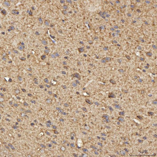
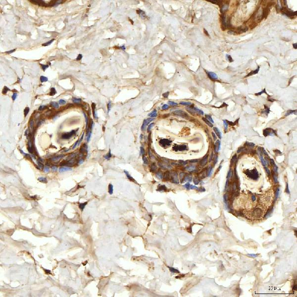
A05492 has real IHC images from paraffin sections of mouse and rat brain and skin (catalog image captions); no IF images are supplied (catalog payload).

Real IHC data IHC analysis of SPTBN2 using anti-SPTBN2 antibody (A05492). SPTBN2 was detected in a paraffin-embedded section of mouse brain tissue. Heat mediated antigen retrieval was performed in EDTA buffer (pH 8.0, epitope retrieval solution). The tissue section was blocked with 10% goat serum. The tissue section was then incubated with 2 μg/ml rabbit anti-SPTBN2 Antibody (A05492) overnight at 4°C. Peroxidase Conjugated Goat Anti-rabbit IgG was used as secondary antibody and incubated for 30 minutes at 37°C. The tissue section was developed using HRP Conjugated Rabbit IgG Super Vision Assay Kit (Catalog # SV0002) with DAB as the chromogen.
Anti-SPTBN2 Antibody ®
Cat # A05492

A05492 is listed for IHC and reacts with human, mouse and rat (catalog applications and reactivity). Its IHC images show paraffin sections of mouse and rat brain and skin (A05492 image captions).

Which to pick: Choose A05492 for tissue IHC in paraffin sections: its captions document 2 μg/ml antibody with EDTA retrieval at pH 8.0 (A05492 image captions); the fixative is unreported (A05492 image captions). No IF/ICC validated SKU is listed (catalog applications and IF images). A05492 lists human, mouse and rat reactivity, but its IHC images demonstrate only mouse and rat tissue (catalog reactivity; A05492 image captions).

Each figure is that product's own IHC / IF validation image from its datasheet.

References

  1. UniProt Consortium. UniProt entry O15020 (SPTN2_HUMAN, Spectrin beta chain, non-erythrocytic 2).
  2. Human Protein Atlas. SPTBN2 tissue IHC expression (reliability: Enhanced).
  3. Human Protein Atlas. SPTBN2 subcellular location (ICC-IF): Mainly localized to the plasma membrane and cytosol. In addition localized to vesicles..
  4. Human Protein Atlas. SPTBN2 antibody validation summary (3 antibodies).
  5. Integrated multi-omics characterization of SPTBN2 overexpression reveals its pro-tumorigenic role and immune microenvironment remodeling in colorectal cancer. Frontiers in cell and developmental biology 2026 — PMC13226874.
  6. Decreased SPTBN2 expression regulated by the ceRNA network is associated with poor prognosis and immune infiltration in low‑grade glioma. Experimental and therapeutic medicine 2023 — PMC10161196.
  7. SPTBN2 suppresses ferroptosis in NSCLC cells by facilitating SLC7A11 membrane trafficking and localization. Redox biology 2024 — PMC10825533.
  8. SPTBN2, a New Biomarker of Lung Adenocarcinoma. Frontiers in oncology 2021 — PMC8563792.
  9. PubMed PMID:9205841 — UniProt-cited evidence.
  10. PubMed PMID:16554811 — UniProt-cited evidence.
  11. PubMed PMID:9826670 — UniProt-cited evidence.