This website uses cookies to ensure you get the best experience on our website.


- Table of Contents
This guide maps expected cytoplasmic SRPK1 staining in paraffin sections (HPA tissue IHC). Use appendix glandular cells as a high-staining reference (HPA tissue IHC) and the IHC-validated antibody at 5 μg/ml (datasheet).

Expected localisation, validated protocols, controls and antibodies — the at-a-glance facts below, then the full design guide.
| Expected localisation | Predominantly cytoplasmic tissue staining (HPA tissue IHC) | |
| Staining pattern | Broad cytoplasmic staining across tissue cell types (HPA tissue IHC) | |
| Antigen retrieval | Tris-EDTA pH 9.0 HIER, 95–98 °C, 20 min (rule: nuclear antigen) | |
| Positive control | Appendix+4 more · see all | |
| Negative control | None in HPA (detected in all 45 tissues); use no-primary + isotype controls |
| Fixation | Matched tissue-IHC evidence does not establish the fixation claim. Validate the specimen-specific method before use. (selected-SKU IHC image A01937-1) | |
| Caveat | Stress or Hsp90 inhibition can shift SRPK1 into nuclei (UniProt) | |
| Regulation | Isoform 2 predominates in testis (UniProt) | |
| Isoform / epitope | 3 isoforms; verify epitope coverage across variants (UniProt) |
The catalog antibody’s IHC-P protocol is accompanied by three published SRPK1 IHC protocols (PMC4351909; PMC3842442; PMC9609466).
| Sample | Tissue sections; selected-image fixative not specified (standard IHC workflow) |
| Fixation | Image fixative and duration unreported (datasheet A01937-1); verify before use. |
| Sectioning | 4–5 µm sections on charged slides (standard) |
| Deparaffinisation | Xylene, graded ethanol series to water (standard) |
| Antigen retrieval | Heat-induced epitope retrieval in Tris-EDTA buffer, pH 9.0, 20 min at 95–98 °C (standard rule: nuclear antigen) |
| Peroxidase block | 3% H2O2, 10 min, room temperature (standard) |
| Blocking | 10% normal serum of the secondary host, 30 min, room temperature (standard) |
| Primary antibody | Rabbit anti-SRPK1, 5 μg/ml (datasheet A01937-1) |
| Primary incubation | Overnight at 4 °C (standard) |
| Detection | HRP-polymer secondary, DAB chromogen 5–10 min (standard) |
| Counterstain | Hematoxylin, blue, dehydrate and mount (standard) |
| Expected result | SRPK1-positive staining in glandular cells of appendix (HPA tissue IHC: High). HPA tissue profile: Ubiquitous cytoplasmic expression. No signal in the no-primary control. |
SRPK1 staining in paraffin sections should be widespread and predominantly cytoplasmic, including strong staining in glandular, respiratory epithelial and neuronal cells (HPA: ubiquitous cytoplasmic expression; High in the listed cells). Nuclear signal can occur because SRPK1 shuttles between cytoplasm and nucleus (UniProt Q96SB4: subcellular location). SRPK1 has no transmembrane segment (UniProt Q96SB4: topology). HPA rates the tissue IHC profile Approved, with medium consistency between staining and RNA expression (HPA: tissue IHC reliability).
| Cytoplasmic staining is strong in colon or breast glandular cells, or bronchial respiratory epithelial cells. | This matches reported high staining in those cell types (HPA: High in colon and breast glandular cells; High in bronchial respiratory epithelial cells). Score the cells and compartment together; intensity alone does not establish specificity (general IHC practice). |
| Nuclear staining accompanies cytoplasmic staining in otherwise plausible cells. | Nuclear signal is biologically plausible because SRPK1 shuttles and is reported in the nucleoplasm and nuclear speckles (UniProt Q96SB4: subcellular location). HPA tissue IHC describes a predominantly cytoplasmic pattern, so interpret a nuclear-only result cautiously (HPA: tissue IHC profile). |
| Staining is confined to an unexpected compartment, such as extracellular material or gland lumina. | Recheck its specificity and section morphology (general IHC practice). UniProt lists intracellular locations and no transmembrane segment or signal peptide; HPA tissue IHC reports cytoplasmic expression (UniProt Q96SB4: location and topology; HPA: tissue IHC profile). |
| The strongest signal is in a cell population reported as low, while nearby reported high cells are unstained. | Possible explanations include cross-reactivity, endogenous chromogenic detection activity, or a technical failure; the pattern alone cannot distinguish them (general IHC practice). Compare cell identities with HPA levels before calling it SRPK1: cholangiocytes and cardiomyocytes are Low, while several glandular cell populations are High (HPA: tissue IHC). |
| Haze covers cells and stroma, or a known high-staining tissue has no interpretable signal. | Diffuse haze prevents a compartment call; a blank positive control prevents a reliable negative call (general IHC practice). The supplied HPA profile lists high-staining cell populations but no negative tissue control (HPA: tissue IHC positive and negative lists). |
| Tissue and cell identity | HPA reports low tissue specificity and ubiquitous cytoplasmic expression, with High staining in selected glandular, epithelial and neuronal cells (HPA: tissue IHC). Compare like cell types; a whole-section average can hide their different staining levels (general IHC practice). |
| Intracellular distribution | Cytoplasmic and nuclear staining are plausible because SRPK1 shuttles between them (UniProt Q96SB4: subcellular location). A predominantly cytoplasmic tissue pattern is the HPA IHC expectation; nuclear intensity need not match it in every cell (HPA: tissue IHC profile; UniProt Q96SB4: nuclear shuttling). |
| Isoforms and tissue context | UniProt reports isoform 2 predominantly in testis and at lower levels in several other tissues; isoform 1 is reported only in testis (UniProt Q96SB4: tissue specificity). Those expression notes do not identify which isoform an IHC antibody detects; its epitope is unspecified here (UniProt Q96SB4: isoforms; supplied antibody data). |
| IHC evidence and interpretation limit | HPA016431 is Approved for IHC; the tissue profile has medium agreement between staining and RNA expression (HPA: antibody validation and tissue IHC reliability). These labels support using the reported pattern as a reference, while discordant staining still merits control review (general IHC practice). |
| IF/ICC Q: What pattern is expected? | A: Mainly nucleoplasm and cytosol, with additional plasma-membrane localization reported (HPA: subcellular ICC-IF, supported main locations and approved additional location). This ICC-IF observation informs compartment interpretation; it does not establish a paraffin-section IHC protocol (HPA: subcellular ICC-IF). |
| Situation | Likely cause | Next action |
|---|---|---|
| No staining in colon glandular cells or bronchial respiratory epithelial cells. | These are reported High populations, so an empty result may reflect a failed IHC run; it does not establish absence of SRPK1 (HPA: tissue IHC; general IHC practice). | Check tissue preservation and a positive control, then review the catalog antibody's IHC-P retrieval, dilution and detection conditions (general IHC practice). No SRPK1-specific retrieval requirement is supplied. |
| Most of the section is uniformly brown, obscuring cell boundaries. | Excess chromogen, overly strong antibody signal, or endogenous detection activity can produce diffuse background (general IHC practice). | Compare a no-primary control, assess endogenous activity blocking, and adjust antibody or detection conditions using the validated IHC workflow (general IHC practice). |
| Only nuclei stain in tissue that otherwise resembles a reported High population. | Nuclear SRPK1 is possible, but HPA describes tissue staining as ubiquitously cytoplasmic (UniProt Q96SB4: nuclear shuttling; HPA: tissue IHC profile). | Check cytoplasmic visibility, counterstain and controls; report the compartment explicitly and avoid equating nuclear-only staining with the usual tissue pattern (general IHC practice; HPA: tissue IHC profile). |
| Low-reported cells stain more strongly than adjacent High-reported cells. | Cross-reactivity or endogenous detection activity is possible; cell identity or assay conditions could also explain the mismatch (HPA: tissue IHC levels; general IHC practice). | Verify the cell types and compare no-primary and positive controls before scoring SRPK1 expression (general IHC practice). HPA lists Low cholangiocytes and High glandular cells in several tissues (HPA: tissue IHC). |
| Extracellular or luminal deposits dominate the apparent signal. | That distribution conflicts with the reported intracellular locations and cytoplasmic tissue profile (UniProt Q96SB4: subcellular location; HPA: tissue IHC profile). | Inspect section morphology and controls for trapped chromogen or nonspecific deposits; score cellular staining separately (general IHC practice). |
| Staining differs between sections despite the same cell type being assessed. | Variation in section handling, antigen retrieval or detection can affect general IHC comparability; no target-specific SRPK1 fixation sensitivity is supplied (general IHC practice; supplied UniProt and HPA evidence). | Compare matched controls and recorded processing conditions, then interpret intensity only across comparable runs (general IHC practice). |


Comprehensive Human Protein Atlas IHC scoring per tissue (reliability: Approved — Medium consistency between antibody staining and RNA expression data.). Rows are taken directly from the HPA tissue chart — click any row's HPA link to view the source.
| Tissue | Cell type | Level | Evidence | Source |
|---|---|---|---|---|
| Appendix | Glandular cells | High | Protein (IHC) | HPA → |
| Breast | Glandular cells | High | Protein (IHC) | HPA → |
| Bronchus | Respiratory epithelial cells | High | Protein (IHC) | HPA → |
| Cerebellum | Purkinje cells | High | Protein (IHC) | HPA → |
| Cerebral cortex | Neuronal cells | High | Protein (IHC) | HPA → |
| Tissue | Cell type | Level | Evidence | Source |
|---|---|---|---|---|
| None in HPA: SRPK1 is detected in all 45 scored tissues. Use a no-primary (secondary-only) and an isotype control instead. | ||||
Troubleshoot SRPK1 chromogenic IHC in paraffin sections by checking retrieval, compartment-specific staining, controls and cell-level scoring.
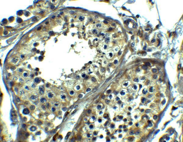
A01937-1 has real IHC data in human testis (image caption: human testis). It lists human, mouse and rat reactivity (catalog: reactivity), with IHC validation reported in human samples (datasheet: validation).
A01937-1 is listed for IHC-P (catalog: applications) and shows human testis staining at 5 μg/ml (image caption: human testis, 5 μg/ml). It lists human, mouse and rat reactivity (catalog: reactivity), while the reported IHC validation is in human samples (datasheet: validation).
Which to pick: Choose A01937-1 for paraffin-section IHC (catalog: IHC-P); its image shows human testis staining at 5 μg/ml (image caption: human testis, 5 μg/ml), and the fixative is unreported (image caption: no fixative specified). No SKU here lists IF/ICC or has an IF image (catalog: applications and image alts). For mouse or rat tissue, A01937-1 lists reactivity with both species (catalog: reactivity), but its reported IHC validation is in human samples (datasheet: validation).