SRSF1 / Serine/arginine-rich splicing factor 1 · IHC design guide

Design Immunohistochemistry for SRSF1

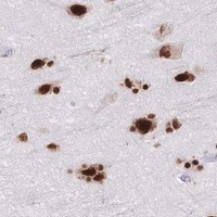
Use nuclear staining as the reference pattern when scoring SRSF1 in paraffin sections (HPA tissue IHC). The catalog antibody was demonstrated at 2 µg/ml with chromogenic detection; keep fixation consistent across comparisons (datasheet PB9404; standard IHC practice).

Evidence assembled Oct 2026 · For research use; verify linked source records and product datasheet before use
Immunohistochemistry protocol sheet for SRSF1 (IHC for SRSF1): expected localisation Nuclear in tissue; speckles are a molecular expectation (HPA tissue IHC; UniProt), antibody PB9404, validated IHC image, and IHC protocol steps
Printable SRSF1 IHC protocol sheet — expected localisation Nuclear in tissue; speckles are a molecular expectation (HPA tissue IHC; UniProt), antibody PB9404, controls and protocol steps. Open the full SRSF1 IHC guide →

SRSF1 Immunohistochemistry Experimental Design Guide

Expected localisation, validated protocols, controls and antibodies — the at-a-glance facts below, then the full design guide.

Must know before staining
Expected localisation Nuclear in tissue; speckles are a molecular expectation (HPA tissue IHC; UniProt)
Staining pattern Many cell types show nuclear staining (HPA tissue IHC)
Antigen retrieval EDTA pH 8.0 HIER, heat-mediated (datasheet PB9404)
Positive control ⓘ Adrenal gland+4 more · see all
Negative control ⓘ None in HPA (detected in all 45 tissues); use no-primary + isotype controls
Important caveats
Reasons your staining may differ from the expected pattern.
Fixation Matched tissue-IHC evidence does not establish the fixation claim. Validate the specimen-specific method before use. (selected-SKU IHC image PB9404)
Caveat Nuclear–cytoplasmic shuttling may alter the staining pattern (UniProt)
Regulation No specific expression regulator annotated (UniProt)
Isoform / epitope 3 isoforms; antibody epitope coverage is unknown (UniProt; datasheet PB9404)
Section 1

Recommended SRSF1 IHC & IF Protocols

The catalog antibody’s IHC-P protocol (datasheet: PB9404) is followed by four published SRSF1 paraffin-section protocols (PMC8123436; PMC8296067; PMC8781844; PMC10326825).

Recommended immunohistochemistry (IHC-P) protocol parameters
SampleParaffin-embedded human placenta tissue; fixative not specified (datasheet PB9404)
FixationImage fixative and duration unreported (datasheet PB9404); verify before use.
Sectioning4–5 µm sections on charged slides (standard)
DeparaffinisationXylene, graded ethanol series to water (standard)
Antigen retrievalHeat retrieval: EDTA pH 8.0 (datasheet PB9404); 20 min, 95–100 °C (standard)
Peroxidase block3% H2O2, 10 min, room temperature (standard)
Blocking10% goat serum (datasheet PB9404)
Primary antibodyRabbit anti-SRSF1, 2-5μg/ml (datasheet PB9404)
Primary incubationOvernight at 4 °C (datasheet PB9404)
DetectionHRP-conjugated secondary, DAB chromogen (datasheet PB9404)
CounterstainHematoxylin, blue, dehydrate and mount (standard)
Expected resultSRSF1-positive staining in glandular cells of adrenal gland (HPA tissue IHC: High). HPA tissue profile: Ubiquitous nuclear expression. No signal in the no-primary control.
💡Decision noteStart with heat-mediated EDTA pH 8.0 retrieval for the catalog antibody (datasheet: PB9404). Published alternatives used citrate pH 6.0 with microwave or pressure heating (PMC8123436; PMC10326825).
Section 2

What Is the Expected SRSF1 Staining Pattern?

SRSF1 is predominantly nuclear, with nuclear speckles reported by UniProt; it can also shuttle to the cytoplasm (UniProt Q07955 subcellular location). Expect staining across many cell types: HPA describes ubiquitous nuclear expression and high staining in several epithelial, hematopoietic, glial and endothelial cell populations (HPA tissue IHC: Supported). SRSF1 has no transmembrane segment (UniProt Q07955 topology).

What am I looking at on my slide?
Clear nuclear staining in glandular cells of adrenal gland, appendix or breast.This matches populations scored High by HPA (HPA tissue IHC). Compare nuclei with nearby background before judging intensity.
Predominantly cytoplasmic staining with little or no nuclear signal.This departs from the expected dominant nuclear pattern (HPA tissue IHC; UniProt Q07955 subcellular location). SRSF1 can shuttle, so limited cytoplasmic signal alone does not prove an artefact (UniProt Q07955).
Strong deposits confined to cells outside the expected nuclear pattern.Consider cross-reactivity or endogenous detection activity. HPA reports ubiquitous nuclear expression; its tissue data do not establish a universally negative cell type (HPA tissue IHC).
Diffuse chromogen across tissue and spaces between cells.A pattern without nuclear contrast is difficult to attribute to SRSF1, whose reported tissue pattern is nuclear (HPA tissue IHC). Assess reagent background and section-wide staining.
No nuclear signal in a known HPA-high population.A negative result in, for example, bone-marrow hematopoietic cells needs technical review before biological interpretation (HPA: High in hematopoietic cells).
💡Expected SRSF1 appearanceCall positive when nuclei stain clearly in an HPA-high cell population, such as bronchial respiratory epithelial cells (HPA: High); cytoplasm-only or diffuse extracellular chromogen is a suspect pattern (HPA tissue IHC: ubiquitous nuclear expression).
How each factor affects the staining
Cellular compartmentNuclear staining is the primary IHC expectation (HPA tissue IHC: ubiquitous nuclear expression). UniProt also places SRSF1 in nuclear speckles and notes nuclear–cytoplasmic shuttling (UniProt Q07955).
Choice of positive tissueAdrenal, appendix and breast glandular cells; bronchial respiratory epithelium; and bone-marrow hematopoietic cells are scored High (HPA tissue IHC). Select a population identifiable on the section.
Antibody validationHPA lists IHC as Supported for HPA061301 and CAB013073 (HPA antibodies). That supports interpreting a matching pattern, but does not validate every antibody or staining run.
Breadth of expressionHPA calls SRSF1 expression ubiquitous and its RNA tissue specificity low (HPA tissue IHC). Its supplied tissue profile lists no negative or low populations, so do not assume a tissue is a true negative control.
Molecular diversityUniProt lists 3 isoforms and multiple modified residues (UniProt Q07955 isoforms; modified residues). Without an antibody epitope, these facts cannot predict which forms the stain detects.
Chromogenic detection backgroundAs general IHC practice, endogenous enzyme activity or nonspecific reagent binding can produce chromogen unrelated to the target. Evaluate detection-only controls; this is not a reported SRSF1-specific effect.
Why is my staining missing, weak or wrong?
SituationLikely causeNext action
Nuclei are blank in the selected positive tissue.The run may have insufficient specific signal; HPA reports High staining in the listed positive populations (HPA tissue IHC).Confirm the expected cells are present, then review antibody use, antigen-retrieval conditions and detection with a positive control. HPA supplies no SRSF1-specific retrieval condition here.
Chromogen is mainly cytoplasmic.A cytoplasm-dominant result conflicts with HPA's ubiquitous nuclear tissue pattern, although SRSF1 can shuttle (HPA tissue IHC; UniProt Q07955).Check whether nuclei retain convincing signal, examine controls and repeat with an IHC-supported antibody if needed (HPA antibodies: HPA061301, CAB013073).
The entire section looks diffusely brown.Section-wide colour may obscure the nuclear pattern reported by HPA (HPA tissue IHC); nonspecific reagent binding is a general IHC possibility.Compare a detection-only control, review blocking and washing, and adjust detection exposure within the assay's validated workflow.
Only isolated, unexpected cells show intense deposits.Cross-reactivity or endogenous detection activity may explain deposits lacking the expected nuclear distribution (HPA tissue IHC: ubiquitous nuclear expression).Inspect cell identity and nuclear counterstain, then compare an appropriate reagent control. Do not call surrounding cells SRSF1-negative solely from this contrast.
Two tissues differ in staining strength.HPA scores several named cell populations High but does not provide a quantitative intensity scale across every tissue (HPA tissue IHC).Score the identified cell population and nuclear localisation in each section; avoid treating intensity differences alone as proof of altered SRSF1 abundance.
IF/ICC Q&A: Is some cytosolic signal compatible with SRSF1?Yes. HPA places SRSF1 mainly in nucleoplasm and additionally in cytosol; UniProt reports shuttling (HPA subcellular ICC-IF; UniProt Q07955).Expect nucleoplasm to remain the main signal (HPA subcellular ICC-IF: approved). HPA marks ICC evidence for CAB013073 and HPA069970 Uncertain (HPA antibodies).

Sample controls for SRSF1 IHC & IF

🧪Run bone marrow first: hematopoietic cells should stain (HPA: High in bone-marrow hematopoietic cells). HPA detects SRSF1 in all 45 scored tissues, so there is no validated negative tissue; use no-primary and isotype controls, and expect anucleate mature erythrocytes on the positive slide to lack nuclear staining without treating them as a validated target-negative control (HPA: no negative rows; standard histology).
Positive control tissue: Adrenal gland (Glandular cells, HPA High)
Negative control tissue: None in HPA: SRSF1 is detected in all 45 scored tissues. Use a no-primary (secondary-only) and an isotype control instead.
ICC-IF cell lines (HPA subcellular resource): HPA ICC-IF images show SRSF1 in A-431, U-251MG, U2OS, HEL, REH, with annotated localisation: Nucleoplasm (approved) (HPA subcellular).
Technical controls: Include a secondary-only slide with the primary antibody omitted, a concentration-matched rabbit IgG isotype control, and an SRSF1-knockout cell block processed alongside the tissue if available (PB9404 tissue-IHC caption: rabbit primary; standard IHC practice). Quench endogenous peroxidase and check the no-primary bone-marrow section for brown background before scoring DAB, particularly around myeloid cells (standard IHC practice).
⚠️Feasibility: No supplied source reports a target-specific fixation window or fixation effect, and the selected PB9404 paraffin-section caption does not state the fixative (PB9404 tissue-IHC caption). Heat-mediated retrieval in EDTA at pH 8.0 is a starting condition from the placenta caption, but retrieval dependence in bone marrow is unreported (PB9404 tissue-IHC caption). The evidence does not establish whether frozen sections or IF are easier; endogenous peroxidase in bone-marrow myeloid cells can complicate chromogenic scoring (standard IHC practice).

HPA tissue IHC evidence for SRSF1

Comprehensive Human Protein Atlas IHC scoring per tissue (reliability: Supported — High consistency between antibody staining and RNA expression data.). Rows are taken directly from the HPA tissue chart — click any row's HPA link to view the source.

Positive expression · recommended positive controls

TissueCell typeLevelEvidenceSource
Adrenal gland Glandular cells High Protein (IHC) HPA →
Appendix Glandular cells High Protein (IHC) HPA →
Bone marrow Hematopoietic cells High Protein (IHC) HPA →
Breast Glandular cells High Protein (IHC) HPA →
Bronchus Respiratory epithelial cells High Protein (IHC) HPA →

Undetected expression · recommended negative controls

TissueCell typeLevelEvidenceSource
None in HPA: SRSF1 is detected in all 45 scored tissues. Use a no-primary (secondary-only) and an isotype control instead.
Section 3

Advanced SRSF1 IHC Tips

Troubleshoot SRSF1 staining in paraffin section IHC by checking retrieval, nuclear localisation, controls and scoring before interpreting biological differences.

How should I optimise retrieval when SRSF1 nuclear staining is weak?
Start with heat mediated retrieval in EDTA at pH 8.0 for paraffin sections (datasheet PB9404). If nuclear staining remains weak, compare heating durations on adjacent sections while holding temperature, antibody concentration and detection constant (standard IHC practice). The documented tissue image used 2 μg/ml primary antibody overnight at 4°C, providing conditions to hold steady during that comparison (caption PB9404). Score signal in intact nuclei alongside tissue morphology, because excessive heating can damage sections and create misleading edge staining (standard IHC practice). SRSF1 localisation includes nuclear speckles, so assess the nuclear pattern as well as overall intensity (UniProt Q07955).
Can fixation explain variable SRSF1 staining across paraffin sections?
Target specific fixation sensitivity is unknown: the PB9404 placenta image shows a paraffin section but does not report its fixative (caption PB9404). Record each specimen’s fixative, fixation duration and processing history before comparing cases (standard IHC practice). Keep retrieval at EDTA pH 8.0 and apply the same staining conditions to study sections and controls (datasheet PB9404; standard IHC practice). If nuclear signal differs, inspect morphology and a separate nuclear control on adjacent sections before assigning the difference to SRSF1 (standard IHC practice). Nuclear shuttling and annotated phosphorylation do not establish a fixation response for this antibody (UniProt Q07955).
Which SRSF1 staining compartment should I score in chromogenic IHC?
Score nuclear staining first: tissue IHC shows ubiquitous nuclear expression, and the approved subcellular location is nucleoplasm (HPA tissue IHC; HPA subcellular). A speckled nuclear pattern is biologically plausible because SRSF1 localises to nuclear speckles (UniProt Q07955). Cytoplasmic signal merits separate recording because SRSF1 shuttles between nucleus and cytoplasm and cytosolic localisation has supporting evidence (UniProt Q07955; HPA subcellular). Compare compartment patterns within the same cell type and section before interpreting a shift (standard IHC practice). Diffuse extracellular or luminal DAB should prompt a background check rather than a localisation call (standard IHC practice).
Can this stain distinguish SRSF1 isoforms or modified epitopes?
Do not assign isoform identity from PB9404 staining without an isoform discriminating epitope validation (standard IHC practice). SRSF1 has 3 listed isoforms, ASF-1, ASF-2 and ASF-3, and contains RRM domains at residues 16–91 and 121–195 (UniProt Q07955). Its annotated modified residues include phosphorylation, acetylation and arginine methylation, which make epitope accessibility a consideration but do not establish this antibody’s modification sensitivity (UniProt Q07955). Check the catalog antibody’s mapped immunogen or epitope before making an isoform claim (standard IHC practice). Where needed, compare staining with an independently validated antibody against a distinct epitope under matched section conditions (standard IHC practice).
How can IF help assess SRSF1 localisation alongside a cell type marker?
For a separate IF experiment, multiplex SRSF1 with a validated marker for the cell type being assessed, such as glandular cells in a relevant tissue (HPA tissue IHC; standard IF practice). Choose a spectrally separated, preferably far red fluorophore when the specimen has strong short wavelength autofluorescence (standard IF practice). Because SRSF1 is intracellular, permeabilise sufficiently for antibody access to cytoplasmic and nuclear epitopes, then check that nuclear morphology remains intact (UniProt Q07955; standard IF practice). Interpret nucleoplasmic signal first while recording cytosolic signal separately (HPA subcellular). Use single colour and no primary controls to assess bleed through and autofluorescence (standard IF practice).
What controls help separate SRSF1 signal from chromogenic background?
The documented IHC example used 10% goat serum blocking before 2 μg/ml primary antibody overnight at 4°C (caption PB9404). Match that blocking and primary incubation when first investigating background, then vary one condition at a time (caption PB9404; standard IHC practice). Include a no primary section to expose secondary reagent or detection background, and check an adjacent section for tissue pigments (standard IHC practice). Peroxidase blocking and careful DAB development are general chromogenic workflow controls, not evidence of SRSF1 specificity (standard IHC practice). Assess nuclear contrast before increasing DAB development, which can obscure compartment boundaries (standard IHC practice).
How should I quantify SRSF1 staining across tissue regions? ⚠ ANSWER MARKED FOR VERIFICATION
Define comparable tissue regions and cell populations before scoring, then exclude folds, necrosis and cut edges (standard IHC practice). For nuclear staining, report the percentage of positive viable nuclei and an H-score using intensity grades 0–3, yielding a range of 0–300 (standard IHC practice). Normalise positive cell counts to all viable nuclei in the scored population, or report positive nuclei per mm² when tissue area is the denominator (standard IHC practice). Record cytoplasmic staining separately because nuclear expression predominates while cytosolic localisation is supported (HPA tissue IHC; HPA subcellular). Keep thresholds, illumination and section selection rules consistent across cases (standard IHC practice).
When is unexpected SRSF1 staining more likely to be artefact?
Convincing staining should include nuclear signal in intact cells, consistent with the ubiquitous nuclear tissue profile and approved nucleoplasmic location (HPA tissue IHC; HPA subcellular). High staining in glandular cells or hematopoietic cells is compatible with the reported tissue observations (HPA tissue IHC). Predominantly extracellular deposits, staining confined to torn edges, or signal over necrotic regions should trigger review of section quality and background controls (standard IHC practice). Check a no primary control for endogenous enzyme or detection signal before interpreting DAB deposits (standard IHC practice). A cytoplasmic component can be plausible, but assess it separately from nuclear staining and against control sections (UniProt Q07955; HPA subcellular; standard IHC practice).
Boster reagents

Best SRSF1 / Serine/arginine-rich splicing factor 1 IHC Antibodies

PB9404 has IHC images from paraffin sections of human placenta, mouse brain and rat brain (PB9404 image captions); M00497 lists IHC and IF/ICC for human, mouse and rat (M00497 catalog).

Real IHC data IHC analysis of SF2/SRSF1 using anti-SF2/SRSF1 antibody (PB9404). SF2/SRSF1 was detected in a paraffin-embedded section of human placenta tissue. Heat mediated antigen retrieval was performed in EDTA buffer (pH 8.0, epitope retrieval solution). The tissue section was blocked with 10% goat serum. The tissue section was then incubated with 2 μg/ml rabbit anti-SF2/SRSF1 Antibody (PB9404) overnight at 4°C. Peroxidase Conjugated Goat Anti-rabbit IgG was used as secondary antibody and incubated for 30 minutes at 37°C. The tissue section was developed using HRP Conjugated Rabbit IgG Super Vision Assay Kit (Catalog # SV0002) with DAB as the chromogen.
Anti-SF2/SRSF1 Antibody ®
Cat # PB9404

The rendered card is PB9404, with its own human placenta paraffin-section IHC figure (PB9404 card caption). Its catalog also documents paraffin-section IHC images from mouse and rat brain (PB9404 catalog image captions).

Which to pick: Choose PB9404 for tissue IHC, especially cross-species work, because its own captions document staining in human, mouse and rat paraffin sections (PB9404 image captions). Choose M00497 when IF/ICC is required: it is a rabbit monoclonal, clone ADGH-19, with IF/ICC listed for human, mouse and rat, though no IF image is supplied (M00497 catalog). PB9404’s captions establish paraffin-section processing; the fixative is unreported (PB9404 image captions).

Each figure is that product's own IHC / IF validation image from its datasheet.

References

  1. UniProt Consortium. UniProt entry Q07955 (SRSF1_HUMAN, Serine/arginine-rich splicing factor 1).
  2. Human Protein Atlas. SRSF1 tissue IHC expression (reliability: Supported).
  3. Human Protein Atlas. SRSF1 subcellular location (ICC-IF): Mainly localized to the nucleoplasm. In addition localized to the cytosol..
  4. Human Protein Atlas. SRSF1 antibody validation summary (3 antibodies).
  5. Diagnostic Utility of the Immunohistochemical Expression of Serine and Arginine Rich Splicing Factor 1 (SRSF1) in the Differential Diagnosis of Adult Gliomas. Cancers 2021 — PMC8123436.
  6. Immunohistochemical Expression of Serine and Arginine-Rich Splicing Factor 1 (SRSF1) in Fluoro-Edenite-Induced Malignant Mesothelioma: A Preliminary Study. International journal of environmental research and public health 2021 — PMC8296067.
  7. The Immunohistochemical Expression of the Serine and Arginine-Rich Splicing Factor 1 (SRSF1) Is a Predictive Factor of the Recurrence of Basal Cell Carcinoma: A Preliminary Study on a Series of 52 Cases. Medicina (Kaunas, Lithuania) 2022 — PMC8781844.
  8. SRSF1 induces glioma progression and has a potential diagnostic application in grading primary glioma. Oncology letters 2023 — PMC10326825.
  9. PubMed PMID:1855257 — UniProt-cited evidence.
  10. PubMed PMID:1830244 — UniProt-cited evidence.
  11. PubMed PMID:14702039 — UniProt-cited evidence.