This website uses cookies to ensure you get the best experience on our website.


- Table of Contents
Plan SSBP1 paraffin IHC around granular cytoplasmic staining, with lung macrophages or testis spermatogonia as high-staining controls (HPA tissue IHC). This guide covers fixation, the IHC-validated antibody at 1–2 μg/ml (datasheet A05166-1), chromogenic detection and cell-aware scoring.

Expected localisation, validated protocols, controls and antibodies — the at-a-glance facts below, then the full design guide.
| Expected localisation | Granular cytoplasm (HPA tissue IHC); mitochondrial matrix and nucleoid expected (UniProt) | |
| Staining pattern | Granular cytoplasmic staining across cell types (HPA tissue IHC) | |
| Antigen retrieval | EDTA pH 8.0 HIER, heat-mediated (datasheet A05166-1) | |
| Positive control | Lung+4 more · see all | |
| Negative control | Adipose tissue+1 more · see all |
| Fixation | Keep fixation consistent across sections (standard IHC practice; not target-specific) | |
| Caveat | High lung macrophage staining may confound tissue scoring (HPA tissue IHC) | |
| Regulation | Specific staining regulator unreported (UniProt) | |
| Isoform / epitope | No isoforms listed; mature chain 17–148 informs epitope choice (UniProt) |
The catalog antibody uses EDTA pH 8.0 retrieval (datasheet: A05166-1). The published IHC protocols below cover gastric tumors, tumor microarrays, cervical tumors, and mouse retina (PMC5762290; PMC10225361; PMC6411633; PMC8855788).
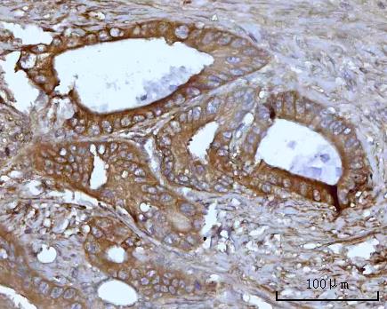
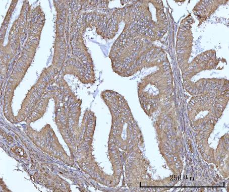
| Sample | Paraffin-embedded human colon adenocarcinoma tissue; fixative not specified (datasheet A05166-1) |
| Fixation | Image fixative and duration unreported (datasheet A05166-1); verify before use. |
| Sectioning | 4–5 µm sections on charged slides (standard) |
| Deparaffinisation | Xylene, graded ethanol series to water (standard) |
| Antigen retrieval | Heat retrieval: EDTA pH 8.0 (datasheet A05166-1); 20 min, 95–100 °C (standard) |
| Peroxidase block | 3% H2O2, 10 min, room temperature (standard) |
| Blocking | 10% goat serum (datasheet A05166-1) |
| Primary antibody | Rabbit anti-SSBP1, 1-2 μg/ml (datasheet A05166-1) |
| Primary incubation | Overnight at 4 °C (datasheet A05166-1) |
| Detection | HRP-conjugated secondary, DAB chromogen (datasheet A05166-1) |
| Counterstain | Hematoxylin, blue, dehydrate and mount (standard) |
| Expected result | SSBP1-positive staining in macrophages of lung (HPA tissue IHC: High). HPA tissue profile: General cytoplasmic expression with a granular pattern. No signal in the no-primary control. |
SSBP1 is a mitochondrial matrix and nucleoid protein with no transmembrane segment (UniProt Q04837). In paraffin IHC, expect granular cytoplasmic staining (HPA: tissue IHC profile), especially in lung macrophages and testicular spermatogonia, where staining is high (HPA: tissue IHC). HPA rates the tissue pattern Supported, with medium consistency between staining and RNA expression (HPA: tissue IHC reliability).
| Granular cytoplasmic stain in lung macrophages or testicular spermatogonia. | This matches the reported IHC pattern and its strongest listed cell populations (HPA: granular cytoplasmic profile; High in macrophages and spermatogonia). Assess granules within identifiable cells, using a counterstain to preserve cell identity (general IHC practice). |
| Predominantly nuclear, membrane-edge, or extracellular stain. | These compartments conflict with the expected mitochondrial matrix and nucleoid location and granular cytoplasmic IHC pattern (UniProt Q04837 localization; HPA: tissue IHC profile). Treat the pattern as suspect and check control sections and detection background before assigning it to SSBP1 (general IHC practice). |
| Strong stain centered on adipocytes or skeletal myocytes. | HPA lists SSBP1 as Not detected in those cells (HPA: adipocytes; skeletal myocytes). Unexpected strong staining warrants investigation for antibody cross-reactivity or endogenous detection activity (general IHC practice); a single discrepant section does not establish either cause. |
| Diffuse color across cells, stroma, or blank areas, with little granular definition. | That distribution is hard to reconcile with HPA's granular cytoplasmic profile (HPA: tissue IHC). Compare a no-primary control and inspect blocking, washes, and chromogen development to distinguish detection background from cell-associated staining (general IHC practice). |
| No detectable stain in a section containing lung macrophages or testicular spermatogonia. | Both are listed as High by HPA, so their absence makes a negative experimental section difficult to interpret (HPA: lung macrophages; testis spermatogonia). Verify that the expected cells are present, then review the antibody and detection run (general IHC practice). |
| Intracellular location | SSBP1 resides in the mitochondrial matrix and nucleoid (UniProt Q04837); its observed paraffin IHC appearance is granular cytoplasmic staining (HPA: tissue IHC profile). These sources support compartment interpretation, not a target-specific retrieval setting. |
| Cell-specific reference levels | Macrophages and spermatogonia are High; several glandular and neuronal cell groups are Medium, while adipocytes and skeletal myocytes are Not detected (HPA: tissue IHC). Score the identified cell population, not the whole tissue indiscriminately. |
| Protein processing and topology | UniProt lists a mature chain at residues 17–148, no transmembrane segment, and no glycosylation sites (UniProt Q04837). The supplied record gives no antibody epitope, so it cannot establish whether processing affects this antibody's staining. |
| Antibody evidence | HPA002866 is rated Supported for IHC, and the overall tissue profile has medium staining-to-RNA consistency (HPA: antibody validation; tissue IHC reliability). Use that support with morphology and controls; it does not validate every unexpected compartment. |
| Fixation and retrieval evidence | Target-specific fixation sensitivity and an SSBP1 retrieval condition are unreported in the supplied UniProt and HPA records (UniProt Q04837; HPA: tissue IHC). Retrieval optimization, if needed, is a general paraffin IHC workflow decision (general IHC practice). |
| Situation | Likely cause | Next action |
|---|---|---|
| Expected positive cells are unstained. | A negative result conflicts with HPA's High staining in lung macrophages and testicular spermatogonia, but the record does not identify a cause (HPA: tissue IHC). | Confirm those cells are in the section; inspect the IHC-validated antibody, detection reagents, and a concurrent positive control before interpreting other negatives (general IHC practice). |
| Brown signal fills most of the section or appears outside cells. | Broad, poorly defined color differs from the reported granular cytoplasmic pattern (HPA: tissue IHC profile). Detection background is one possible explanation (general IHC practice). | Compare a no-primary control; review blocking, wash steps, and chromogen development, then reassess whether signal remains cell-associated (general IHC practice). |
| Strong staining appears in adipocytes or skeletal myocytes. | Those cell types are listed as Not detected (HPA: tissue IHC); cross-reactivity or endogenous detection activity is possible, but neither is proven by appearance alone (general IHC practice). | Confirm cell identity on the counterstained section and compare controls before scoring this as SSBP1 expression (general IHC practice). |
| Nuclei or cell borders dominate the stain. | That location disagrees with mitochondrial matrix and nucleoid localization and HPA's granular cytoplasmic pattern (UniProt Q04837; HPA: tissue IHC). | Recheck morphology and control sections; treat the compartment assignment as unresolved until the granular cytoplasmic pattern can be demonstrated (general IHC practice). |
| Only weak staining is seen in a proposed control. | Some listed populations are Low, including kidney tubular cells and liver cholangiocytes; tissue-wide RNA specificity is Low (HPA: tissue IHC). | Choose a section containing HPA High lung macrophages or testicular spermatogonia for a clearer positive reference, and score the relevant cells (HPA: tissue IHC). |
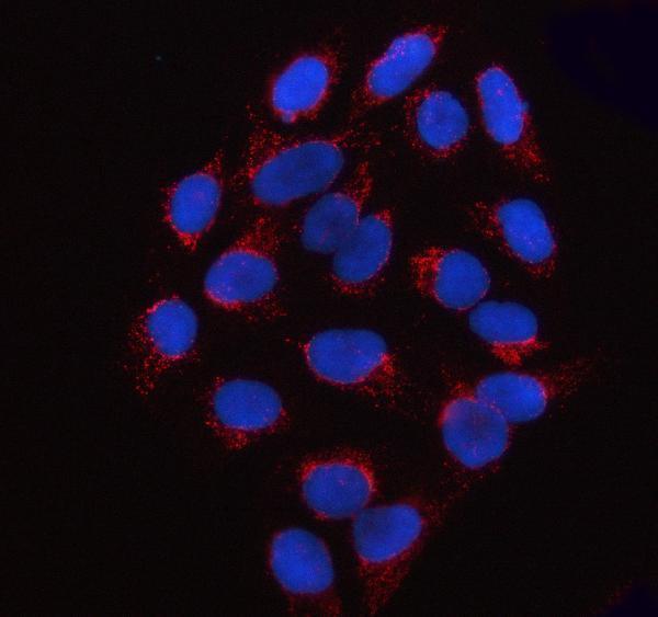
| IF/ICC Q: What localization should an image show? | HPA reports mitochondria as the main supported ICC-IF location; calyx and principal piece are additional approved locations (HPA: subcellular ICC-IF). | Expect predominantly mitochondrial signal when interpreting IF/ICC images (HPA: subcellular ICC-IF). Use the separate IF/ICC guide for that application. |
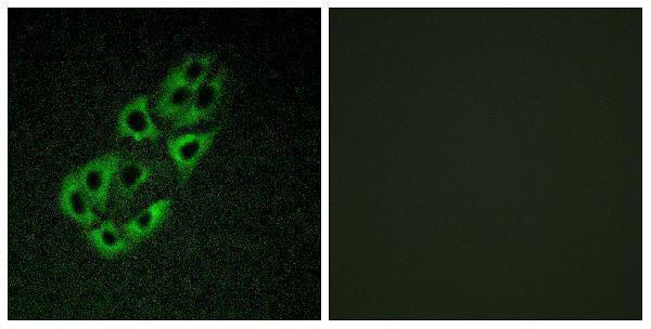
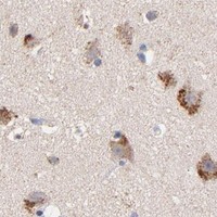


Comprehensive Human Protein Atlas IHC scoring per tissue (reliability: Supported — Medium consistency between antibody staining and RNA expression data.). Rows are taken directly from the HPA tissue chart — click any row's HPA link to view the source.
| Tissue | Cell type | Level | Evidence | Source |
|---|---|---|---|---|
| Lung | Macrophages | High | Protein (IHC) | HPA → |
| Testis | Spermatogonia cells | High | Protein (IHC) | HPA → |
| Adrenal gland | Glandular cells | Medium | Protein (IHC) | HPA → |
| Breast | Glandular cells | Medium | Protein (IHC) | HPA → |
| Caudate | Neuronal cells | Medium | Protein (IHC) | HPA → |
Troubleshoot chromogenic SSBP1 staining in paraffin sections using the catalog antibody’s tissue example, mitochondrial localisation, and cell-specific reference patterns.
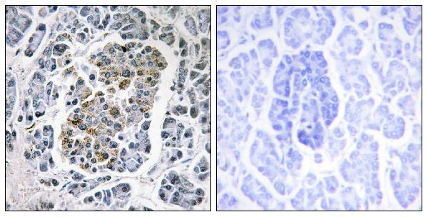
The IHC-validated SSBP1 antibodies have paraffin-section images from human tissues (catalog IHC captions) and IF images from human tissue or cells (catalog IF captions); one also lists mouse, rat and cow reactivity (A30475 catalog).
A05166-1 has IHC images from human colon, endometrioid, liver and lung cancers and IF images from U2OS cells and human colon and ovarian cancers (A05166-1 image captions). A30475 has an IHC image from human pancreas and an IF image from A549 cells; its listed reactivity is human, mouse, rat and cow (A30475 image captions; A30475 catalog).
Which to pick: For human tissue IHC, choose A05166-1 if you want a documented starting condition: its paraffin-section caption specifies EDTA retrieval at pH 8.0 and 2 μg/ml primary antibody (A05166-1 IHC caption). For IF/ICC, A05166-1 has U2OS cell and tissue IF examples (A05166-1 IF captions); for work across listed species, consider the polyclonal A30475, while recognizing that its pictured IHC and IF examples are human only (A30475 catalog; A30475 image captions). Both IHC captions describe paraffin sections, and neither reports the fixative (A05166-1 IHC captions; A30475 IHC caption).