This website uses cookies to ensure you get the best experience on our website.


- Table of Contents
Plan SSX1 chromogenic IHC on paraffin sections using nuclear staining in testis pachytene spermatocytes as the expected tissue pattern (HPA tissue IHC). The human-reactive catalog antibody A07363 has an IHC dilution of 1:100 (datasheet); interpret staining with the reported cross-gene caution (HPA tissue IHC).

Expected localisation, validated protocols, controls and antibodies — the at-a-glance facts below, then the full design guide.
| Expected localisation | Nuclear staining observed in testis (HPA tissue IHC) | |
| Staining pattern | Nuclear staining in pachytene spermatocytes (HPA tissue IHC) | |
| Antigen retrieval | Citrate pH 6.0 HIER, 95–98 °C, 20 min (rule: cytoplasmic / membrane antigen) | |
| Positive control | Testis | |
| Negative control | Adipose tissue+4 more · see all |
| Fixation | Keep fixation consistent across sections. (standard IHC practice; not target-specific) | |
| Caveat | Staining may detect proteins from more than one gene (HPA tissue IHC) | |
| Regulation | High in testis; low in thyroid (UniProt) | |
| Isoform / epitope | No annotated isoforms; one 1–188 chain (UniProt) |
The catalog antibody’s IHC-P protocol is paired with one published SSX1 chromogenic IHC protocol for paraffin sections (PMC10088888).
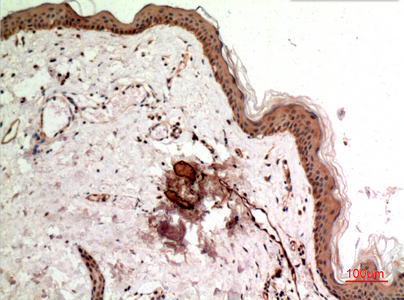
| Sample | Paraffin-embedded human skin tissue; fixative not specified (datasheet A07363) |
| Fixation | Image fixative and duration unreported (datasheet A07363); verify before use. |
| Sectioning | 4–5 µm sections on charged slides (standard) |
| Deparaffinisation | Xylene, graded ethanol series to water (standard) |
| Antigen retrieval | Heat-induced epitope retrieval in citrate buffer, pH 6.0, 20 min at 95–98 °C (standard rule: cytoplasmic / membrane antigen) |
| Peroxidase block | 3% H2O2, 10 min, room temperature (standard) |
| Blocking | 10% normal serum of the secondary host, 30 min, room temperature (standard) |
| Primary antibody | Rabbit anti-SSX1, 1:100 (datasheet A07363) |
| Primary incubation | Overnight at 4 °C (standard) |
| Detection | HRP-polymer secondary, DAB chromogen 5–10 min (standard) |
| Counterstain | Hematoxylin, blue, dehydrate and mount (standard) |
| Expected result | SSX1-positive staining in pachytene spermatocytes of testis (HPA tissue IHC: High). HPA tissue profile: Nuclear expression in testis. No signal in the no-primary control. |
SSX1 staining is expected mainly in the nuclei of pachytene spermatocytes in testis, where HPA reports high staining and a Supported tissue IHC pattern (HPA: tissue IHC). UniProt describes SSX1 as lacking a transmembrane segment and also annotates cytoplasm, cytoskeleton and flagellum axoneme locations (UniProt Q16384). HPA cautions that its staining evidence includes antibodies targeting proteins from more than one gene (HPA: tissue IHC).
| Strong nuclear staining in pachytene spermatocytes, with little staining in surrounding cells (HPA: tissue IHC). | This matches HPA’s high testis signal and nuclear tissue pattern (HPA: tissue IHC). Score the named cell population and compartment, rather than calling the whole section uniformly positive. HPA rates the tissue pattern Supported, while warning that the antibody evidence can include other gene products (HPA: tissue IHC). |
| Predominantly cytoplasmic or diffuse staining replaces the expected nuclear testis pattern (HPA: tissue IHC). | Treat this as a possible artefact and review controls and morphology. UniProt also annotates cytoplasm, cytoskeleton and flagellum axoneme, so compartment alone cannot prove an artefact; the slide-level comparison is with HPA’s observed nuclear tissue pattern (UniProt Q16384; HPA: tissue IHC). |
| Strong staining appears in cells outside the expected pachytene spermatocyte population (HPA: tissue IHC). | Check for cross-reactivity or endogenous detection activity before assigning SSX1 expression. HPA’s tissue result identifies pachytene spermatocytes as high and cautions that antibodies can target proteins from multiple genes (HPA: tissue IHC). Distribution across cell types is therefore part of the interpretation. |
| Haze, widespread weak color, or staining that obscures nuclei spans the section. | This is nonspecific background until controls show otherwise (general IHC practice). Compare the slide with a negative reagent control and the expected cell-specific nuclear pattern in testis (HPA: tissue IHC). Background that follows section edges or covers many cell types should not be scored as SSX1. |
| No convincing nuclear signal appears in morphologically intact testis pachytene spermatocytes (HPA: tissue IHC). | First assess the run using a known-positive testis section and detection controls (general IHC practice). Absence of the HPA high-staining cell population calls the assay result into question; it does not alone establish SSX1 absence in the specimen (HPA: tissue IHC). |
| Cell and tissue distribution | HPA identifies high staining in testis pachytene spermatocytes and no detected staining in the listed negative cell populations, including adipocytes and adrenal glandular cells (HPA: tissue IHC). Use those reported patterns to judge cell-specific signal; they do not guarantee that every section or antibody will behave identically. |
| Compartment evidence | HPA tissue IHC reports nuclear expression in testis, whereas UniProt annotates cytoplasm, cytoskeleton and flagellum axoneme (HPA: tissue IHC; UniProt Q16384). Base the paraffin-section decision on the observed tissue IHC pattern and document discordant staining for review. |
| Antibody interpretation | The HPA tissue assessment is Supported for consistency with RNA data, but HPA warns that antibodies target proteins from more than one gene (HPA: tissue IHC). A matching pattern supports the result without proving SSX1-specific binding in each stained cell. |
| Topology and processing | UniProt reports no transmembrane segment, no signal peptide or propeptide, and one 1–188 protein chain (UniProt Q16384). These annotations do not establish an SSX1-specific retrieval condition or fixation sensitivity; optimize those assay settings by general IHC practice. |
| Situation | Likely cause | Next action |
|---|---|---|
| Testis positive control has no nuclear staining in pachytene spermatocytes (HPA: tissue IHC). | The run may have failed, or the expected cells may be absent from the section; a blank slide alone cannot distinguish these possibilities (general IHC practice). | Confirm the cells by morphology, then check antibody, retrieval and detection steps against the assay’s validated settings; repeat with a known-positive section (general IHC practice). |
| Only cytoplasmic staining is visible in testis. | It differs from HPA’s nuclear tissue IHC pattern, although UniProt lists cytoplasmic and cytoskeletal locations (HPA: tissue IHC; UniProt Q16384). | Check nuclear morphology, counterstain and negative reagent control; record the compartment before interpreting the signal (general IHC practice). Do not call this the expected tissue IHC pattern (HPA: tissue IHC). |
| Many unrelated cell types stain strongly. | Cross-reactivity or endogenous detection activity is possible (general IHC practice); HPA cautions that its antibody evidence can include multiple gene products (HPA: tissue IHC). | Compare with the reported pachytene spermatocyte pattern and negative reagent control; review blocking and detection controls before scoring unrelated cells as positive (HPA: tissue IHC; general IHC practice). |
| Diffuse brown haze makes individual nuclei hard to assess. | Nonspecific reagent binding or incomplete blocking can produce broad background (general IHC practice). | Review negative controls, blocking, washes and detection development; score only resolved cellular signal after the background is controlled (general IHC practice). |
| A reported negative tissue shows staining. | HPA reports no detected staining in several sampled cell populations, such as adipocytes and adrenal glandular cells; an unexpected signal needs verification (HPA: tissue IHC). | Verify the stained cell type and compare matched controls. Investigate background and detection activity before treating the difference as biological expression (general IHC practice). |
| Q: How should an IF/ICC nuclear signal be read here? | HPA reports approved nucleoplasm and nucleoli locations, with a multiple-gene antibody caution, in ICC-IF (HPA: subcellular). | A: Use that observation as context for compartment interpretation; assess IF/ICC on its own guide page. For this paraffin IHC result, use HPA’s nuclear testis pattern in pachytene spermatocytes (HPA: subcellular; HPA: tissue IHC). |


Comprehensive Human Protein Atlas IHC scoring per tissue (reliability: Supported — High consistency between antibody staining and RNA expression data. Caution, targets protein from more than one gene.). Rows are taken directly from the HPA tissue chart — click any row's HPA link to view the source.
| Tissue | Cell type | Level | Evidence | Source |
|---|---|---|---|---|
| Testis | Pachytene spermatocytes | High | Protein (IHC) | HPA → |
| Tissue | Cell type | Level | Evidence | Source |
|---|---|---|---|---|
| Adipose tissue | Adipocytes | Not detected | Protein (IHC) | HPA → |
| Adrenal gland | Glandular cells | Not detected | Protein (IHC) | HPA → |
| Appendix | Glandular cells | Not detected | Protein (IHC) | HPA → |
| Bone marrow | Hematopoietic cells | Not detected | Protein (IHC) | HPA → |
| Breast | Adipocytes | Not detected | Protein (IHC) | HPA → |
Use compartment and cell-type controls when optimizing SSX1 staining; the available tissue and subcellular evidence requires careful interpretation.
A07363 is a human-reactive SSX1 antibody listed for IHC and IF (catalog: applications and reactivity). Its IHC image shows paraffin-embedded human skin (catalog: IHC image caption).
A07363 will render with an IHC image of paraffin-embedded human skin at 1:100 (catalog: IHC image caption). A07363 is also listed for human IF, but no IF image is supplied (catalog: applications, reactivity, and image captions).
Which to pick: For tissue IHC, choose A07363: its own image documents paraffin-embedded human skin, while the fixative is unreported (catalog: IHC image caption). For IF, A07363 is listed at 1:50, but the payload supplies no IF image or separate ICC validation (catalog: applications, IF dilution, and image captions). Cross-species use is unverified because A07363 lists human reactivity only (catalog: reactivity).