SSX2IP / Afadin- and alpha-actinin-binding protein · IHC design guide

Design Immunohistochemistry for SSX2IP

Plan chromogenic SSX2IP IHC on paraffin sections using the catalog antibody’s documented conditions (datasheet A05918-1). Compare cytoplasmic and nuclear staining with high staining in colon glandular cells (HPA tissue IHC).

Evidence assembled Oct 2026 · For research use; verify linked source records and product datasheet before use
Immunohistochemistry protocol sheet for SSX2IP (IHC for SSX2IP): expected localisation Cytoplasmic and nuclear staining (HPA tissue IHC), antibody A05918-1, validated IHC image, and IHC protocol steps
Printable SSX2IP IHC protocol sheet — expected localisation Cytoplasmic and nuclear staining (HPA tissue IHC), antibody A05918-1, controls and protocol steps. Open the full SSX2IP IHC guide →

SSX2IP Immunohistochemistry Experimental Design Guide

Expected localisation, validated protocols, controls and antibodies — the at-a-glance facts below, then the full design guide.

Must know before staining
Expected localisation Cytoplasmic and nuclear staining (HPA tissue IHC)
Staining pattern Across cell types: general cytoplasmic and nuclear staining (HPA tissue IHC)
Antigen retrieval EDTA pH 8.0 HIER, heat-mediated (datasheet A05918-1)
Positive control ⓘ Appendix+4 more · see all
Negative control ⓘ Adipose tissue+1 more · see all
Important caveats
Reasons your staining may differ from the expected pattern.
Fixation Keep fixation conditions consistent across sections (standard IHC practice; not target-specific)
Caveat Staining and RNA show medium consistency (HPA tissue IHC)
Regulation Expression varies by tissue (UniProt)
Isoform / epitope 3 isoforms; effect on epitope recognition unknown (UniProt)
Section 1

Recommended SSX2IP IHC & IF Protocols

The catalog antibody protocol uses EDTA pH 8.0 heat retrieval (datasheet A05918-1). Published IHC protocols below cover gastric cancer sections (PMC3691322) and ovarian tumors (PMC12985276).

Recommended immunohistochemistry (IHC-P) protocol parameters
SampleParaffin-embedded human cervix squamous cell carcinoma tissue; fixative not specified (datasheet A05918-1)
FixationImage fixative and duration unreported (datasheet A05918-1); verify before use.
Sectioning4–5 µm sections on charged slides (standard)
DeparaffinisationXylene, graded ethanol series to water (standard)
Antigen retrievalHeat retrieval: EDTA pH 8.0 (datasheet A05918-1); 20 min, 95–100 °C (standard)
Peroxidase block3% H2O2, 10 min, room temperature (standard)
Blocking10% goat serum (datasheet A05918-1)
Primary antibodyRabbit anti-SSX2IP, 2-5 μg/ml (datasheet A05918-1)
Primary incubationOvernight at 4 °C (datasheet A05918-1)
DetectionHRP-conjugated secondary, DAB chromogen (datasheet A05918-1)
CounterstainHematoxylin, blue, dehydrate and mount (standard)
Expected resultSSX2IP-positive staining in glandular cells of appendix (HPA tissue IHC: High). HPA tissue profile: General cytoplasmic and nuclear expression in all tissues. No signal in the no-primary control.
💡Decision noteTry EDTA pH 8.0 heat retrieval first with the catalog antibody (datasheet A05918-1); neither published excerpt specifies a retrieval method (PMC3691322; PMC12985276).
Section 2

What Is the Expected SSX2IP Staining Pattern?

SSX2IP is an intracellular protein with cytoplasmic and nuclear tissue staining, and centrosome, centriolar satellite and basal body localization (HPA tissue IHC; HPA ICC-IF; UniProt Q9Y2D8). In paraffin-section IHC, assess staining in the specific cells identified by HPA, including appendix glandular cells and caudate neurons, while accounting for HPA's Supported reliability rating and medium consistency with RNA data (HPA tissue IHC). SSX2IP has no transmembrane segment (UniProt Q9Y2D8 topology).

What am I looking at on my slide?
Cytoplasmic and nuclear staining appears in appendix glandular cells or caudate neurons, with clearly identifiable tissue structure.This fits the broad tissue compartment pattern; HPA rates staining High in both named cell populations (HPA tissue IHC). Judge the identified cells and compartments together: a brown deposit in a tissue section alone does not establish SSX2IP specificity (general IHC practice).
Signal is confined to the extracellular space or outlines only the cell surface, without convincing intracellular staining.That distribution conflicts with the reported cytoplasmic and nuclear IHC profile and intracellular localization (HPA tissue IHC; UniProt Q9Y2D8). Consider nonspecific deposit or a staining artefact, then compare with a suitable control section before interpreting it as SSX2IP (general IHC practice).
Strong staining appears in adipocytes or skeletal-muscle myocytes, especially if the intended positive cells are faint.HPA lists SSX2IP as Not detected in those specific cell populations (HPA tissue IHC). Unexpected signal may reflect cross-reactivity or endogenous detection activity; inspect tissue morphology and detection controls before assigning its cause (general IHC practice). These cell-level observations do not make every cell in either tissue a negative control (HPA tissue IHC).
Brown color spreads across the section, including spaces between cells, and cell boundaries are hard to distinguish.Diffuse deposit cannot be assigned confidently to the reported cytoplasmic or nuclear compartments (HPA tissue IHC). Check background and detection controls, blocking, and wash quality as general chromogenic-IHC troubleshooting steps (general IHC practice); diffuse color by itself does not establish SSX2IP expression.
A known-positive cell population is unstained, such as appendix glandular cells or caudate neurons.HPA rates these cell populations High, so an absent signal warrants a technical check before a biological conclusion (HPA tissue IHC). Verify that the expected cells are present, then review antibody suitability, retrieval, dilution and detection using the applicable IHC protocol (general IHC practice).
💡Expected SSX2IP appearanceCall a section positive when identifiable HPA High cells, such as appendix glandular cells or caudate neurons, show interpretable intracellular cytoplasmic and/or nuclear chromogen; extracellular-only deposit or strong staining of HPA Not detected adipocytes is suspect (HPA tissue IHC; general IHC practice).
How each factor affects the staining
Tissue and cell selectionHPA reports High staining in appendix glandular cells, bone-marrow hematopoietic cells, breast myoepithelial cells, bronchial respiratory epithelial cells, caudate neurons, cerebral-cortex glia, and colon and duodenum glandular cells (HPA tissue IHC). Select and score a named cell population rather than treating every cell in a tissue as equivalent (general IHC practice).
Compartment and imaging methodTissue IHC has a general cytoplasmic and nuclear profile, while ICC-IF mainly resolves centrosomes, centriolar satellites and basal bodies (HPA tissue IHC; HPA ICC-IF). IF/ICC Q&A: Should an IHC section show distinct centrosomal puncta? That fine detail may be easier to assess by ICC-IF; do not require visible puncta to call a chromogenic tissue section positive (HPA ICC-IF; general IHC practice).
Antibody evidenceHPA075946 has Supported IHC validation; HPA027306 has Supported ICC validation with no IHC status listed (HPA antibodies). Keep validation application-specific when choosing an antibody or interpreting a disagreement between tissue IHC and cell-based IF (HPA antibodies; general IHC practice).
Isoforms and epitope coverageUniProt lists three SSX2IP isoforms, but the supplied record does not locate an IHC antibody epitope or show which isoforms it detects (UniProt Q9Y2D8; HPA antibodies). An unexpected staining difference cannot be attributed to isoform selectivity from these data alone.
Processing, topology and fixation evidenceTarget-specific fixation effects are not established by the supplied assay evidence. Verify with a matched IHC source before attributing a result to fixation.
Why is my staining missing, weak or wrong?
SituationLikely causeNext action
The positive-control cell population has no chromogen.Possible workflow failure or unsuitable antibody conditions; HPA expects High staining in appendix glandular cells and caudate neurons (HPA tissue IHC; general IHC practice).Confirm the named cells are present, then check the IHC antibody's applicable instructions, retrieval conditions, dilution and detection controls one step at a time (general IHC practice). The supplied sources give no SSX2IP-specific retrieval setting.
Only a sharp cell-surface rim or extracellular deposit is visible.The pattern does not match reported intracellular staining and localization (HPA tissue IHC; UniProt Q9Y2D8); nonspecific deposit is a possibility (general IHC practice).Compare with a detection control and examine the same cells at higher magnification; require interpretable intracellular signal before recording the section as positive (general IHC practice; HPA tissue IHC).
Adipocytes or skeletal-muscle myocytes stain strongly.Both named cell populations are Not detected in HPA tissue IHC; cross-reactivity or endogenous chromogenic activity may explain unexpected color (HPA tissue IHC; general IHC practice).Check cell identity and run an appropriate detection control. Reassess specificity before scoring those cells as SSX2IP positive; do not extend their Not detected rating to surrounding cell types (general IHC practice; HPA tissue IHC).
Background obscures nuclei and cytoplasm throughout the section.Diffuse background prevents compartment-based interpretation; inadequate blocking, washing or detection control is worth checking as general IHC practice (HPA tissue IHC; general IHC practice).Review blocking and washes, compare with the detection control, and adjust routine staining conditions according to the applicable antibody protocol before scoring (general IHC practice).
A slide lacks discrete centrosomal dots despite plausible intracellular staining.HPA's punctate centrosome, satellite and basal-body localization comes from ICC-IF; its tissue IHC summary describes general cytoplasmic and nuclear staining (HPA ICC-IF; HPA tissue IHC).Interpret the paraffin-section chromogen against the tissue IHC profile and named positive cells. Reserve fine puncta assessment for the separate IF/ICC workflow (HPA tissue IHC; HPA ICC-IF).
Two antibodies or staining methods give different distributions.Validation is application-specific: HPA075946 is Supported for IHC, while HPA027306 is Supported for ICC and has no listed IHC rating (HPA antibodies).Compare results within the same application and named cell population first. Document antibody identity and controls; the supplied data do not establish epitope coverage or a fixation-based explanation (HPA antibodies; UniProt Q9Y2D8; general IHC practice).

Sample controls for SSX2IP IHC & IF

🧪Run appendix first: glandular cells should stain (HPA: High in appendix glandular cells). Use adipose tissue as the negative tissue; adipocytes are expected to be unstained (HPA: Not detected in adipocytes). On the appendix slide, use neighboring cells without staining to judge background, but do not assume they lack SSX2IP (standard IHC practice).
Positive control tissue: Appendix (Glandular cells, HPA High)
Negative control tissue: Adipose tissue (HPA Not detected)
ICC-IF cell lines (HPA subcellular resource): HPA ICC-IF images show SSX2IP in A-431, U-251MG, ASC52telo, RPTEC/TERT1, hTERT-RPE1 (serum starved), Sperm, with annotated localisation: Centriolar satellite (supported), Centrosome (supported), Basal body (supported) (HPA subcellular).
Technical controls: Include a no-primary, secondary-only control and a concentration-matched rabbit isotype control matched to the primary antibody’s clonality (selected-SKU caption: rabbit primary; standard IHC practice). A validated SSX2IP knockout specimen or peptide competition can test target specificity (standard IHC practice). Quench endogenous peroxidase and check appendix inflammatory cells for residual DAB signal (standard chromogenic IHC practice).
⚠️Feasibility: No target-specific fixation window or fixation effect is reported, and the selected-SKU paraffin-section caption does not state the fixative (selected-SKU tissue-IHC caption). That caption reports heat retrieval in EDTA at pH 8.0, but does not establish whether retrieval is required or whether frozen sections are easier (selected-SKU tissue-IHC caption). IF/ICC may help assess the expected centrosome and centriolar-satellite pattern, but the supplied evidence does not establish that it is easier than paraffin IHC (HPA: supported subcellular locations; selected-SKU tissue-IHC caption).

HPA tissue IHC evidence for SSX2IP

Comprehensive Human Protein Atlas IHC scoring per tissue (reliability: Supported — Medium consistency between antibody staining and RNA expression data.). Rows are taken directly from the HPA tissue chart — click any row's HPA link to view the source.

Positive expression · recommended positive controls

TissueCell typeLevelEvidenceSource
Appendix Glandular cells High Protein (IHC) HPA →
Bone marrow Hematopoietic cells High Protein (IHC) HPA →
Breast Myoepithelial cells High Protein (IHC) HPA →
Bronchus Respiratory epithelial cells High Protein (IHC) HPA →
Caudate Neuronal cells High Protein (IHC) HPA →

Undetected expression · recommended negative controls

TissueCell typeLevelEvidenceSource
Adipose tissue Adipocytes Not detected Protein (IHC) HPA →
Skeletal muscle Myocytes Not detected Protein (IHC) HPA →
Section 3

Advanced SSX2IP IHC Tips

Troubleshoot chromogenic SSX2IP IHC in paraffin sections using the catalog antibody’s tissue result and independent localisation evidence (datasheet A05918-1; HPA subcellular).

Which retrieval conditions should I start with for SSX2IP in paraffin sections?
Start with heat mediated retrieval in EDTA at pH 8.0 (datasheet A05918-1). The catalog antibody detected SSX2IP in a paraffin section after this retrieval, followed by 2 μg/ml primary antibody overnight at 4°C (datasheet A05918-1). If staining is weak, vary heating duration on adjacent sections while holding antibody concentration and detection constant (standard IHC practice). Compare signal in intact cells with a no primary control, because stronger DAB alone does not establish better antigen recovery (standard IHC practice). Record retrieval temperature and duration so a useful result can be reproduced (standard IHC practice).
Could fixation explain weak or uneven SSX2IP staining?
Target specific fixation sensitivity is unknown: the paraffin section caption does not state its fixative (datasheet A05918-1). Do not infer fixation tolerance from SSX2IP phosphorylation, lack of a transmembrane segment, or tissue staining patterns (UniProt Q9Y2D8; HPA tissue; datasheet A05918-1). For a controlled comparison, document fixative, fixation duration, section thickness and storage for each specimen (standard IHC practice). Apply the documented EDTA pH 8.0 retrieval and 2 μg/ml overnight primary incubation consistently across sections (datasheet A05918-1). Interpret differences only after checking morphology and a no primary control, since damaged tissue can complicate chromogenic scoring (standard IHC practice).
Should SSX2IP stain nuclei, junctions or small cytoplasmic puncta?
Small perinuclear puncta are plausible because SSX2IP localises to centrosomes, centriolar satellites and basal bodies (UniProt Q9Y2D8; HPA subcellular). Nuclear and general cytoplasmic staining are also reported, while UniProt places the protein at cell–cell adherens junctions and excludes cell–matrix adherens junctions (HPA tissue; UniProt Q9Y2D8). Assess staining within identifiable cells at matched magnification, with a nuclear counterstain to distinguish nuclear signal from adjacent cytoplasm (standard IHC practice). The catalog image establishes detection in paraffin tissue, but its caption does not validate every compartment (datasheet A05918-1). Treat an isolated, diffuse extracellular DAB deposit as suspect and compare it with no primary staining (standard IHC practice).
Can this stain distinguish SSX2IP isoforms or phosphorylation states?
SSX2IP has 3 annotated isoforms and several reported phosphoserines, including positions 290, 293 and 312 (UniProt Q9Y2D8). The supplied catalog caption gives no epitope map or isoform specificity, so a positive section cannot be assigned to one isoform (datasheet A05918-1; UniProt Q9Y2D8). It also cannot establish whether the detected molecules carry a particular phosphorylation (datasheet A05918-1; UniProt Q9Y2D8). Keep EDTA retrieval at pH 8.0 and the 2 μg/ml primary concentration fixed while comparing specimens (datasheet A05918-1). If isoform or modification identity matters, obtain epitope information and an independently validated assay before making that claim (standard IHC practice).
How should I assess SSX2IP by multiplex IF alongside this IHC guide?
Treat IF as a separate optimisation: the supplied catalog evidence documents chromogenic staining in a paraffin section, with no IF procedure specified (datasheet A05918-1). Pair SSX2IP with a marker that identifies the cell population under study, and check single channel controls for bleed through (standard IF practice). Select fluorophores after inspecting tissue autofluorescence, favouring a cleaner spectral channel for the weaker target signal (standard IF practice). SSX2IP has no transmembrane segment and includes intracellular centrosomal, cytoplasmic and nuclear locations; permeabilisation must permit access to the compartment being tested (UniProt Q9Y2D8; HPA subcellular; standard IF practice). Confirm puncta against a no primary control before translating IF localisation into an IHC interpretation (standard IF/IHC practice).
How can I reduce widespread DAB staining without losing SSX2IP signal?
Begin with a no primary control and inspect whether DAB accumulates in the same structures as the test section (standard IHC practice). The catalog tissue procedure used 10% goat serum block, 2 μg/ml primary overnight at 4°C, and a peroxidase conjugated secondary for 30 minutes at 37°C (datasheet A05918-1). A peroxidase block and careful washes are general chromogenic IHC controls, particularly when background persists across cell types (standard IHC practice). Titrate primary exposure or concentration while retaining EDTA retrieval at pH 8.0 as the documented starting condition (datasheet A05918-1; standard IHC practice). Compare intact cellular staining with control sections before calling faint diffuse colour SSX2IP positive (standard IHC practice).
What is a defensible way to score SSX2IP IHC across sections? ⚠ ANSWER MARKED FOR VERIFICATION
Choose a defined cell population and region before scoring, then report percentage of positive cells and staining intensity or an H-score (standard IHC practice). Record nuclear and cytoplasmic signal separately when both appear, because both compartments occur in tissue staining reports (HPA tissue; standard IHC practice). For discrete puncta, count positive cells or puncta per mm² within a specified tissue area rather than pooling unlike compartments (HPA subcellular; standard IHC practice). Normalise counts to evaluable cells or tissue area, and exclude folds, necrosis and section edges by a prespecified rule (standard IHC practice). Hold retrieval at EDTA pH 8.0 and detection conditions constant across the comparison (datasheet A05918-1; standard IHC practice).
How do I separate true SSX2IP staining from tissue artefact?
Look for signal within intact cells in plausible compartments: cytoplasm or nucleus, with possible centrosomal or satellite associated puncta (HPA tissue; HPA subcellular). Cell identity matters because high staining is reported in appendix glandular cells and bronchial respiratory epithelial cells, whereas adipocytes are reported as not detected (HPA tissue). An edge only pattern, necrotic deposit or colour present without primary antibody warrants investigation before assigning positivity (standard IHC practice). Check endogenous peroxidase with appropriate controls when DAB appears in unexpected cells or tissue spaces (standard IHC practice). Interpret weak staining cautiously: HPA rates tissue evidence as Supported with medium agreement between antibody staining and RNA expression (HPA tissue).
Boster reagents

Best SSX2IP / Afadin- and alpha-actinin-binding protein IHC Antibodies

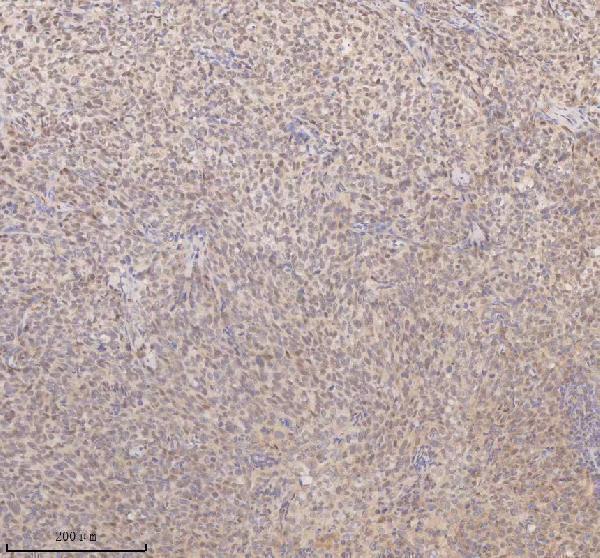
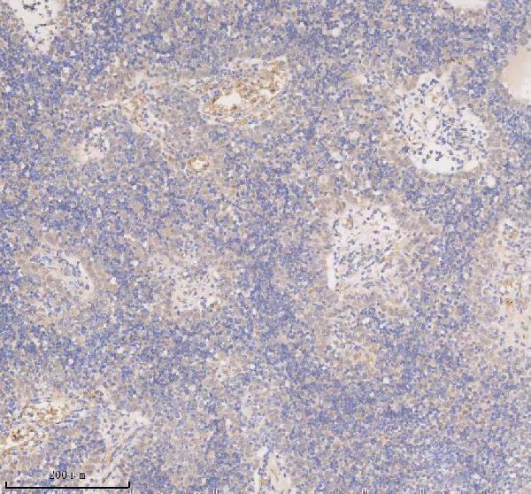
The catalog lists human-reactive SSX2IP IHC antibodies, with paraffin-section images for human cervix squamous cell carcinoma, human spleen and rat brain using A05918-1 (catalog applications; A05918-1 image captions). No IF data are shown (catalog image alts).

Real IHC data IHC analysis of SSX2IP using anti-SSX2IP antibody (A05918-1). SSX2IP was detected in a paraffin-embedded section of human cervix squamous cell carcinoma tissue. Heat mediated antigen retrieval was performed in EDTA buffer (pH 8.0, epitope retrieval solution). The tissue section was blocked with 10% goat serum. The tissue section was then incubated with 2 μg/ml rabbit anti-SSX2IP Antibody (A05918-1) overnight at 4°C. Peroxidase Conjugated Goat Anti-rabbit IgG was used as secondary antibody and incubated for 30 minutes at 37°C. The tissue section was developed using HRP Conjugated Rabbit IgG Super Vision Assay Kit (Catalog # SV0002) with DAB as the chromogen.
Anti-SSX2IP Antibody ®
Cat # A05918-1

A05918-1 has IHC images from paraffin sections of human cervix squamous cell carcinoma, human spleen and rat brain (A05918-1 image captions). M05918-1 is listed for human IHC, but has no IHC image in the payload (M05918-1 catalog applications and reactivity; image alts).

Which to pick: For tissue IHC, choose A05918-1 when an imaged paraffin-section protocol is useful: its captions specify EDTA retrieval at pH 8.0 and 2 μg/ml primary antibody, with no fixative reported (A05918-1 image captions). For human IHC, M05918-1 is a rabbit monoclonal option, although no tissue image is provided (M05918-1 catalog clone, host, applications and image alts). Neither SKU lists IF/ICC validation; for human and rat IHC, choose A05918-1 based on its stated reactivity and imaged sections (catalog applications, reactivity and image captions).

Each figure is that product's own IHC / IF validation image from its datasheet.

References

  1. UniProt Consortium. UniProt entry Q9Y2D8 (ADIP_HUMAN, Afadin- and alpha-actinin-binding protein).
  2. Human Protein Atlas. SSX2IP tissue IHC expression (reliability: Supported).
  3. Human Protein Atlas. SSX2IP subcellular location (ICC-IF): Mainly localized to the centrosome, centriolar satellites and basal body. In addition localized to the acrosome, equatorial segment and principal piece..
  4. Human Protein Atlas. SSX2IP antibody validation summary (2 antibodies).
  5. Epigenetic silencing of miR-338-3p contributes to tumorigenicity in gastric cancer by targeting SSX2IP. PloS one 2013 — PMC3691322.
  6. MicroRNA-625-3p Increases Chemosensitivity in Ovarian Cancer Cells Through Decreasing SSX2IP-Mediated Cisplatin Export in Extracellular Vesicles. Cancers 2026 — PMC12985276.
  7. MiRNA-181b-5p Modulates Cell Proliferation, Cell Cycle, and Apoptosis by Targeting SSX2IP in Acute Lymphoblastic Leukemia. Turkish journal of haematology : official journal of Turkish Society of Haematology 2022 — PMC9421343.
  8. PubMed PMID:12007189 — UniProt-cited evidence.
  9. PubMed PMID:10231032 — UniProt-cited evidence.
  10. PubMed PMID:14702039 — UniProt-cited evidence.