This website uses cookies to ensure you get the best experience on our website.


- Table of Contents
Plan chromogenic IHC for ST3GAL3 using the cytoplasmic tissue staining profile (HPA tissue IHC) and Golgi localization as a molecular expectation (UniProt). Compare high-staining liver hepatocytes with adipocytes reported as undetected, while treating the tissue staining profile as pending external verification (HPA tissue IHC).

Expected localisation, validated protocols, controls and antibodies — the at-a-glance facts below, then the full design guide.
| Expected localisation | Cytoplasmic staining in most tissues (HPA tissue IHC) | |
| Staining pattern | Cytoplasmic staining across most tissue types (HPA tissue IHC) | |
| Antigen retrieval | Citrate pH 6.0 HIER, 95–98 °C, 20 min (rule: cytoplasmic / membrane antigen) | |
| Positive control | Adrenal gland+4 more · see all | |
| Negative control | Adipose tissue+4 more · see all |
| Fixation | Matched tissue-IHC evidence does not establish the fixation claim. Validate the specimen-specific method before use. (selected-SKU IHC image A08054-1) | |
| Caveat | A secreted form may complicate cellular staining (UniProt) | |
| Regulation | Expression varies by tissue and cell type (HPA tissue IHC) | |
| Isoform / epitope | 26 isoforms; check cytoplasmic vs lumenal epitope coverage (UniProt) |
The catalog antibody’s IHC-P protocol is paired with one published ST3GAL3 gastric biopsy protocol (PMC9577379).
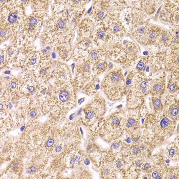
| Sample | Paraffin-embedded human liver damage tissue; fixative not specified (datasheet A08054-1) |
| Fixation | Image fixative and duration unreported (datasheet A08054-1); verify before use. |
| Sectioning | 4–5 µm sections on charged slides (standard) |
| Deparaffinisation | Xylene, graded ethanol series to water (standard) |
| Antigen retrieval | Heat-induced epitope retrieval in citrate buffer, pH 6.0, 20 min at 95–98 °C (standard rule: cytoplasmic / membrane antigen) |
| Peroxidase block | 3% H2O2, 10 min, room temperature (standard) |
| Blocking | 10% normal serum of the secondary host, 30 min, room temperature (standard) |
| Primary antibody | Rabbit anti-ST3GAL3, 1:50-1:200 (datasheet A08054-1) |
| Primary incubation | Overnight at 4 °C (standard) |
| Detection | HRP-polymer secondary, DAB chromogen 5–10 min (standard) |
| Counterstain | Hematoxylin, blue, dehydrate and mount (standard) |
| Expected result | ST3GAL3-positive staining in glandular cells of adrenal gland (HPA tissue IHC: High). HPA tissue profile: Cytoplasmic expression in most tissues. No signal in the no-primary control. |
ST3GAL3 is a Golgi stack membrane protein with a short cytoplasmic tail and a lumenal catalytic region; a secreted form is also reported (UniProt Q11203). In tissue IHC, expect predominantly cytoplasmic staining in selected glandular cells, hepatocytes, germinal center cells, and pancreatic endocrine cells (HPA tissue IHC). Treat these expectations as provisional: HPA rates its tissue IHC evidence Uncertain, pending external verification (HPA tissue IHC).
| Cytoplasmic staining in appendix or duodenal glandular cells, liver hepatocytes, lymph node germinal center cells, or pancreatic endocrine cells (HPA tissue IHC). | These cell populations are scored High by HPA and provide provisional positive comparators (HPA tissue IHC). A cytoplasmic pattern is compatible with Golgi localization, although routine chromogenic IHC cannot establish the stained organelle on its own (UniProt Q11203; standard IHC interpretation). |
| Predominantly nuclear staining, or an isolated crisp plasma membrane outline without convincing cytoplasmic staining. | Neither pattern is the expected tissue IHC profile (HPA tissue IHC). UniProt places the membrane bound form in the Golgi stack, so check whether the signal is reproducible and antibody dependent before assigning it to ST3GAL3 (UniProt Q11203 topology; standard IHC interpretation). |
| Strong signal in adipocytes, skeletal myocytes, or placental trophoblastic cells, especially when the expected positive cells are faint. | HPA reports these cells as Not detected in its tissue IHC dataset (HPA tissue IHC). Investigate antibody cross reactivity or endogenous detection activity; skeletal muscle needs particular caution because UniProt reports high adult expression there (UniProt Q11203 tissue specificity). |
| Diffuse color across multiple cell types, extracellular spaces, and the section background. | A widespread haze does not resemble HPA's cell selective cytoplasmic profile (HPA tissue IHC). Assess nonspecific antibody binding, incomplete blocking, and endogenous detection activity with appropriate control sections (standard chromogenic IHC practice). |
| No convincing signal in a selected HPA high cell population while tissue morphology and counterstain remain interpretable. | Check the staining run before calling the sample ST3GAL3 negative: the HPA high designation is provisional, and its antibody HPA051102 has Uncertain IHC validation (HPA tissue IHC; HPA antibodies). A second positive tissue can help distinguish a sample result from a failed run (standard IHC practice). |
| Golgi topology and interpretation | Residues 9–28 form a transmembrane segment, with residues 29–375 lumenal (UniProt Q11203 topology). This supports an intracellular membrane associated expectation, but a chromogenic cytoplasmic signal alone does not resolve Golgi cisternae (standard IHC interpretation). |
| Tissue and cell choice | HPA scores glandular cells in adrenal gland, appendix, duodenum, parathyroid gland, and seminal vesicle High; it also scores hepatocytes, germinal center cells, and pancreatic endocrine cells High (HPA tissue IHC). Select the scored cell population, not the entire organ, for comparison. |
| Evidence disagreement | UniProt reports high expression in adult skeletal muscle, while HPA scores skeletal myocytes Not detected by tissue IHC (UniProt Q11203 tissue specificity; HPA tissue IHC). Interpret a negative muscle section as assay specific evidence, not proof that the protein is absent. |
| Processing and molecular diversity | UniProt reports a secreted form, two glycosylation sites at residues 80 and 171, and 26 isoforms (UniProt Q11203). Without an antibody epitope or isoform coverage record, these annotations cannot predict which forms a particular IHC stain detects. |
| Validation strength | HPA describes its tissue IHC reliability as Uncertain, pending external verification, and lists HPA051102 IHC as Uncertain (HPA tissue IHC; HPA antibodies). Use matched positive and negative comparators when judging a new staining pattern (standard IHC practice). |
| Situation | Likely cause | Next action |
|---|---|---|
| A selected HPA high population is blank (HPA tissue IHC). | A failed staining run, low effective antibody signal, or an unsupported epitope assumption could explain the result (standard IHC practice). HPA's high score is provisional (HPA tissue IHC). | Check the run with another HPA high population, then review the catalog antibody's IHC-P instructions and the run's antigen retrieval, dilution, and detection controls (standard IHC practice). |
| Nuclei dominate the stain. | Nuclear localization does not match the reported cytoplasmic tissue profile or Golgi membrane localization (HPA tissue IHC; UniProt Q11203). | Compare with a no primary control and a separate positive tissue; reassess antibody specificity if nuclear staining persists (standard IHC practice). |
| HPA not detected cell types stain as strongly as positive comparators (HPA tissue IHC). | Cross reactivity or endogenous chromogenic detection activity is plausible (standard IHC practice); HPA's tissue calls remain uncertain (HPA tissue IHC). | Check no primary and detection controls, compare cell types within the same section, and seek independent antibody evidence before reclassifying the pattern (standard IHC practice). |
| The whole section has diffuse color. | Nonspecific binding or residual endogenous detection activity can obscure a cell selective cytoplasmic pattern (standard IHC practice; HPA tissue IHC). | Review blocking, washes, antibody dilution, and detection controls; retain a counterstain that lets the scored cell population be identified (standard chromogenic IHC practice). |
| Skeletal muscle is negative while a liver comparator is positive. | This resembles HPA's myocyte Not detected and hepatocyte High IHC scores, despite UniProt's adult skeletal muscle expression report (HPA tissue IHC; UniProt Q11203). | Report the disagreement with the assay and cell type specified; avoid treating either source as a validated sensitivity test for the other (HPA tissue IHC; UniProt Q11203). |
| Can IF/ICC confirm the subcellular pattern? | HPA summarizes ST3GAL3 as membrane localized but provides no ICC-IF images or main location call; UniProt places the membrane bound form in Golgi trans cisternae (HPA subcellular; UniProt Q11203). | Treat IF/ICC localization as a separate validation question. Compare any result with a Golgi reference and appropriate controls; do not infer an IF protocol or confirmed image pattern from these records (standard IF practice; HPA subcellular). |


Comprehensive Human Protein Atlas IHC scoring per tissue (reliability: Uncertain — Pending external verification.). Rows are taken directly from the HPA tissue chart — click any row's HPA link to view the source.
| Tissue | Cell type | Level | Evidence | Source |
|---|---|---|---|---|
| Adrenal gland | Glandular cells | High | Protein (IHC) | HPA → |
| Appendix | Glandular cells | High | Protein (IHC) | HPA → |
| Duodenum | Glandular cells | High | Protein (IHC) | HPA → |
| Liver | Hepatocytes | High | Protein (IHC) | HPA → |
| Lymph node | Germinal center cells | High | Protein (IHC) | HPA → |
| Tissue | Cell type | Level | Evidence | Source |
|---|---|---|---|---|
| Adipose tissue | Adipocytes | Not detected | Protein (IHC) | HPA → |
| Oral mucosa | Squamous epithelial cells | Not detected | Protein (IHC) | HPA → |
| Placenta | Trophoblastic cells | Not detected | Protein (IHC) | HPA → |
| Skeletal muscle | Myocytes | Not detected | Protein (IHC) | HPA → |
| Smooth muscle | Smooth muscle cells | Not detected | Protein (IHC) | HPA → |
Troubleshooting paraffin section chromogenic IHC for ST3GAL3, with one question on adapting the assay to IF.
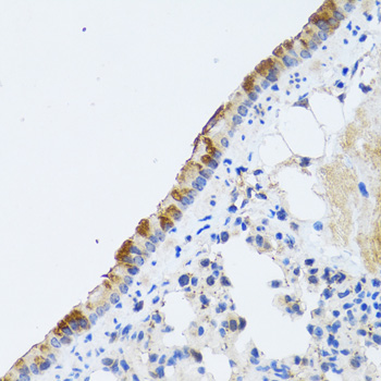
A08054-1 has paraffin-section IHC images from human liver damage, rat lung, and mouse spinal cord (catalog IHC captions). IF/ICC is listed, but no IF image is supplied (catalog applications and image alts).
A08054-1 has IHC images of paraffin-embedded human liver damage, rat lung, and mouse spinal cord (catalog IHC captions). A08054-1 lists IF/ICC and Human, Mouse, and Rat reactivity; no IF image is supplied (catalog applications, reactivity, and image alts).
Which to pick: Choose A08054-1 for paraffin-section IHC: its captions document 1:100 staining after microwave retrieval in 10 mM PBS, pH 7.2; the fixative is unreported (catalog IHC captions). For IF/ICC, A08054-1 lists those applications and a 1:50–1:200 dilution, but provides no IF image; clonality is unreported (catalog applications, IF dilution, image alts, and clone field). For work across human, mouse, and rat, A08054-1 lists reactivity with all three and has a paraffin-section IHC caption for each (catalog reactivity and IHC captions).