This website uses cookies to ensure you get the best experience on our website.


- Table of Contents
Plan paraffin-section IHC for STARD3NL using its widespread cytoplasmic tissue profile (HPA tissue IHC). Compare high-staining cell types with reported unstained cells, and interpret the pattern alongside its late-endosome membrane location (UniProt).

Expected localisation, validated protocols, controls and antibodies — the at-a-glance facts below, then the full design guide.
| Expected localisation | Cytoplasmic tissue staining (HPA tissue IHC) | |
| Staining pattern | Widespread cytoplasmic staining across cell types (HPA tissue IHC) | |
| Antigen retrieval | Citrate pH 6.0 HIER, 95–98 °C, 20 min (rule: cytoplasmic / membrane antigen) | |
| Positive control | Breast+4 more · see all | |
| Negative control | Adipose tissue+1 more · see all |
| Fixation | Keep fixation consistent across sections (standard IHC practice; not target-specific). Selected-image fixative and duration unreported (datasheet A11121); verify before use. | |
| Caveat | Adipocytes and esophageal squamous cells may be unstained (HPA tissue IHC) | |
| Regulation | No expression regulator annotated (UniProt) | |
| Isoform / epitope | Two isoforms; epitope differences are unspecified (UniProt) |
The catalog antibody’s IHC-P protocol (datasheet) is accompanied by a published chromogenic protocol for hepatocellular carcinoma tissue microarrays (PMC13485826).
| Sample | Paraffin-embedded Human colon carcinoma tissue; fixative not specified (datasheet A11121) |
| Fixation | Image fixative and duration unreported (datasheet A11121); verify before use. |
| Sectioning | 4–5 µm sections on charged slides (standard) |
| Deparaffinisation | Xylene, graded ethanol series to water (standard) |
| Antigen retrieval | Heat-induced epitope retrieval in citrate buffer, pH 6.0, 20 min at 95–98 °C (standard rule: cytoplasmic / membrane antigen) |
| Peroxidase block | 3% H2O2, 10 min, room temperature (standard) |
| Blocking | 10% normal serum of the secondary host, 30 min, room temperature (standard) |
| Primary antibody | Rabbit anti-STARD3NL, 1:50-1:200 (datasheet A11121) |
| Primary incubation | Overnight at 4 °C (standard) |
| Detection | HRP-polymer secondary, DAB chromogen 5–10 min (standard) |
| Counterstain | Hematoxylin, blue, dehydrate and mount (standard) |
| Expected result | STARD3NL-positive staining in myoepithelial cells of breast (HPA tissue IHC: High). HPA tissue profile: Ubiquitous cytoplasmic expression. No signal in the no-primary control. |
STARD3NL is a four-pass late-endosome membrane protein that also contacts the endoplasmic reticulum (UniProt O95772 topology and function). In paraffin-section IHC, expect cytoplasmic staining, especially in breast myoepithelial cells, cerebellar Purkinje cells, cortical and hippocampal neurons, epididymal glandular cells, and testicular Leydig cells (HPA: High in each). HPA describes the overall tissue pattern as ubiquitous cytoplasmic expression and rates its IHC reliability Enhanced, with medium consistency between staining and RNA data (HPA tissue IHC).
| Cytoplasmic signal in the listed High cell populations, with tissue structures still discernible. | This fits the reported IHC pattern (HPA: High in those cell populations; ubiquitous cytoplasmic profile). The late-endosome membrane assignment supports an intracellular location (UniProt O95772). Score the specified cells rather than treating every cell in a positive tissue as equally positive (HPA tissue IHC). |
| A predominantly nuclear, extracellular, or continuous cell-surface pattern replaces cytoplasmic staining. | That compartment disagrees with the reported cytoplasmic IHC profile and late-endosome membrane location (HPA tissue IHC; UniProt O95772). Treat it as a possible staining artefact; review morphology, the negative control, and the validated antibody before assigning it to STARD3NL (standard IHC practice). |
| Strong staining appears in adipocytes or esophageal squamous epithelial cells. | Both cell populations are reported as Not detected (HPA tissue IHC). Check whether the signal is specific: cross-reactivity or endogenous detection activity are possibilities, not established causes (standard IHC practice). Compare the negative control and a High cell population on the same run. |
| Diffuse color covers cells or tissue regions without a discernible cellular pattern. | A uniform haze cannot establish the reported cytoplasmic localization (HPA tissue IHC). It may reflect nonspecific background or detection activity (standard IHC practice). Judge it against a reagent negative control and the morphology before scoring positive cells. |
| No staining is visible in a chosen High cell population. | An absent signal conflicts with that population's reported High staining (HPA tissue IHC), but one section does not establish biological absence. Confirm that the relevant cells are present, then examine controls and the staining workflow (standard IHC practice). |
| Cell population selected for scoring | HPA reports High staining in six specified populations, Medium in adrenal and appendix glandular cells, and Not detected in adipocytes and esophageal squamous cells (HPA tissue IHC). Compare like cells. |
| Membrane topology and compartment | Four transmembrane segments place STARD3NL in the late-endosome membrane (UniProt O95772 topology). HPA's tissue-level readout is cytoplasmic; it does not establish puncta resolution in paraffin sections (HPA tissue IHC). |
| Antibody evidence | The listed rabbit polyclonal HPA019204 is IHC Enhanced (HPA antibodies). That supports the reported pattern, while HPA notes only medium consistency between antibody staining and RNA expression data (HPA tissue IHC). |
| Isoforms and epitope | UniProt lists two isoforms and cytoplasmic plus extracellular segments (UniProt O95772). No epitope position is supplied, so these records cannot predict isoform coverage or an optimal permeabilisation choice. |
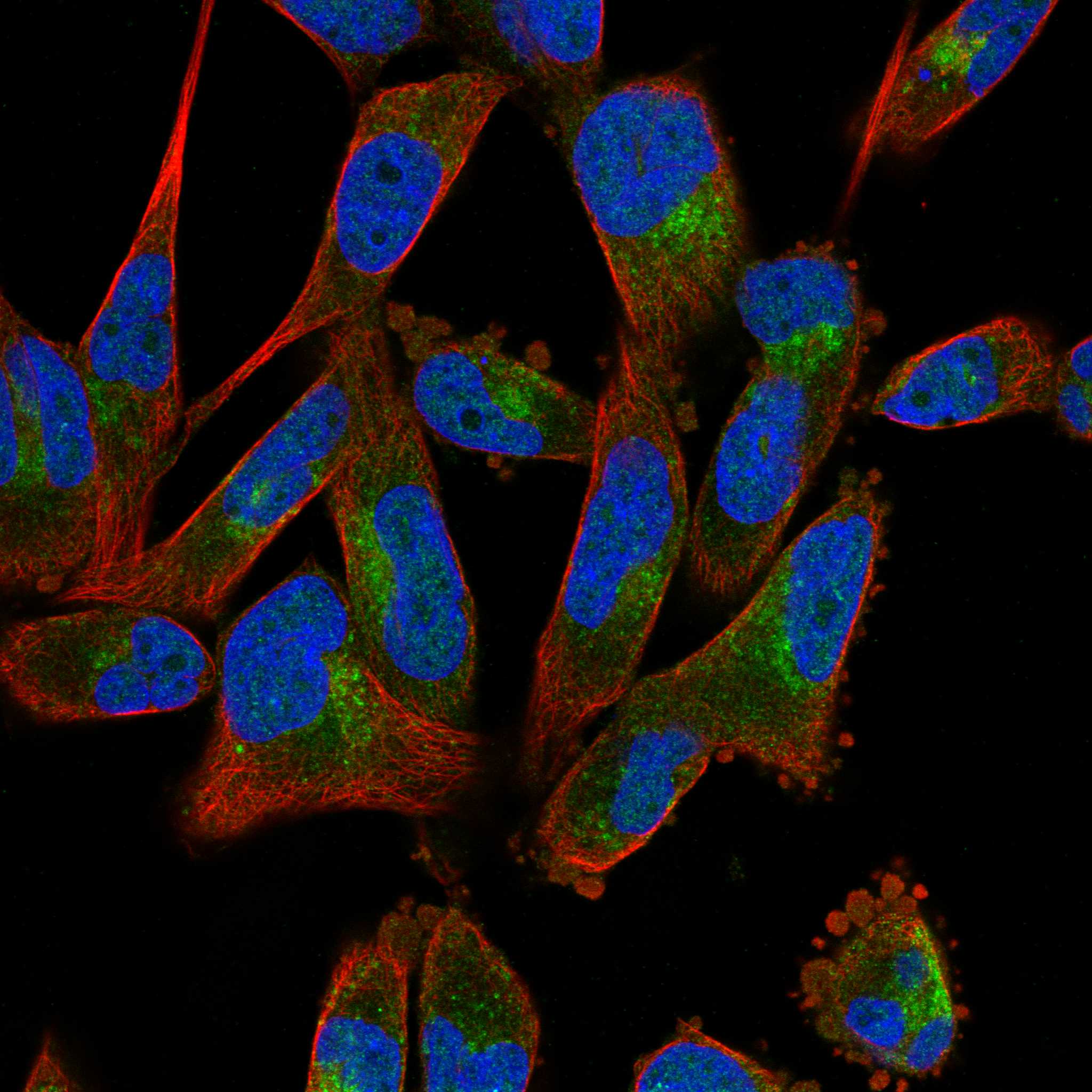
| What should IF/ICC show? | Vesicular staining is the supported ICC-IF location (HPA subcellular); HPA019204 is ICC Supported (HPA antibodies). Use the separate IF/ICC guide for its workflow. |
| Situation | Likely cause | Next action |
|---|---|---|
| High cell populations show no chromogenic signal. | The expected cells may be absent from the section, or the staining run may have failed; the cause is unresolved (HPA: High in the specified populations; standard IHC practice). | Locate the relevant cells morphologically and check a positive control, primary-antibody step, and detection reagents (standard IHC practice). |
| Color appears in reagent negative controls or across many unrelated structures. | Endogenous detection activity or nonspecific reagent background is possible (standard IHC practice); it does not confirm STARD3NL localization. | Use the matching negative control to identify the affected step; review blocking and detection conditions before scoring (standard IHC practice). |
| Signal is mainly nuclear or outlines the entire cell surface. | The pattern conflicts with cytoplasmic IHC and late-endosome membrane localization (HPA tissue IHC; UniProt O95772). | Inspect the negative control and a known High cell population; reassess antibody specificity and staining conditions (standard IHC practice). |
| Adipocytes or esophageal squamous cells stain strongly. | These cells are reported Not detected (HPA tissue IHC); cross-reactivity or detection background is possible (standard IHC practice). | Compare a reagent negative control and the run's positive cells; avoid calling that signal specific without supporting controls (standard IHC practice). |
| A Medium population stains less than a High population. | Different reported levels can explain the contrast: adrenal and appendix glandular cells are Medium, while the listed positive populations are High (HPA tissue IHC). | Score the same cell types and compartments separately; do not require equal intensity across tissues (HPA tissue IHC; standard IHC practice). |
| The expected cytoplasmic pattern remains weak after routine setup checks. | This record supplies no target-specific fixation sensitivity or antigen-retrieval condition; the cause cannot be assigned from HPA or UniProt. | Review the catalog antibody's validated IHC conditions and run controls, then optimize routine retrieval and dilution variables empirically (standard IHC practice). |



Comprehensive Human Protein Atlas IHC scoring per tissue (reliability: Enhanced — Medium consistency between antibody staining and RNA expression data.). Rows are taken directly from the HPA tissue chart — click any row's HPA link to view the source.
| Tissue | Cell type | Level | Evidence | Source |
|---|---|---|---|---|
| Breast | Myoepithelial cells | High | Protein (IHC) | HPA → |
| Cerebellum | Purkinje cells | High | Protein (IHC) | HPA → |
| Cerebral cortex | Neuronal cells | High | Protein (IHC) | HPA → |
| Epididymis | Glandular cells | High | Protein (IHC) | HPA → |
| Hippocampus | Neuronal cells | High | Protein (IHC) | HPA → |
Troubleshoot STARD3NL staining in paraffin-section IHC by checking retrieval, cellular distribution, controls, and scoring before interpreting chromogenic signal.
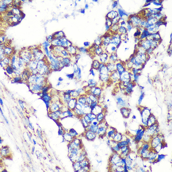
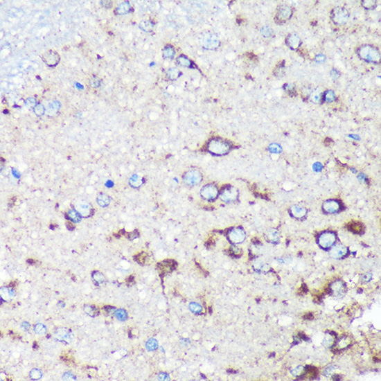
A11121 has real paraffin-section IHC images from human colon carcinoma and rat brain (catalog image captions). Mouse is listed as reactive, but no IF/ICC data are provided (catalog reactivity; applications; image list).
A11121 is the sole card and lists IHC for human, mouse and rat (catalog applications; reactivity). Its images show staining of paraffin-embedded human colon carcinoma and rat brain at 1:100, with microwave retrieval in 10 mM PBS, pH 7.2 (A11121 image captions).
Which to pick: Choose A11121 for paraffin-section IHC: it is a rabbit polyclonal antibody with those two tissue images and a listed IHC dilution of 1:50–1:200 (A11121 image captions; catalog IHC dilution). Its listed reactivity covers human, mouse and rat, although the images document human and rat tissue only (catalog reactivity; A11121 image captions). No listed SKU has IF/ICC data, and the fixative used for the imaged paraffin sections is unreported (catalog applications; IF image list; A11121 image captions).