STAT2 / Signal transducer and activator of transcription 2 · IHC design guide

Design Immunohistochemistry for STAT2

Plan chromogenic STAT2 IHC in paraffin sections with an IHC-validated antibody starting at 2–5 μg/mL (A01360 datasheet). Use cytoplasmic tissue staining as the baseline (HPA tissue IHC), and consider IFN-induced nuclear entry when interpreting the pattern (UniProt).

Evidence assembled Oct 2026 · For research use; verify linked source records and product datasheet before use
Immunohistochemistry protocol sheet for STAT2 (IHC for STAT2): expected localisation Cytoplasmic in most tissues (HPA tissue IHC); nuclear entry after IFN activation (UniProt), antibody M01360, validated IHC image, and IHC protocol steps
Printable STAT2 IHC protocol sheet — expected localisation Cytoplasmic in most tissues (HPA tissue IHC); nuclear entry after IFN activation (UniProt), antibody M01360, controls and protocol steps. Open the full STAT2 IHC guide →

STAT2 Immunohistochemistry Experimental Design Guide

Expected localisation, validated protocols, controls and antibodies — the at-a-glance facts below, then the full design guide.

Must know before staining
Expected localisation Cytoplasmic in most tissues (HPA tissue IHC); nuclear entry after IFN activation (UniProt)
Staining pattern Mostly cytoplasmic; high in tonsil squamous cells (HPA tissue IHC)
Antigen retrieval Tris-EDTA pH 9.0 HIER, 95–98 °C, 20 min (rule: nuclear antigen)
Positive control ⓘ Tonsil+4 more · see all
Negative control ⓘ Adipose tissue+2 more · see all
Important caveats
Reasons your staining may differ from the expected pattern.
Fixation Keep fixation consistent across paraffin sections. (standard IHC practice; not target-specific)
Caveat IFN activation can shift STAT2 into the nucleus (UniProt)
Regulation Type I IFN activates STAT2 (UniProt)
Isoform / epitope 2 isoforms; intracellular target, epitope differences unknown (UniProt)
Section 1

Recommended STAT2 IHC & IF Protocols

The catalog antibody’s IHC-P protocol (datasheet) appears alongside two published chromogenic STAT2 protocols: paraffin sections (PMC6480560) and frozen sections (PMC4206267).

Recommended immunohistochemistry (IHC-P) protocol parameters
SampleParaffin-embedded human lung cancer tissue; fixative not specified (datasheet M01360)
FixationImage fixative and duration unreported (datasheet M01360); verify before use.
Sectioning4–5 µm sections on charged slides (standard)
DeparaffinisationXylene, graded ethanol series to water (standard)
Antigen retrievalHeat-induced epitope retrieval in Tris-EDTA buffer, pH 9.0, 20 min at 95–98 °C (standard rule: nuclear antigen)
Peroxidase block3% H2O2, 10 min, room temperature (standard)
Blocking10% normal serum of the secondary host, 30 min, room temperature (standard)
Primary antibodyRabbit monoclonal (clone HCB-19) anti-STAT2, 1:50 (datasheet M01360)
Primary incubationOvernight at 4 °C (standard)
DetectionHRP-polymer secondary, DAB chromogen 5–10 min (standard)
CounterstainHematoxylin, blue, dehydrate and mount (standard)
Expected resultSTAT2-positive staining in squamous epithelial cells of tonsil (HPA tissue IHC: High). HPA tissue profile: Cytoplasmic expression in most tissues. No signal in the no-primary control.
💡Decision noteStart with Tris-EDTA pH 9.0 retrieval at 95–98 °C for 20 min (page retrieval); citrate pH 6 was used for paraffin sections (PMC6480560).
Section 2

What Is the Expected STAT2 Staining Pattern?

STAT2 is a cytoplasmic and nuclear protein with no transmembrane segment; type I interferon activation drives nuclear entry (UniProt P52630). In tissue IHC, expect mainly cytoplasmic staining across many cell types, including strong staining in tonsillar squamous epithelium and bladder urothelium (HPA tissue IHC). HPA rates its tissue staining Approved, with medium consistency between staining and RNA data (HPA tissue IHC).

What am I looking at on my slide?
Cytoplasmic staining in tonsillar squamous epithelial cells or bladder urothelial cells.This matches two HPA High examples and its broader cytoplasmic profile (HPA tissue IHC). Compare signal within the named cell populations, since a positive tissue need not stain every cell equally (standard IHC practice).
Nuclear staining accompanies cytoplasmic staining.Nuclear entry can follow IFN-alpha/beta activation (UniProt P52630). A nuclear signal alone does not establish activation: confirm compartment boundaries and, if activation is the question, use an independently validated activation readout (standard IHC practice).
Strong membrane-only or extracellular chromogen replaces the expected cell-body pattern.This departs from the mainly cytoplasmic tissue profile (HPA tissue IHC). Check section edges, precipitate and secondary-only background before assigning it to STAT2 (standard IHC practice); IF localization reports do not validate that tissue IHC pattern (HPA subcellular).
Prominent signal appears in adipocytes, cardiomyocytes or cholangiocytes.Those specific populations were Not detected in HPA tissue IHC (HPA tissue IHC). Recheck cell identity and controls; unexpected staining may reflect cross-reactivity or endogenous detection activity (standard IHC practice), but HPA's result is not a universal biological absence.
Diffuse chromogen obscures tissue structure, or known-positive epithelium lacks discernible signal.The pattern cannot be scored confidently (standard IHC practice). Tonsillar squamous cells and bladder urothelial cells are High HPA examples (HPA tissue IHC); review the positive control and detection background before interpreting the sample as negative.
💡Expected STAT2 appearanceA convincing positive is mainly cytoplasmic staining in the named epithelial cells, potentially High in tonsillar squamous epithelium or bladder urothelium (HPA tissue IHC); widespread cell-free deposit or dominant membrane-only chromogen is suspect (standard IHC practice).
How each factor affects the staining
IFN-alpha/beta response (UniProt P52630)STAT2 can move into the nucleus after activation (UniProt P52630). Record nuclear and cytoplasmic staining separately; nuclear localization by itself does not identify the stimulus or prove phospho-STAT2 (standard IHC practice).
Cell population and reference intensity (HPA tissue IHC)HPA reports High tonsillar squamous and bladder urothelial staining, Medium adrenal and appendix glandular staining, and Not detected adipocytes, cardiomyocytes and cholangiocytes (HPA tissue IHC). Use the named cells when comparing fields.
Antibody validation (HPA antibodies)HPA018888 and CAB003858 are each IHC Approved (HPA antibodies). HPA calls the overall tissue result Approved with medium staining–RNA consistency (HPA tissue IHC); approval supports a reference pattern, not guaranteed performance for another antibody or staining run.
Isoforms and phospho-state (UniProt P52630)UniProt lists two STAT2 isoforms and phosphorylation sites including Tyr-690 (UniProt P52630). The supplied records give no catalog-antibody epitope or phospho-specificity, so do not infer which isoform or activation state its chromogen detects.
IF/ICC Q&A: should it match tissue IHC? (HPA subcellular)HPA ICC-IF mainly places STAT2 in cytosol and sperm mid piece, with additional plasma-membrane and principal-piece localization (HPA subcellular). Those observations come from a different assay and sampled cells; assess tissue IHC against its own cytoplasmic profile (HPA tissue IHC).
Why is my staining missing, weak or wrong?
SituationLikely causeNext action
No staining in tonsillar squamous epithelium or bladder urothelium (HPA tissue IHC).A missed positive may reflect a run or detection failure (standard IHC practice); HPA's High designation describes its observations, not a guaranteed result with every antibody (HPA tissue IHC).Confirm the named cells are present, then review the positive control, antigen-retrieval run, primary-antibody application and detection reagents (standard IHC practice). Do not call the test specimen negative until the control works.
Strong nuclear staining conflicts with an expected resting, mainly cytoplasmic profile (UniProt P52630; HPA tissue IHC).Type I interferon activation can drive nuclear entry (UniProt P52630); nonspecific nuclear deposit remains possible (standard IHC practice).Score both compartments, compare the negative reagent control and inspect nuclear boundaries (standard IHC practice). If activation matters, corroborate it independently rather than using total-STAT2 location as proof.
Adipocytes, cardiomyocytes or cholangiocytes stain prominently (HPA tissue IHC).HPA recorded these populations as Not detected (HPA tissue IHC); misidentified cells, cross-reactivity or endogenous detection activity can create an apparent exception (standard IHC practice).Verify cell identity and repeat with appropriate negative detection controls; evaluate a second IHC-validated antibody if available (standard IHC practice). Treat HPA absence as a reference observation, not an absolute exclusion.
Diffuse chromogen masks cytoplasmic detail (standard IHC practice).Background from blocking, reagent concentration, washing or endogenous activity can obscure cell boundaries (standard IHC practice). HPA's cytoplasmic profile cannot be assessed in an unreadable field (HPA tissue IHC).Inspect a no-primary control, check the detection chemistry and optimize blocking, washes and reagent concentration under the assay's standard procedure (standard IHC practice). Reassess localization only after the cell bodies are clear.
Only weak signal appears in a cell population expected to be High (HPA tissue IHC).HPA's intensity is a reference observation, while assay conditions and antibody choice affect visible chromogen (standard IHC practice). HPA reports medium staining–RNA consistency, so the reference has limits (HPA tissue IHC).Compare the same named cell type in a working positive control, then review retrieval, primary incubation and detection steps (standard IHC practice). Report observed intensity rather than forcing the HPA category onto the slide.
Membrane or mid-piece IF signal is used to justify membrane-dominant tissue IHC (HPA subcellular).HPA's ICC-IF location summary includes plasma membrane and sperm mid piece, whereas its tissue IHC profile is cytoplasmic in most tissues (HPA subcellular; HPA tissue IHC).Interpret each assay against its own HPA reference and check tissue IHC background controls (HPA subcellular; HPA tissue IHC; standard IHC practice). Keep the IF/ICC result as contextual evidence, not an IHC scoring rule.

Sample controls for STAT2 IHC & IF

🧪Run tonsil first: its squamous epithelial cells should stain for STAT2 (HPA: High in tonsil squamous epithelial cells). Use adipose tissue adipocytes as the negative comparison (HPA: Not detected in adipocytes); on the tonsil slide, treat unstained neighboring cells, if present, as a background reference without assuming a particular cell type is STAT2-negative (HPA: tonsil row identifies squamous epithelial cells only).
Positive control tissue: Tonsil (Squamous epithelial cells, HPA High)
Negative control tissue: Adipose tissue (HPA Not detected)
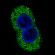
ICC-IF cell lines (HPA subcellular resource): HPA ICC-IF images show STAT2 in A-431, U-251MG, U2OS, hTERT-RPE1 (serum starved), Sperm, with annotated localisation: Cytosol (supported), Mid piece (approved) (HPA subcellular).
Technical controls: Include a no-primary, secondary-only control and an irrelevant isotype control matched to the primary antibody’s host species and clonality; use STAT2 knockout material as a biological specificity control (standard IHC practice). For chromogenic detection in tonsil, quench endogenous peroxidase if using HRP and block endogenous biotin if using biotin-based detection (standard IHC practice).
⚠️Feasibility: A target-specific fixation window and antigen-retrieval dependency are unreported in the supplied evidence; optimize retrieval empirically (supplied evidence; standard IHC practice). The selected M01360 paraffin-section lung-cancer caption does not report a fixative, so it does not establish fixation (M01360 caption). ICC-IF images show that IF has been performed for STAT2, but the supplied evidence does not establish whether IF or frozen sections are easier than paraffin IHC; check tonsil sections for endogenous-peroxidase background when using HRP (HPA: subcellular ICC-IF images; standard IHC practice). The selected M01360 tissue-IHC caption documents paraffin sections, but does not specify the fixative (selected-SKU IHC image M01360).

HPA tissue IHC evidence for STAT2

Comprehensive Human Protein Atlas IHC scoring per tissue (reliability: Approved — Medium consistency between antibody staining and RNA expression data.). Rows are taken directly from the HPA tissue chart — click any row's HPA link to view the source.

Positive expression · recommended positive controls

TissueCell typeLevelEvidenceSource
Tonsil Squamous epithelial cells High Protein (IHC) HPA →
Urinary bladder Urothelial cells High Protein (IHC) HPA →
Adrenal gland Glandular cells Medium Protein (IHC) HPA →
Appendix Glandular cells Medium Protein (IHC) HPA →
Bone marrow Hematopoietic cells Medium Protein (IHC) HPA →

Undetected expression · recommended negative controls

TissueCell typeLevelEvidenceSource
Adipose tissue Adipocytes Not detected Protein (IHC) HPA →
Heart muscle Cardiomyocytes Not detected Protein (IHC) HPA →
Liver Cholangiocytes Not detected Protein (IHC) HPA →
Section 3

Advanced STAT2 IHC Tips

Troubleshoot STAT2 staining in paraffin sections by checking retrieval, cellular distribution and controls before interpreting signal (UniProt P52630; HPA tissue IHC).

What retrieval should I try first when STAT2 staining is weak in paraffin sections?
Use Tris-EDTA at pH 9.0 for heat-induced retrieval at 95–98 °C for 20 min (page retrieval specification). Allow sections to cool in retrieval buffer, then compare a positive control and the study section using the same staining run (standard IHC practice). Tonsil squamous epithelial cells offer a reported high-staining comparator, although the HPA tissue result has medium RNA concordance (HPA tissue IHC). If signal remains weak, test an alternative retrieval buffer on adjacent sections while holding antibody concentration and detection constant (standard IHC practice). Score both staining intensity and tissue damage, since stronger color alone does not establish improved STAT2 detection (standard IHC practice).
How should I troubleshoot weak STAT2 staining when fixation details are incomplete?
STAT2-specific fixation sensitivity is unknown; the M01360 lung cancer caption identifies paraffin embedding but does not report a fixative (caption M01360). Record the available fixation history and compare sections with similar processing before attributing a staining difference to STAT2 biology (standard IHC practice). For newly prepared material, use a documented fixation schedule and keep specimen thickness and processing consistent across comparison groups (standard IHC practice). Apply the specified Tris-EDTA pH 9.0, 95–98 °C, 20 min retrieval consistently when testing those groups (page retrieval specification). If staining varies, check morphology and a positive tissue control before changing fixation conditions (standard IHC practice; HPA tissue IHC).
Should STAT2 staining be cytoplasmic or nuclear in an IHC section?
Expect substantial cytoplasmic staining in many tissues, consistent with the reported tissue profile and supported cytosolic localisation (HPA tissue IHC; HPA subcellular). Nuclear STAT2 is biologically plausible after type I interferon activation, when phosphorylated STAT2 joins STAT1 and IRF9 in a transcriptional complex (UniProt P52630). Score nuclear and cytoplasmic compartments separately rather than treating any brown signal as one positive category (standard IHC practice). Compare similarly processed sections and examine cell borders under a counterstain before assigning faint signal to nuclei (standard IHC practice). Nuclear staining alone does not prove activation without a relevant comparison or orthogonal activation readout (UniProt P52630; standard IHC practice).
Could an isoform or modified epitope explain discordant STAT2 staining?
STAT2 has 2 annotated isoforms, so check whether the antibody's documented immunogen spans a sequence shared by both before comparing specimens (UniProt P52630; standard IHC practice). Its SH2 domain spans residues 572–667, and annotated modifications include Tyr 690 phosphorylation and Ser 753 phosphorylation (UniProt P52630). The supplied caption gives no epitope sequence, so these annotations cannot establish which isoform or modified state M01360 detects (caption M01360; UniProt P52630). Review the available antibody documentation, then test a second epitope or orthogonal readout if staining remains discordant (standard IHC practice). Interpret any nuclear shift alongside the expected type I interferon response, not as proof of phospho-specific recognition (UniProt P52630).
How can I check STAT2 localisation by IF alongside the chromogenic IHC result?
Use the IF/ICC guide's separately validated conditions; the supplied M01360 evidence is a paraffin-section IHC caption and does not establish IF performance (caption M01360). Multiplex STAT2 with a marker identifying the cell population under study, and include a nuclear counterstain to distinguish cytosolic from nuclear signal (standard IF practice; UniProt P52630). Choose spectrally separated fluorophores after checking tissue autofluorescence in unstained sections, reserving the cleaner channel for weaker signal (standard IF practice). STAT2 has no transmembrane segment, so assess cytoplasmic or nuclear access with a mild permeabilisation trial, such as 0.1% Triton X-100, rather than assuming a surface epitope (UniProt P52630 topology; standard IF practice). Compare single-color controls before interpreting overlap as colocalisation (standard IF practice).
What should I change when STAT2 IHC has diffuse brown background?
Run a no-primary control to identify detection-system or tissue background, then inspect whether the signal follows cell boundaries and plausible compartments (standard IHC practice; UniProt P52630). For peroxidase and DAB detection, an endogenous-peroxidase block such as 3% hydrogen peroxide for 10 min is a general workflow step, not STAT2-specific evidence (standard IHC practice). Check blocking, wash stringency and antibody concentration one variable at a time, using the same positive control in each run (standard IHC practice). Diffuse cytoplasmic color can be real because HPA reports cytoplasmic expression in most tissues, but uniform stromal haze warrants scrutiny (HPA tissue IHC; standard IHC practice). Reject interpretations driven by folds, drying or damaged edges (standard IHC practice).
How should I score STAT2 when cells differ in intensity and localisation? ⚠ ANSWER MARKED FOR VERIFICATION
Define the cell population and compartment before scoring, because STAT2 can be cytoplasmic and can enter nuclei after type I interferon activation (HPA tissue IHC; UniProt P52630). Report the percentage of positive cells and an H-score from intensity categories 0–3, giving a possible range of 0–300 (standard IHC scoring practice). Score nuclear and cytoplasmic staining separately, using fixed thresholds and matched exposure or viewing conditions across sections (standard IHC practice). Normalise positive counts to evaluable cells of the same type, or report density per mm² of viable, defined tissue area (standard IHC practice). Exclude necrosis and section edges consistently, and record the number of regions assessed (standard IHC practice).
When is an apparent STAT2-positive IHC result likely to be artefactual?
Look for cytoplasmic staining in intact cells as a plausible baseline, while interpreting nuclear signal in the context of type I interferon activation (HPA tissue IHC; UniProt P52630). Check whether the stained cell type fits the comparison tissue: HPA reports high staining in tonsil squamous epithelium and no detection in heart cardiomyocytes (HPA tissue IHC). Brown precipitate concentrated at cut edges, folds or necrotic areas is suspect, especially if it lacks a coherent cellular pattern (standard IHC practice). A no-primary control helps reveal endogenous enzyme activity or detection background in a peroxidase workflow (standard IHC practice). HPA rates its tissue data Approved with medium staining-to-RNA consistency, so corroborate consequential claims independently (HPA tissue IHC).
Boster reagents

Best STAT2 / Signal transducer and activator of transcription 2 IHC Antibodies

M01360 and A01360 have IHC/IF catalog support; M01360 lists human, mouse and rat reactivity, while A01360 lists human reactivity (catalog: applications, reactivity).

Real IHC data Immunohistochemical analysis of paraffin-embedded human lung cancer, using STAT2 Antibody.
Anti-STAT2 Rabbit Monoclonal Antibody
Cat # M01360
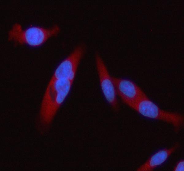
Real IF data IF analysis of STAT2 using anti-STAT2 antibody (A01360). STAT2 was detected in an immunocytochemical section of PC-3 cells. Enzyme antigen retrieval was performed using IHC enzyme antigen retrieval reagent (AR0022) for 15 mins. The cells were blocked with 10% goat serum. And then incubated with 5 μg/mL rabbit anti-STAT2 Antibody (A01360) overnight at 4°C. Cy3 Conjugated Goat Anti-Rabbit IgG (BA1032) was used as secondary antibody at 1:500 dilution and incubated for 30 minutes at 37°C. The section was counterstained with DAPI. Visualize using a fluorescence microscope and filter sets appropriate for the label used.
Anti-STAT2 Antibody ®
Cat # A01360

M01360 has an IHC image of paraffin-embedded human lung cancer and lists IHC/IF applications (M01360 image caption; catalog: applications). A01360 has an IF image in PC-3 cells and lists IF/ICC and IHC applications for human samples (A01360 image caption; catalog: applications, reactivity).

Which to pick: For tissue IHC, choose M01360: its own image shows paraffin-embedded human lung cancer, and its listed IHC dilution is 1:50; the fixative is unreported (M01360 image caption; catalog: IHC dilution). For human IF/ICC, A01360 has a PC-3 cell IF image at 5 μg/ml, while its IHC listing has no accompanying IHC image (A01360 image caption; catalog: IF dilution, applications, IHC image alts). For mouse or rat samples, M01360 is the listed option because its reactivity includes both species; it is a monoclonal antibody, and its IHC image depicts a human sample (catalog: M01360 reactivity, clone; M01360 image caption).

Each figure is that product's own IHC / IF validation image from its datasheet.

References

  1. UniProt Consortium. UniProt entry P52630 (STAT2_HUMAN, Signal transducer and activator of transcription 2).
  2. Human Protein Atlas. STAT2 tissue IHC expression (reliability: Approved).
  3. Human Protein Atlas. STAT2 subcellular location (ICC-IF): Mainly localized to the cytosol and mid piece. In addition localized to the plasma membrane and principal piece..
  4. Human Protein Atlas. STAT2 antibody validation summary (2 antibodies).
  5. Interferon-Stimulated Genes-Mediators of the Innate Immune Response during Canine Distemper Virus Infection. International journal of molecular sciences 2019 — PMC6480560.
  6. IL-36 promotes anti-viral immunity by boosting sensitivity to IFN-α/β in IRF1 dependent and independent manners. Nature communications 2019 — PMC6795910.
  7. Decreased signal transducers and activators of transcription (STAT) protein expression in lymphatic organs during EAE development in mice. Immunology innovation 2013 — PMC4206267.
  8. The role of Tec kinase signaling pathways in the development of Mallory Denk Bodies in balloon cells in alcoholic hepatitis. Experimental and molecular pathology 2017 — PMC5680109.
  9. PubMed PMID:1502204 — UniProt-cited evidence.
  10. PubMed PMID:7885841 — UniProt-cited evidence.
  11. PubMed PMID:14702039 — UniProt-cited evidence.