This website uses cookies to ensure you get the best experience on our website.


- Table of Contents
Plan STAT5B paraffin IHC around cytoplasmic and nuclear tissue staining (HPA tissue IHC). Compare high staining in testis Leydig cells with undetected staining in adipocytes, and interpret the HPA pattern cautiously because its antibody may detect more than one gene (HPA tissue IHC).

Expected localisation, validated protocols, controls and antibodies — the at-a-glance facts below, then the full design guide.
| Expected localisation | Cytoplasmic and nuclear tissue staining (HPA tissue IHC) | |
| Staining pattern | General cytoplasmic and nuclear staining (HPA tissue IHC) | |
| Antigen retrieval | Tris-EDTA pH 9.0 HIER, 95–98 °C, 20 min (rule: nuclear antigen) | |
| Positive control | Testis+4 more · see all | |
| Negative control | Adipose tissue+4 more · see all |
| Fixation | Matched tissue-IHC evidence does not establish the fixation claim. Validate the specimen-specific method before use. (selected-SKU IHC image M00681) | |
| Caveat | HPA antibody may detect more than one gene (HPA tissue IHC) | |
| Regulation | Activation shifts STAT5B to nuclei (UniProt) | |
| Isoform / epitope | No isoforms; one unprocessed, intracellular chain (UniProt) |
The catalog antibody's IHC-P protocol is followed by published STAT5B chromogenic IHC methods for pancreatic cancer, lymphoma, breast cancer, and placental tissue (PMC4869940; PMC7012494; PMC4053108; PMC9131539).
| Sample | Paraffin-embedded human breast tissue; fixative not specified (datasheet M00681) |
| Fixation | Image fixative and duration unreported (datasheet M00681); verify before use. |
| Sectioning | 4–5 µm sections on charged slides (standard) |
| Deparaffinisation | Xylene, graded ethanol series to water (standard) |
| Antigen retrieval | Heat-induced epitope retrieval in Tris-EDTA buffer, pH 9.0, 20 min at 95–98 °C (standard rule: nuclear antigen) |
| Peroxidase block | 3% H2O2, 10 min, room temperature (standard) |
| Blocking | 10% normal serum of the secondary host, 30 min, room temperature (standard) |
| Primary antibody | Rabbit monoclonal (clone HDF-19) anti-STAT5B, 1:50 (datasheet M00681) |
| Primary incubation | Overnight at 4 °C (standard) |
| Detection | HRP-polymer secondary, DAB chromogen 5–10 min (standard) |
| Counterstain | Hematoxylin, blue, dehydrate and mount (standard) |
| Expected result | STAT5B-positive staining in leydig cells of testis (HPA tissue IHC: High). HPA tissue profile: General cytoplasmic and nuclear expression. No signal in the no-primary control. |
STAT5B is found in the cytoplasm and nucleus, with nuclear translocation after phosphorylation (UniProt P51692 subcellular location). In paraffin-section IHC, expect staining in Leydig cells and several glandular, tubular, and other listed cell populations; staining is not uniform across tissues (HPA tissue IHC). HPA calls its tissue profile Approved but cautions that it may detect protein from more than one gene and awaits external verification (HPA tissue IHC). STAT5B has no transmembrane segment (UniProt P51692 topology).
| Leydig cells stain strongly; hepatocytes or kidney tubule cells stain more moderately. | This follows the reported High testis and Medium liver and kidney patterns (HPA tissue IHC). Compare cell types within the section; those levels describe HPA observations, not a guaranteed intensity for every specimen or IHC run (HPA tissue IHC; general IHC practice). |
| Staining is cytoplasmic, nuclear, or both in an expected cell population. | Both compartments are compatible with STAT5B; phosphorylation can drive nuclear translocation (UniProt P51692 subcellular location). Nuclear staining alone does not establish pathway activation without an activation-specific assay (UniProt P51692 modified residues; general IHC interpretation). |
| A crisp membrane rim dominates the staining. | Membrane localisation is inconsistent with the supplied cytoplasm/nucleus annotation and absence of a transmembrane segment (UniProt P51692 subcellular location and topology). Review morphology, antibody specificity, and detection controls before assigning that rim to STAT5B (general IHC practice). |
| Adipocytes or skeletal-muscle myocytes stain as strongly as the expected positive cells. | HPA reports STAT5B as Not detected in those cell types (HPA tissue IHC). Unexpected staining warrants checks for cross-reactivity or endogenous detection activity; HPA itself cautions that its tissue profile may detect protein from more than one gene (HPA tissue IHC; general IHC practice). |
| Color covers most of the section, including areas without expected positive cells. | Diffuse deposit obscures the cell-level pattern and can reflect nonspecific binding or detection background (general IHC practice). It cannot be scored as STAT5B solely because HPA reports general cytoplasmic and nuclear expression (HPA tissue IHC; general IHC interpretation). |
| Tissue and cell selection | Testis Leydig cells offer a reported High reference; liver hepatocytes and kidney tubule cells are Medium references, while adipocytes are Not detected (HPA tissue IHC). Score the specified cells, since a tissue name alone does not identify the stained population (general IHC practice). |
| Activation and compartment | STAT5B is annotated in cytoplasm and nucleus and moves into the nucleus in response to phosphorylation (UniProt P51692 subcellular location). A change in nuclear fraction may merit follow-up, but total-STAT5B IHC does not identify which modified residue is present (UniProt P51692 modified residues; general IHC interpretation). |
| Specificity of the tissue reference | HPA assigns Approved reliability while warning that the profile may target protein from more than one gene and remains pending external verification (HPA tissue IHC). Treat a match to its cell pattern as supporting evidence, not proof that every colored cell contains STAT5B (HPA tissue IHC; general IHC interpretation). |
| IF/ICC: Where should the signal appear? | The HPA ICC/IF summary identifies cytosol as the approved main location (HPA subcellular ICC/IF). UniProt also allows nuclear localisation following phosphorylation, so assess the compartment in the experiment's biological context (UniProt P51692 subcellular location). |
| Situation | Likely cause | Next action |
|---|---|---|
| No stain appears in Leydig cells of an otherwise intact testis section. | HPA reports High Leydig-cell staining, making this a useful positive-reference failure; weak detection, retrieval, or staining workflow may be involved (HPA tissue IHC; general IHC practice). | Check section integrity, primary-antibody application, detection reagents, and chromogen development; then review retrieval against the antibody's IHC instructions (general IHC practice). Do not infer a STAT5B-specific fixation effect from this result. |
| A membrane-like outline is stronger than cytoplasmic or nuclear staining. | A dominant membrane pattern conflicts with the annotated compartments and no-transmembrane topology (UniProt P51692 subcellular location and topology). | Inspect adjacent morphology and a primary-omission control, then compare expected positive and Not detected cell types on the same run (general IHC practice; HPA tissue IHC). |
| Adipocytes show strong brown deposit. | Adipocytes are reported Not detected; cross-reactivity or endogenous detection activity may explain the discordance (HPA tissue IHC; general IHC practice). | Check a primary-omission control and the relevant endogenous-activity blocking step; compare the pattern with Leydig cells or another reported positive population (general IHC practice; HPA tissue IHC). |
| All cells have hazy color that hides their boundaries. | The pattern is consistent with detection background or nonspecific binding, rather than a scorable cell-specific result (general IHC practice). | Review blocking, washing, antibody concentration, and detection-control staining; rescore only when cell boundaries and compartments are visible (general IHC practice). |
| Nuclear staining appears, but the study expects mostly cytoplasmic staining. | Nuclear STAT5B can occur after phosphorylation; total staining alone does not identify the activation state (UniProt P51692 subcellular location and modified residues; general IHC interpretation). | Confirm nuclear location against the counterstain, compare matched biological conditions, and use an activation-specific assay if phosphorylation is the question (general IHC practice; UniProt P51692 subcellular location). |
| The HPA-like tissue pattern is present, but target identity remains uncertain. | HPA marks the tissue profile Approved yet cautions that it may detect protein from more than one gene and awaits external verification (HPA tissue IHC). | Interpret the pattern alongside independent specificity evidence before making a STAT5B-specific claim; an HPA pattern match alone does not resolve the stated caveat (HPA tissue IHC; general IHC interpretation). |


Comprehensive Human Protein Atlas IHC scoring per tissue (reliability: Approved — Caution, targets protein from more than one gene. Pending external verification.). Rows are taken directly from the HPA tissue chart — click any row's HPA link to view the source.
| Tissue | Cell type | Level | Evidence | Source |
|---|---|---|---|---|
| Testis | Leydig cells | High | Protein (IHC) | HPA → |
| Adrenal gland | Glandular cells | Medium | Protein (IHC) | HPA → |
| Kidney | Cells in tubules | Medium | Protein (IHC) | HPA → |
| Liver | Hepatocytes | Medium | Protein (IHC) | HPA → |
| Lung | Alveolar cells | Medium | Protein (IHC) | HPA → |
| Tissue | Cell type | Level | Evidence | Source |
|---|---|---|---|---|
| Adipose tissue | Adipocytes | Not detected | Protein (IHC) | HPA → |
| Caudate | Glial cells | Not detected | Protein (IHC) | HPA → |
| Hippocampus | Glial cells | Not detected | Protein (IHC) | HPA → |
| Ovary | Follicle cells | Not detected | Protein (IHC) | HPA → |
| Pancreas | Exocrine glandular cells | Not detected | Protein (IHC) | HPA → |
Troubleshoot STAT5B staining in paraffin-section chromogenic IHC by checking retrieval, compartment, cellular context and controls before interpreting signal.
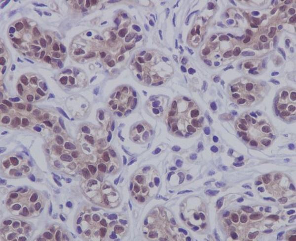
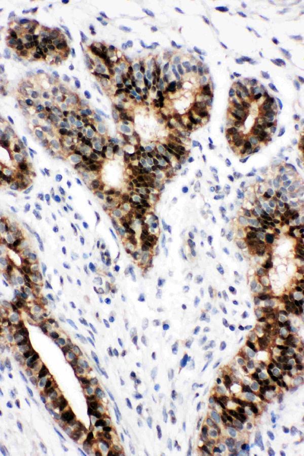
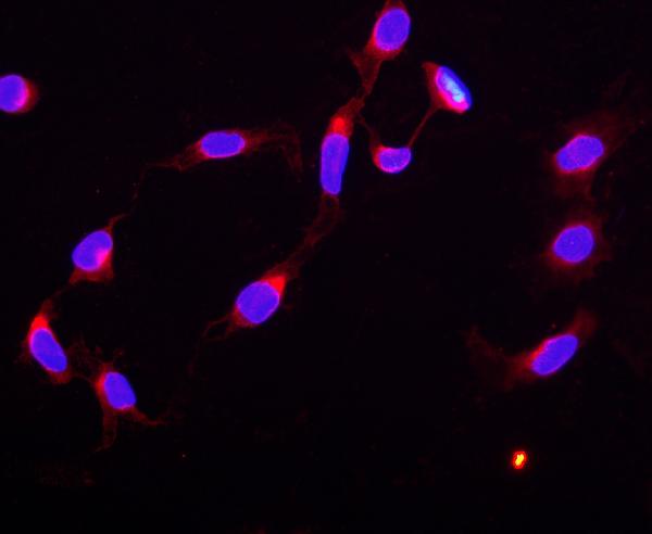
Anti-STAT5B antibodies have IHC images from human breast and human mammary cancer tissue (image captions), and an IF/ICC image from U20S cells (image caption).
M00681 shows IHC on paraffin-embedded human breast (image caption); PA1841 shows IHC(P) on human mammary cancer tissue (image caption). PB9512 shows IF/ICC in U20S cells (image caption) and lists Human, Mouse and Rat reactivity (catalog reactivity).
Which to pick: For tissue IHC, choose M00681 for paraffin-embedded human breast (image caption) or PA1841 for IHC(P) of human mammary cancer tissue (image caption); fixation is unreported in both captions. For IF/ICC, PB9512 has an IF image from U20S cells (image caption). For work spanning Human, Mouse and Rat, M00681 lists all three species and both IHC and IF/ICC applications, and is rabbit monoclonal clone HDF-19 (catalog reactivity, applications and clone).